Complex Coronary Intervention Using Coronary Orbital Atherectomy (Diamondback 360˚) via a Transradial Approach
Case presentation
A 68-year-old male patient with a past medical history of hypertension, dyslipidemia, and coronary artery disease status post coronary artery bypass surgery (CABG) in 2002 was referred for cardiac catheterization. The patient complained of angina on exertion with an associated large area of inducible inferolateral ischemia on stress Myoview imaging. A diagnostic cardiac catheterization was performed via a femoral approach, secondary to a history of harvesting the radial artery for CABG and unknown graft anatomy. Diagnostic angiography revealed a subtotal occlusion of the distal left main artery and patent left internal mammary artery (LIMA) to left anterior descending coronary artery (LAD) graft. The left circumflex (LCX) artery had a proximal 100% occlusion. The right coronary artery (RCA) was heavily calcified, with multiple, sequential 95 to 99% severe stenoses (Figure 1). The radial graft and saphenous vein grafts appeared to be occluded on aortography.
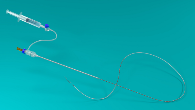
The patient was brought back for an elective intervention of the RCA secondary to his symptoms. After obtaining a 6 French (Fr) access in the right radial artery with a hydrophilic Glidesheath (Terumo), an intra-arterial cocktail was injected. Over an .035” J wire, the 6 Fr hydrophilic sheath was exchanged for a 7 Fr non-hydrophilic sheath in the right radial artery. Right common femoral vein access was used for transvenous pacemaker placement. A 7 Fr Amplatz Left (AL)1 guide was selectively engaged in the RCA. An over-the-wire (OTW) 1.25 mm x 10 mm balloon was advanced over a 300 cm .014” Kinetix wire (Boston Scientific) past the first lesion. Due to a long area of very heavy calcification and severe angulation within the diseased segment (Figure 2), several different wires, including the Asahi Fielder XT (Abbott Vascular), Confianza (Abbott Vascular), and different tip shapes, failed to cross the lesions. Ultimately, a Fielder FC wire (Abbott Vascular) was able to pass through the last lesion. Unfortunately, the 1.25 mm OTW balloon failed to cross the last two lesions, despite solid support from the AL1 guide catheter. A Corsair microcatheter (Asahi Intecc) passed successfully through the lesion and the Fielder FC wire was exchanged for a 300 cm Viper wire (.012” core guide wire with a .014” spring tip) [Cardiovascular Systems, Inc. (CSI)] (Figure 3). An orbital
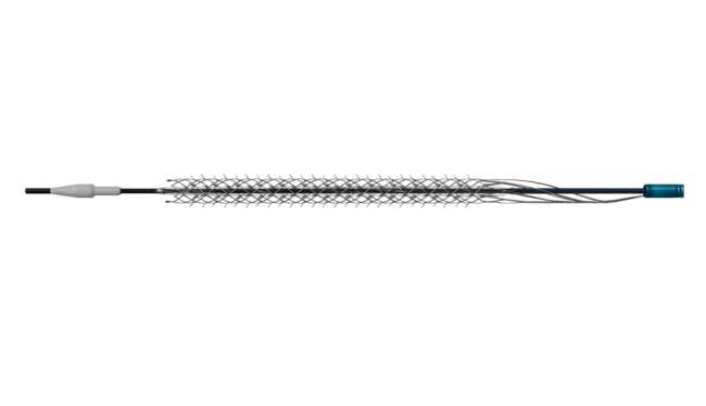
 atherectomy system (Diamondback 360˚ coronary classic crown, CSI) was advanced over the Viper wire (Figure 4). Orbital atherectomy was performed in the entire proximal and mid severely calcified RCA with multiple 25- to 30-second passes. The initial six passes were performed at low revolution speed (80K rpm). The final three passes were at high revolution speed (120K rpm). Subsequent Promus drug-eluting stents (3.5 x 38 mm and 3.5 x 18 mm) (Boston Scientific) were deployed over the Viper wire in overlapping fashion, covering the entire diseased segment. An excellent angiographic result was achieved with TIMI-3 flow (Figure 5).
 The patient was discharged home after 12 hours on dual antiplatelet agents, along with other secondary cardiovascular preventive care measures.
Discussion
With increased experience and reduced equipment size, use of the transradial approach for coronary intervention has grown over the past 5 years. The advantages of the radial approach include fewer access-related bleeding complications, improved patient comfort, early ambulation, shorter hospital stays, and reduced procedure cost.1,2 These advantages translate to a mortality benefit in higher risk interventional procedures, although historically, arguments have been made against using radial approach for complex coronary interventions.1,2 This case illustrates where coronary orbital atherectomy can be easily utilized via a radial approach to modify the plaque and allow for successful stenting of an extremely calcified long lesion.
Historically, we know that calcified lesions are difficult to treat and lead to increased complications. In the pooled analysis of the ACUITY and HORIZONS trial, target lesion coronary calcium was associated with increased rates of major bleeding.3 The ORBIT II trial demonstrated the primary safety and efficacy of Diamondback orbital atherectomy in severely calcified coronary artery disease, with an 89% success rate and 89% freedom from 30-day major adverse cardiac events (MACE).4 Orbital atherectomy may increase treatment options for this difficult-to-treat patient population.
The following tips may be helpful for utilizing the transradial approach for an orbital atherectomy procedure:
- Very good coaxial guide support is crucial for procedural success. Consider an extra back-up guide for the left coronary system and MAC 3.0 (Medtronic) or Amplatz Left curve for the right coronary artery.
- Ultrasound measurement of the radial artery with sublingual nitroglycerin can be helpful to determine the sheath and guide size that can be used without increasing the incidence of spasm or asymptomatic radial artery occlusion. A 6 Fr system can be used when the radial artery size is < 2.2 mm and a 7 Fr system when the diameter is > 2.2 mm. Orbital atherectomy can be used through a 6 or 7 Fr guide catheter.
- Another option is the use of a sheathless guide system (which has limited availability in the United States). Since the outer diameter of a 5 Fr sheath (2.28 mm) parallels that of a 7 Fr guide (2.31 mm), the sheathless approach may reduce the incidence of associated
 spasm.
 - In the absence of a sheathless guide catheter, it is possible to use a 7 Fr guide catheter in many patients (Figure 6) by inserting only one cm of sheath into the radial artery to allow for atraumatic entry of the 7 Fr guide into the radial artery.
- Eligible anatomy for orbital atherectomy includes severe calcified disease to facilitate balloon angioplasty and stenting of the coronary artery, and vessel diameter of 2-4 mm.
- Contraindication for orbital atherectomy includes failure to cross the lesion with the wire, bypass graft of previously stented segment, single remaining conduit, dissection at the treatment site, thrombus at the treatment site, and excessive tortuosity.
- Consider placing a transvenous pacemaker for the RCA or dominant LCX, or in high-risk patients.
- Each run (spin time) should be limited to 30 seconds. Longer lesions require additional runs, not longer runs. The rest time between each run should be equal to or longer than the run time.
- Avoid use of the Viper wire in small branches and keep the radio-opaque portion of the wire at least 0.5 cm distal to the most distal target treatment segment.
- Avoid closing the hemostatic valve on the Y connector too tightly.
- Keep the crown tip at least 5 mm proximal to the lesion when starting and finishing a run and avoid orbital atherectomy with ostial lesions.
- Use the “pecking” motion for the initial treatment of the lesion. Advance the knob a maximum of 1cm/second and do not end the run in the middle of a lesion. Never force the crown through the lesion. Increase the rotation speed after the initial low-speed run passes through the lesion without audible, high-pitched resistance from the device.

Disclosure: Dr. Trayer and Dr. Sanghvi report no conflicts of interest regarding the content herein.
The authors can be contacted via Kintur Sanghvi, MD, at sanghvik@deborah.org.
References
- Caputo RP, Tremmel JA, Rao S, Gilchrist IC, Pyne C, Pancholy S, Frasier D, Gulati R, Skelding K, Bertrand O, Patel T. Transradial arterial access for coronary and peripheral procedures: Executive summary by the transradial committee of the SCAI. Catheter Cardiovasc Interv. 2011 Nov 15;78(6):823-839. doi: 10.1002/ccd.23052.
- Hamon M, Pristipino C, Di Mario C, Nolan J, Ludwig J, Tubaro M, Sabate M, Mauri-Ferré J, Huber K, Niemelä K, Haude M, Wijns W, Dudek D, Fajadet J, Kiemeneij F; European Association of Percutaneous Cardiovascular Interventions; Working Group on Acute Cardiac Care of the European Society of Cardiology; Working Group on Thrombosis on the European Society of Cardiology. Consensus document on the radial approach in percutaneous cardiovascular interventions: position paper by the European Association of Percutaneous Cardiovascular Interventions and Working Groups on Acute Cardiac Care** and Thrombosis of the European Society of Cardiology. EuroIntervention. 2013 Mar;8(11): 1242-1251. doi: 10.4244/EIJV8I11A192.
- Genereux P, Maehara A, Kirtane A, et al. Association between coronary calcification and bleeding after PCI in ACS: pooled analysis from HORIZONS-AMI and ACUITY trials. J Am Coll Cardiol. 2013;61(10_S):. doi:10.1016/S0735-1097(13)60144-8.
- Hambers JJ, Feldman RL, Himmelstein SI, et al. Pivotal trial to evaluate the safety and efficacy of the orbital atherectomy system in treating de novo, severely calcified coronary lesions (ORBIT II). J Am Coll Cardiol Intv. Provisionally accepted.