VEITHSymposium: 1. The European History with BTK Lesions; 2. Decreasing Access Site Complications with Routine Ultrasound Guidance
1. The European history with BTK lesions.
How have Europeans contributed to treatment of below-the-knee lesions?
As a group, the Europeans have pioneered much of the critical limb ischemia (CLI) work now being done in multiple countries. Many of the tools, ideas and approaches that we have now were first attempted and then further developed in Europe before transferring to other areas. The primary countries where CLI investigations are occurring include Germany, Belgium, and Italy. CLI is being treated everywhere, certainly, but there are some specific developments from Europe. The long, low profile, below-the-knee (BTK) balloon, which has become the standard of care, was developed in Italy. The concept of crossing long BTK lesions was being done routinely around Europe well before it was being done in other places. Retrograde access using the pedal or tibial arteries for a bi-directional approach was popularized by the group in Leipzig, Germany. Angioplasty of the foot arteries was developed in Italy, and now many programs around the world practice this.
New technologies, like drug-coated balloons or drug-eluting stents, are routinely tested,
evaluated, developed and deployed in Europe before they get to the United States. The research that is going on is tremendous, and it is much needed in the area of BTK lesions, which usually means CLI patients. In this area, research has been very focused in Europe, looking at bare metal stents versus drug-eluting stents, or drug-eluting stents versus percutaneous transluminal angioplasty. Three European CLI drug-eluting stent trials, YUKON, DESTINY, and ACHILLES, have driven a great deal of research. In the area of below-the-knee treatments, the vast majority of clinical trials have come from Europe, especially Belgium, Switzerland, Germany and Italy. Specific techniques, specific concepts, and specific research studies have all helped to answer certain questions. I focus on “specific” because there are concepts and ideas that work well in other vascular beds, but this is a different vascular bed and CLI is a different disease process. Not everything can be transferred. Some of it can be adapted, but it still has to be developed for that specific indication.
How does the infrastructure of Europe contribute to their success with CLI research?
Many of the countries have regionalized care and access to new devices much earlier than we do in the United States. For example, each country is only going to have so many regional
centers, and thus have a very nice opportunity to share concepts across their country. Maybe if the specific country is not so sizable, all of its CLI experts can get together in the same room at the same time. It is an economy of scale. In Belgium, several of the busiest sites actually formed a research group so that when they take on a study, they are entering patients into multiple sites at once. It is a huge advantage. By teaming up, hospitals can enter more patients into a trial and complete it in a more reasonable period of time. We don’t typically do that in the United States – trial enrollment is site by site, or hospital by hospital. CLI is extremely complicated. The whole question can’t be answered at once. You can answer a piece of the puzzle with a reasonable-sized study, and that is what the Europeans have done, studying one technique versus another, or one technology versus another, and studying it in a specific group of patients. It’s focused research.
How has your own practice changed as a result of what you have learned?
My practice has been profoundly influenced by the opportunity and time I have spent in Europe.
For example, I visited Leipzig several times and have observed and learned pedal and tibial access, and that is now a routine procedure for me. I went to Brescia, Italy and spent time with Dr. Lanfroi Graziani. He performs a great deal of pedal angioplasty, reconstitution of the pedal loop or angioplasty of foot lesions. That is now a routine part of my practice. With regard to research, we have to be responsive to our own structure, so I can’t truly change the structure of our research program. I did have opportunities to visit Dr. Marc Bosiers in Belgium where he has set up a program to evaluate these focused research questions that I mentioned previously. These developing concepts and techniques make you think differently about how to approach patients. If anything, it has made me more enthusiastic about adding tools to our treatment modalities for our below-the-knee population.
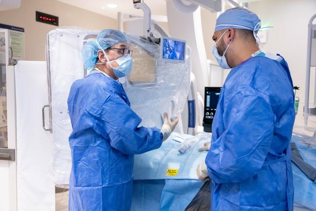
2. Decreasing access site complications with routine ultrasound guidance.
Do you get the most benefit from using ultrasound guidance in femoral (versus any other site) access?
Femoral access, in my mind, is where the real opportunity lies. There are obviously multiple
places where you can gain access to the vascular system using ultrasound guidance, such as brachial or radial access, superficial femoral artery access, or pedal or tibial access. We were routinely using ultrasound for brachial artery access for many years, but we were not using it routinely for femoral access, perhaps because we felt great confidence with femoral access. Eighty percent of our cases are femoral access. We felt we had it figured out: we know the anatomy and we know how to do the correct puncture. Well, that is probably true 90 or 95% of the time you do a femoral access, but some of the time, when the puncture site is not optimal, there is a much higher chance of having a complication. It turns out that when standard femoral access is guided by ultrasound, there is more benefit, since this is such a large volume of cases, and many complications can be prevented.
How much time does ultrasound use add to the access procedure?
Once you become proficient, it doesn’t take extra time. In our lab, it is now routine to have the ultrasound at the bedside with a sterile sleeve, so all preparation is done by staff, because they know ahead of time what to expect. It probably takes about 30 seconds to get it ready, although that part of it is invisible to the operator, because it is part of the routine set up by staff. It is not a specific request; they just know I want it. With regard to the actual access time, it takes less than a minute to look at the femoral artery and decide where to puncture. Ultrasound also reveals where there may be calcium collections in the wall of the artery and if there are lesions in the lumen to avoid. It also means the potential for a first-pass success is extremely high. There is probably an 80 or 90% chance that the first time I put in the needle, I will place it where I want it to be. That is just not true with a standard femoral access. No matter how good you are at understanding the anatomy, patients have some variability. There is value in avoiding sticking the needle distal to the bifurcation, avoiding sticking the needle through the back wall, into a side branch, or some other suboptimal location, and the cost is spending the extra minute to use ultrasound. The key is to use it on every case. If you just do it every so often, then you will only use ultrasound when it is most difficult to gain access. This approach makes it much more challenging to develop facility and speed, as well as achieve a comfort level where it is not an extra task. It is routine use that makes ultrasound guidance an added value.
You mentioned a reduced rate of second puncture with ultrasound guidance. Have you tracked data?
Yes. There have also been a few studies. There was a prospective, randomized trial by Seto that was published in JACC Interventions in 2010.1 He looked at ultrasound-guided versus fluoroscopy- or palpation-guided femoral access. There were 500 patients in each group, and the complication rate in the fluoroscopy-guided group was about 3.5%, and the complication rate in the ultrasound-guided was about 1.5%. That is more than a 50% reduction in complications with ultrasound guidance. Data from our own site showed a reduction in complications that was greater than 50% as well. (Our data uses consecutive cases from one year before we used ultrasound routinely versus consecutive cases after we started ultrasound routinely.) We had a 6% complication rate prior to ultrasound use. With routine ultrasound use, our complication rate decreased from 6% overall to 1.8%.
From a cost standpoint, that would be beneficial as well.
Yes, we did not do a cost analysis, but it would be reasonable to do that. In most ORs and cath labs and angio suites, portable ultrasound is already available.
Any final thoughts?
First, there have been a number of studies looking at what happens when you get the needle in the optimal puncture site, versus what happens when the needle is in a suboptimal site. Patients with an optimal puncture site have dramatically fewer complications, blood transfusions, and need for surgical repair. The other thing I should share – and I do this routinely; I don’t know if everybody does – is that when I put the needle in under ultrasound, I always get a spot film, using x-ray, of the needle in the artery. I want to confirm that the needle is not placed too proximally along the common femoral artery. If it is too proximal, and the tip goes into the external iliac artery, then the potential risk is retroperitoneal hematoma. That is more than a nuisance complication. Retroperitoneal hematoma is a potentially fatal complication. It’s true that the rates in the literature are low, typically <1%, but when a patient suffers a fatal outcome, it is usually going to be because of a retroperitoneal hematoma. So while this complication isn’t very common, the results can be catastrophic. Getting that plain film, I think, is important. Because of the variability from patient to patient in the inguinal ligament, you cannot always distinguish exactly where it is located with ultrasound.
Dr. Peter Schneider can be contacted at peterschneidermd@aol.com.
Reference
- Seto AH, Abu-Fadel MS, Sparling JM, Zacharias SJ, Daly TS, Harrison AT, et al. Real-time ultrasound guidance facilitates femoral arterial access and reduces vascular complications: FAUST (Femoral Arterial Access With Ultrasound Trial). JACC Cardiovasc Interv. 2010 Jul; 3(7): 751-758. doi: 10.1016/j.jcin.2010.04.015.