What Does the New 2021 Chest Pain Guideline Say to the Cath Lab?
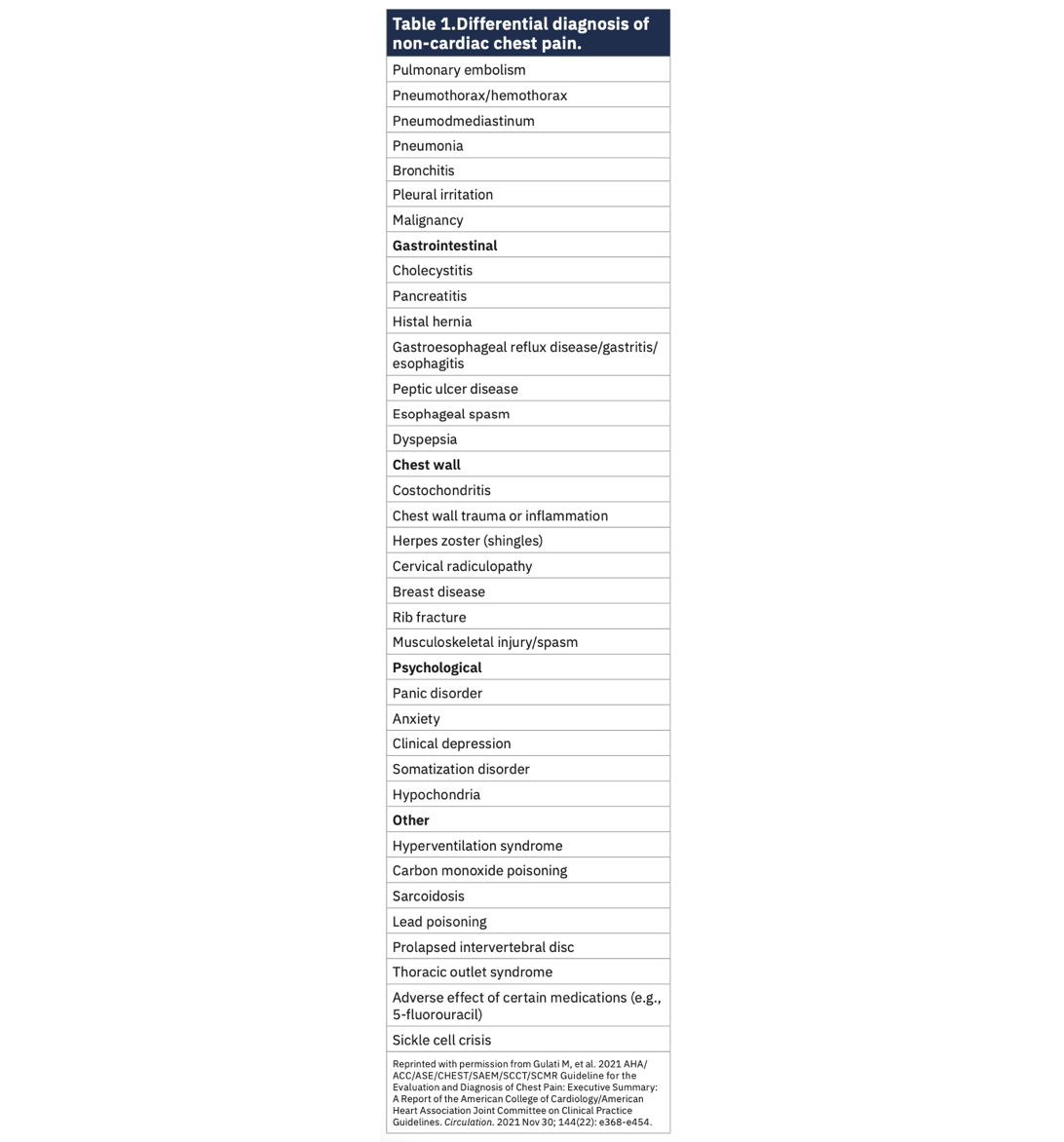
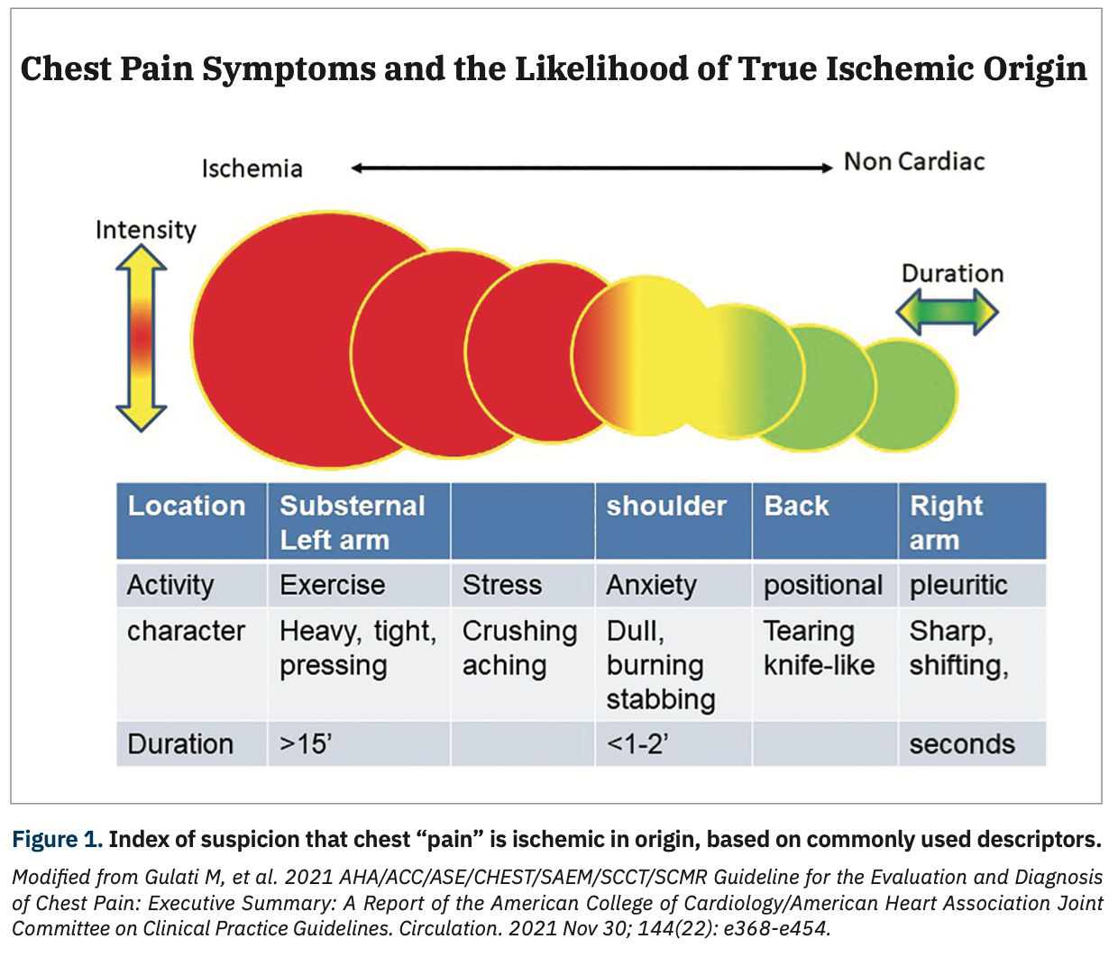
Chest pain is the symptom most related to heart disease. However, there are many conditions other than the heart that cause chest pain (Table 1). Moreover, clinicians have a wide variation in their approach to making a correct diagnosis relating the chest pain symptom to actual heart involvement. For example, just think of how many normal coronary angiograms have been done in your lab in patients with typical anginal chest pain or similar complaints? Would better screening or testing have made these procedures unnecessary? Many of the patients we see in the cath lab have some symptom, but it may not meet the classic description of angina (exertional substernal chest pressure, burning, aching) (Figure 1), but was considered atypical (now called non-cardiac, see below) and could include almost any complaint around the chest or abdomen, or shortness of breath alone. Dyspnea is often considered by some as an ‘anginal-equivalent’ symptom. What should be our approach to the patient’s palpitations? Is there an ischemic cause?
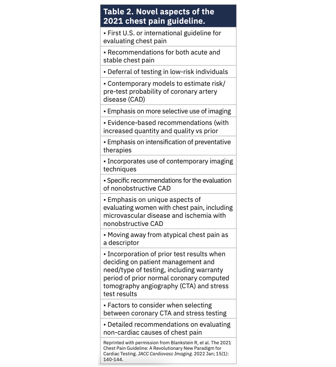
The recently released 2021 American Heart Association (AHA)/American College of Cardiology (ACC) Guideline for the Evaluation and Diagnosis of Chest Pain1 provides some perspective around this group of patients. Clinical practice guidelines present the consensus of experts in the field to help clinicians and allied health care personal deliver the best evidence-based approach to taking care of our patients. The 2021 chest pain guideline, chaired by Dr. Martha Gulati of the University of Arizona – Phoenix, describes the clinical scenarios around chest pain, and how patients should be risk stratified and have their diagnostic testing done before coming for their invasive procedures. The novel aspects of the 2021 chest pain guideline are summarized in Table 2.2
Why is This Guideline Pertinent to the Work We Do in the Cardiac Cath Lab?
The guideline helps us understand what the patient’s symptoms mean, the variety of presentations patients with coronary artery disease (CAD) may have, and how clinicians should select appropriate diagnostic testing before coming into the cath lab. An appropriate pre-cath evaluation makes our invasive procedures more precise, allows us to understand the symptom presentation, and eliminates questions about whether the cath procedure was indeed indicated.
Where Should You Start the Review of a Large Guideline Document?
To begin your review of this extensive work, start with the introductory section’s top 10 take-home messages, which encapsulate the high points of the guideline. Among the most important aspects of decision-making is the concept of risk stratification, meaning how likely is it that the patient’s presentation means they could experience an adverse event like death, myocardial infarction, or urgent hospitalization. The guideline makes it clear that a patient at low risk can undergo outpatient stress testing, while a patient at high risk may need urgent angiography and intervention. Risk stratification by age and sex leads to a logical and cost-effective diagnostic pathway for evaluating chest pain (see below, item “i.”, and Figure 2).
Top 10 Take-Home Messages for the Evaluation and Diagnosis of Chest Pain (Reorganized1) Chest Pain Syndromes
a. Chest pain means more than pain in the chest. Pain, pressure, tightness, or discomfort in the chest, shoulders, arms, neck, back, upper abdomen, or jaw, as well as shortness of breath and fatigue should all be considered anginal equivalents. [MK: When asking a patient about symptoms, I suggest to our fellows not to use the term “chest pain”, but rather “chest discomfort” to let the patient describe what he/she is experiencing. Asking about what started the discomfort, what relieves it, where it radiates to, and whether this is recurrent are all standard questions to get a better feeling for a cardiac or non-cardiac source of discomfort.]
b. The term “non-cardiac” chest pain is in. “Atypical” is out. The term “atypical chest pain” is vague and potentially misleading. It does not focus on the main issue, but is always assumed to be cardiac in some form. The term “non-cardiac” should be used if heart disease is not suspected. Use of “atypical” as a descriptor is now discouraged.
c. Accompanying symptoms may be non-specific but important. Chest pain is the dominant and most frequent symptom for both men and women ultimately diagnosed with acute coronary syndrome. Women may be more likely to present with accompanying symptoms such as nausea and shortness of breath. Figure 1 depicts an index of suspicion that chest “pain” is ischemic in origin, based on commonly used descriptors. Table 1 lists several non-cardiac causes of chest pain.
Approach to Acute Symptom Onset
d. Call 9-1-1 early. Patients with acute chest pain or equivalent symptoms should immediately call 9-1-1. Although most patients will not have a cardiac cause, the evaluation should focus on the early identification or exclusion of life-threatening causes.
e. Use clinical decision pathways routinely for chest pain in the emergency department and outpatient settings.
f. High-sensitivity troponin testing is preferred. High-sensitivity cardiac troponins are the preferred biomarker diagnosis of acute myocardial infarction, offering more accurate detection and exclusion of myocardial injury.
Testing for Diagnosis
g. Use a structured risk assessment. For patients presenting with acute or stable chest pain, risk for coronary artery disease and adverse events should be estimated using evidence-based diagnostic protocols.
h. Testing is not needed routinely for low-risk patients. For patients with acute or stable chest pain determined to be low risk, urgent diagnostic testing for suspected coronary artery disease is not needed.
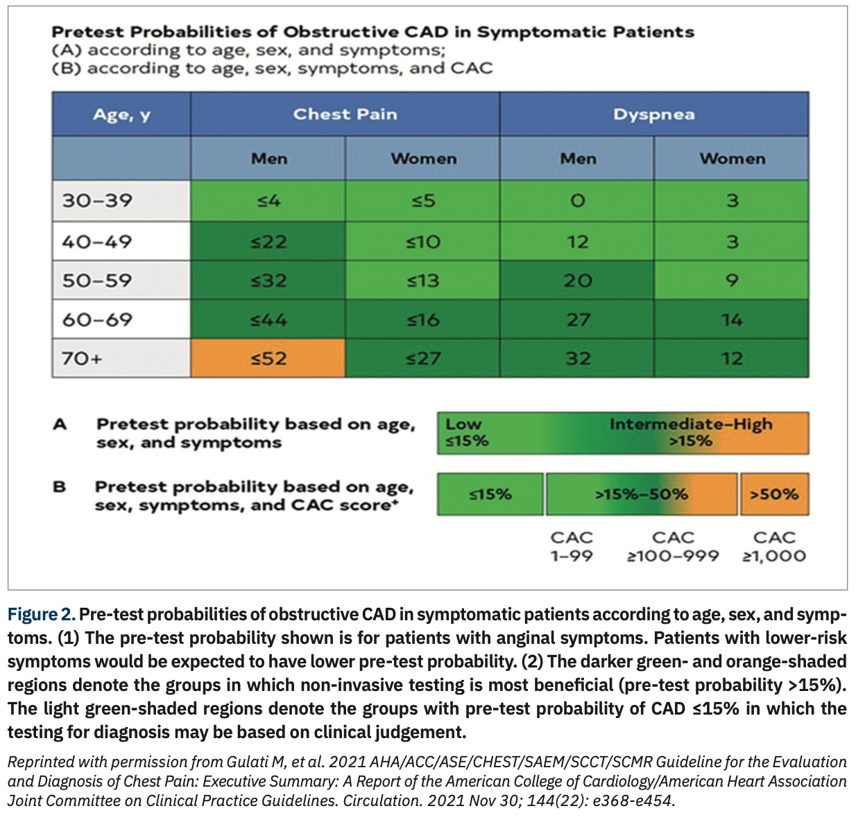
i. Identify the patients most likely to benefit from further testing. Patients with acute or stable chest pain who are at intermediate risk or intermediate to high pre-test risk of obstructive CAD, respectively, will benefit the most from cardiac imaging and testing. Figure 2 provides the pre-test probabilities of obstructive CAD in symptomatic patients according to age, sex, and symptoms. On the grid, the pre-test probability for patients with anginal symptoms shows that patients with lower-risk symptoms are expected to have lower pretest probability. The darker green- and orange-shaded regions denote the groups in which noninvasive testing is most beneficial (pre-test probability >15%). The light green-shaded regions denote the groups with pretest probability of CAD at ≤15%; in these groups, the testing for diagnosis may be based on clinical judgement.
j. Share the decision making. Clinically stable patients presenting with chest pain should be included in decision making; information about risk of adverse events, radiation exposure, costs, and options should be provided to facilitate the discussion [MK: We should already be doing this for all clinical care decisions.]
What’s the Best Diagnostic Testing Before Coming to the Cath Lab? The Role for Coronary Computed Tomography Angiography (CCTA)
CCTA is a great tool that can diagnose the extent and severity of CAD, as well as atherosclerotic plaque composition and high-risk features (e.g., positive remodeling, low attenuation plaque). CCTA with the added determination of angiographically derived fractional flow reserve (FFRCT) provides an estimation of lesion-specific ischemia and is more informative than CCTA alone. It may be superior (sensitivity/specificity) to many of the other non-invasive test modalities.
CCTA contraindications include allergy to iodinated contrast media, inability to cooperate for breath holding, clinical instability including cardiac rhythm, and an inability to take beta blockers or nitroglycerin. Although CCTA can image the heart, lungs, and aorta, in general, CCTA should focus on the question at hand and avoid the “triple rule-out” approach [MK: I think CCTA with FFR is highly underutilized as a high-quality, one-stop test, giving the clinician both the anatomy and the ischemic potential of stenosis with a higher sensitivity/specificity than most non-invasive imaging tests.]
Anatomic Testing in the Cath Lab: Invasive Coronary Angiography
Working every day in the cath lab, we know invasive coronary angiography (ICA) defines the presence, extent, and severity of the epicardial coronary artery, as well as a limited assessment of coronary blood flow. The primary goal of coronary angiography is to identify any high-grade stenosis with the potential for revascularization by percutaneous coronary intervention (PCI) or coronary artery bypass graft (CABG) surgery. The use of in-lab physiologic testing (hyperemic [FFR] and non-hyperemic indices [e.g., instantaneous wave-free ratio [iFR], diastolic pressure ratio [dPR], resting full-cycle ratio [RFR], or soon, angiographically derived FFR, etc.]) remains important to couple a functional assessment to the anatomy. Normal or non-obstructive coronary angiography does not exclude abnormal coronary vascular function. For patients with non-obstructive CAD, assessment of the coronary microcirculation may have additional value.
The guideline supports and reiterates the longstanding clinical recommendations that before ICA, all patients with known CAD should be on guideline-directed medical therapy (GDMT) and optimized when the patient is symptomatic. For the intermediate-risk patients with known CAD, ICA is reasonable for patients presenting with frequent symptoms or for those known to have high-risk CAD (left main or proximal left anterior descending, or multivessel CAD).
The guideline also suggests that repeat coronary angiography for non-obstructive CAD is not necessary. Patients can have a CCTA. CCTA has been shown to effectively document progressive CAD, including more extensive atherosclerotic plaque or the presence of high-risk plaque features, or new obstructive stenosis ≥50%. However, for patients with extensive plaque, a stress test is preferred. Patients with acute chest pain who have known coronary artery stenosis ranging from 40% to 90% on CCTA will benefit from measurement of FFRCT.
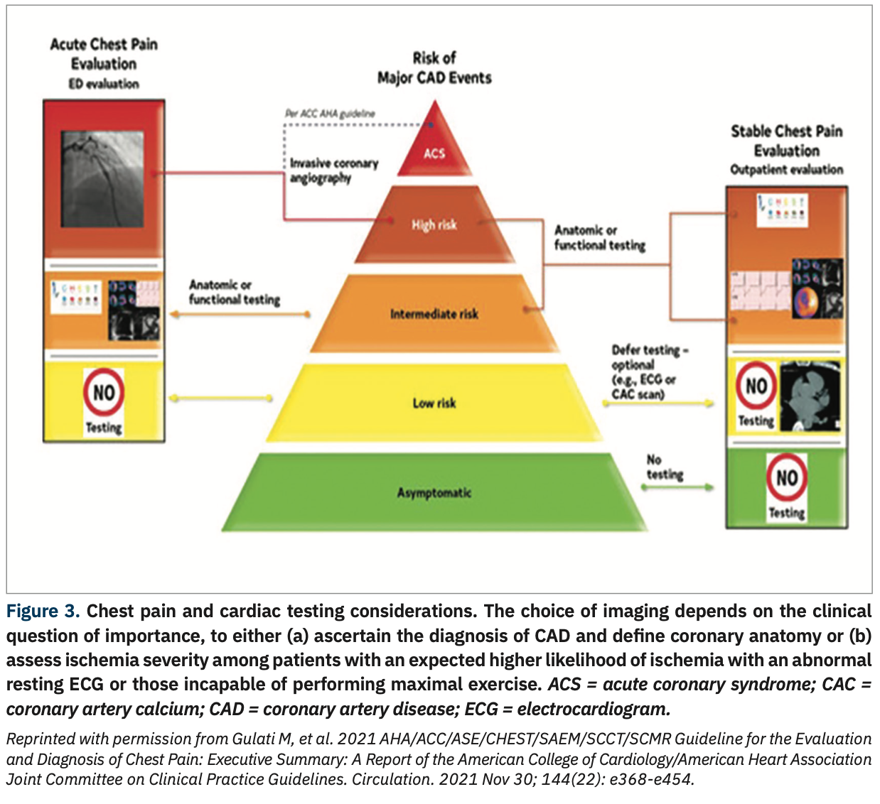
The considerations around which type of cardiac testing for which patient are summarized in Figure 3. The choice of imaging depends on the clinical question of importance to either (a) establish a diagnosis of CAD anatomy; or (b) assess ischemia in patients with a high likelihood of ischemia (e.g., abnormal resting electrocardiogram [ECG] or those incapable of performing maximal exercise). Note that invasive coronary angiography is primarily associated with acute coronary syndromes (ACS) and high ischemic-risk patients, and incorporates functional testing for the intermediate-risk patient. Low-risk and asymptomatic patients should rarely require ICA.
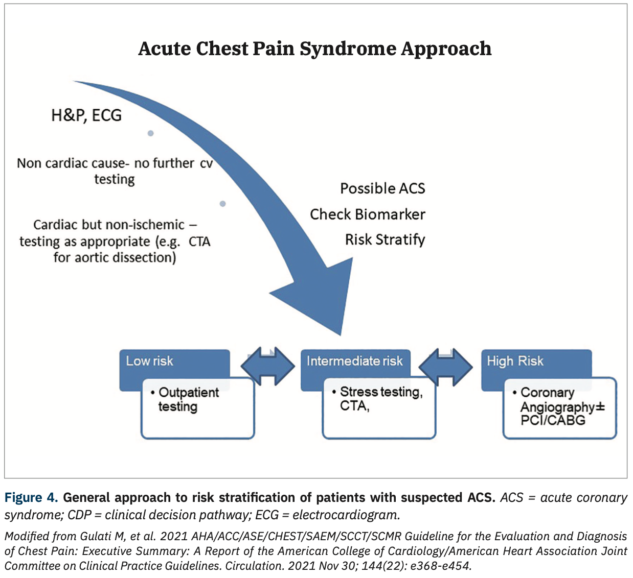
For patients with acute chest pain in the presence of moderate to severe ischemia on a current or prior (>1 year) stress test, or in the presence of high-risk CAD (left main >50% or 3-vessel >70% stenosis), or obstructive CAD (>50%) and frequent angina, invasive angiography is recommended (Class 1; level of evidence C, expert opinion). Invasive angiography is also suggested if there is moderate to severe ischemia on an imaging stress test or FFRCT with inconclusive stenosis. Figure 4 provides a general approach to risk stratification of patients with a suspected acute coronary syndrome.
The Bottom Line
In conjunction with the 2021 chest pain guideline, I recommend reading the guideline statement on the approach to revascularization in patients with coronary artery disease, which addresses the indications for PCI and CABG,3 a subject for a future editor’s page.
Appreciating the wide spectrum of CAD presentations and the risk stratification of patients with chest pain will increase our awareness of the needs of our patients, the usefulness of invasive coronary angiography, and improve the approach to their evaluation before and after our cath lab procedures.
References
1. Gulati M, Levy PD, Mukherjee D, Amsterdam E, Bhatt DL, Birtcher KK, Blankstein R, Boyd J, Bullock-Palmer RP, Conejo T, Diercks DB, Gentile F, Greenwood JP, Hess EP, Hollenberg SM, Jaber WA, Jneid H, Joglar JA, Morrow DA, O’Connor RE, Ross MA, Shaw LJ. 2021 AHA/ACC/ASE/CHEST/SAEM/SCCT/SCMR guideline for the evaluation and diagnosis of chest pain: executive summary: a report of the American College of Cardiology/American Heart Association Joint Committee on Clinical Practice Guidelines. Circulation. 2021 Nov 30; 144(22): e368-e454. doi: 10.1161/CIR.0000000000001030
2. Blankstein R, Gulati M, Jaber WA, Bullock-Palmer RP, Bhatt DL, Shaw LJ. The 2021 chest pain guideline: a revolutionary new paradigm for cardiac testing. JACC Cardiovasc Imaging. 2022 Jan; 15(1): 140-144. doi: 10.1016/j.jcmg.2021.10.005
3. Writing Committee Members, Lawton JS, Tamis-Holland JE, Bangalore S, et al. 2021 ACC/AHA/SCAI guideline for coronary artery revascularization: executive summary: a report of the American College of Cardiology/American Heart Association Joint Committee on Clinical Practice Guidelines. J Am Coll Cardiol. 2021 Dec 7: S0735-1097(21)06157-X. doi: 10.1016/j.jacc.2021.09.005
Disclosures: Dr. Morton Kern reports he is a consultant for Abiomed, Abbott Vascular, Philips Volcano, ACIST Medical, and Opsens Inc.
Dr. Kern can be contacted at mortonkern2007@gmail.com
On Twitter @drmortkern