EMS Recap: Acute Respiratory Distress Syndrome (ARDS)
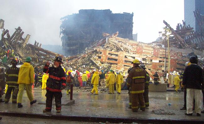
You and your partner respond to the scene of an industrial accident. One of the workers inhaled chlorine vapors when the valve on the tanker he was working on malfunctioned. Your patient was decontaminated by hazmat personnel and delivered to you for treatment and transport to the trauma center. He presents wrapped in a sheet, with a nasal cannula and oxygen flowing at 4 Lpm. He is experiencing difficulty breathing and watering eyes. He describes his throat as raw, and he has chest tightness, a headache, some nausea and admits to vomiting during decontamination. You recognize your patient has inhaled a caustic chemical, and may have also subsequently aspirated stomach contents when he became ill. As a result, the risk for eventually developing acute respiratory distress syndrome has increased. You replace his nasal cannula with a non-rebreather mask, turn the flow up to 15 Lpm and consider applying CPAP for the crackles you start to hear en route to the hospital. Upon arrival, his oxygen perfusion is below 85% and the physician decides to intubate and place him on a ventilator. Your patient is experiencing acute respiratory distress syndrome.
Acute respiratory distress syndrome (ARDS) is a severe inflammatory lung injury to the vascular endothelium and the alveolar epithelium.1–3 The inflammation can be the result of either direct or indirect insult to the lung.2, 4 Direct injury can be from pneumonia or aspiration of gastric contents. Indirect, or extrapulmonary, injury to the lung can happen in the form of sepsis, shock, bacterial pneumonia, multiple traumas or aspiration pneumonia, with sepsis-related ARDS having the greatest overall severity, poorest recovery and highest mortality.4, 5
When a person experiences acute respiratory injury, the coagulation system is activated, the fibrinolysis system is inhibited and pulmonary hypertension occurs as a complication of the resulting abnormal vascular tone, vascular destruction, vascular remodeling and intraluminal clotting.2 Currently, almost two-thirds of ARDS patients admitted to intensive care succumb to sepsis, multiple organ dysfunction syndrome and irreversible respiratory failure.4, 6 Renal failure is the most common organ dysfunction resulting from ARDS.4
Acute respiratory distress syndrome is difficult to treat because with more than 50 recognized trauma conditions and critical illnesses associated as complicating factors there is no typical patient.5, 6 Women are more likely than men to develop ARDS after a critical injury and women hospitalized with COPD have an increased risk of developing ARDS. The mortality rates are the same for both genders.6 Additionally, mechanical ventilation—the mainstay of ARDS treatment—may actually increase inflammation and worsen the lung injury by causing alveolar over-distension and diffusion of bacteria producing bacteremia.4
In the early stage of ARDS development, injury occurs when alveolar macrophages release cytokines, interleukins and tumor necrosis factor. Neutrophils are activated and attracted to the alveoli causing epithelial injury, increased permeability, fluid movement into the alveolar space and reduced ability to clear this fluid.2, 5 Endothelial injury to the alveolar epithelium decreases surfactant production and renders the remaining surfactant non-functional, causing atelectasis, decreased pulmonary compliance and hypoxemia.2, 3, 5 With coagulation cascade activation, clotting causes high pulmonary arterial pressures, right ventricular failure, vascular remodeling and increased alveolar dead-space.2, 5 The resulting hypoxemia does not respond to supplemental oxygen and most ARDS patients die at this point.5 If allowed to progress, myofibroblast proliferation, fibrosis and increased deposition of collagen occurs.2
When assessing your patient look for intercostal retractions, crackles and identify risk factors like shock, sepsis, trauma, severe hypoxemia, decreased pulmonary compliance, bilateral pulmonary infiltrates and lack of cardiogenic pulmonary edema.5 The presence of more than one risk factor increases the probability of developing ARDS.4 Shock is most often associated with acute respiratory distress syndrome.4
If your patient is already on a ventilator it is important to know increased positive end expiratory pressure (PEEP) above 10 cm H2O can produce progressive declines in cardiac output, mean arterial pressure and left ventricular size related to right ventricular systolic afterload. The higher the PEEP, the greater the increase of right ventricular afterload and decrease in right ventricular ejection fraction.1 Limited PEEP does help to increase the amount of lung available for ventilation, so it’s necessary to find the level where PEEP is effective versus causing over-distension of the lung.1 Hypercapnia induces constriction of pulmonary circulation and increases pulmonary vascular resistance, mean pulmonary artery pressure and right ventricular end-diastolic volume. All of these factors lead to a 20% reduction in right ventricular ejection fraction.1 Right ventricular function reflects lung status, so what’s bad for the right ventricle is ultimately bad for the lungs.1
Current treatment for ARDS patients is primarily supportive, with supplemental oxygen used initially as the patient becomes hypoxemic, and eventual intubation and ventilation as the hypoxemia progresses.5 Once on the ventilator, avoid plateau pressures (Pplat) below 28 cm H2O, limit hypercapnia by using heated humidified oxygen with an increased inspiratory rate, and limit PEEP to 7 cm H2O or less. Low PEEP helps limit hypercapnia and lung stress. High PEEP induces hypercapnia and impairs right ventricle function.1
Placing a ventilated patient with ARDS in the prone position has been found to improve compliance, decrease plateau pressure, reduce PaCO2, and improve oxygenation.1 Positioning severely burned patients with sepsis-induced ARDS in a prone position has been used as a treatment mode for those with poor ventilator response. Prone positioning helps to redistribute oxygen to the dorsal areas, better distribute tidal volume subsequent to improved fit of lungs within the chest and provide relief from compression forces of the heart on the lungs.7
Corticosteroids have beneficial short-term effects when given at low or moderate doses sooner than two weeks but appear to be harmful if initiated later and are of unclear benefit if lung protective ventilation is used.2 Corticosteroids and sivelestat, a neutrophil elastase inhibitor, target inflammation.2 Anticoagulants target microvascular thrombosis.2 Vasoactives like inhaled nitric oxide, prostacyclin and almitrine can be used to treat pulmonary artery hypertension and dysregulated ventilation perfusion problems.2 Beta 2 agonists are used to dilate the airways and thus help with fluid clearance and reduce alveolar edema.2 Sedatives can be employed to reduce oxygen consumption, decrease anxiety and allow for full ventilator support. If sedation is not effective, a neuromuscular blocking agent can be used for a short period of time.
To summarize, in the prehospital environment, assess and provide supportive care. Supportive care will be continued in the hospital and pulmonary artery catheterization will be used to definitively determine the diagnosis of pulmonary permeability edema from ARDS versus hemodynamic pulmonary edema from heart failure. Carefully monitor any patient you have on a ventilator and be prepared to intubate a patient experiencing severe hypoxemia that does not resolve with supplemental oxygen therapy.
References
- Bouferrache K, Vieillard-Baron A. Acute respiratory distress syndrome, mechanical ventilation, and right ventricular function. Current Opinion in Critical Care, 2011; 145: 24–33.
- Frank AJ, Thompson BT. Pharmacological treatments for acute respiratory distress syndrome. Current Opinion in Critical Care, 2010; 16: 62–68.
- Isotani E. Pathophysiology of acute respiratory distress syndrome. Critical Care Medicine, 2012 July; 40(7): 2233–34.
- Del Sorbo L, Slutsky AS. Acute respiratory distress syndrome and multiple organ failure. Current Opinion in Critical Care, 2011; 17: 1–6.
- Villar J. What is the acute respiratory distress syndrome? Respiratory Care, 2011 Oct; 56(10): 1539–45.
- Heffernan DS, Dossett LA, Lightfoot MA, Fremont RD, Ware L, Sawyer RG, May AK. Gender and acute respiratory distress syndrome in critically injured adults: A prospective study. The Journal of Trauma: Injury, Infection, and Critical Care, 2011 Oct; 71(4): 878–85.
- Hale DF, Cannon JW, Batchinsky AI, Cancio LC, Aden JK, Blackbourne LH, Chung KK. Prone positioning improves oxygenation in adult burn patients with severe acute respiratory distress syndrome. Journal of Trauma and Acute Care Surgery, 2012; 72(6): 1634–39.
Robert E. Sippel, Major, USAF (Ret.), MS, MAEd, NREMT-P, LP is an assistant professor and clinical coordinator in the Emergency Health Science Department at the University of Texas Health Science Center, San Antonio, Texas.