Not a Jersey Finger – A Case of a Closed Traumatic Rupture of the Small Finger Flexor Digitorum Profundus Tendon at the Lumbrical Origin
© 2024 HMP Global. All Rights Reserved.
Any views and opinions expressed are those of the author(s) and/or participants and do not necessarily reflect the views, policy, or position of ePlasty or HMP Global, their employees, and affiliates.
Abstract
A 55-year-old right-handed male presented acutely with inability to flex at the distal interphalangeal (DIP) joint of the right small finger after feeling a pop while moving heavy furniture. Avulsion of the flexor digitorum profundus (FDP) tendon at its insertion, or "jersey finger," was suspected. During operative exploration, the insertion on the distal phalanx was intact, and the rupture was noted proximally at the level of the lumbrical origin. There are cases in the literature describing attritional ruptures of the tendon proximally due to metacarpophalangeal or intercarpal arthrosis, but this is the first case of a closed, proximal tendon rupture due solely to trauma. Imaging may be helpful to diagnose the level of tendon rupture. We propose surgeons perform an A1 pulley incision to determine the level of rupture prior to planning the surgical exposure for tendon repair.
Introduction
We present a case of small finger flexor digitorum profundus (FDP) avulsion that appeared to be a Leddy and Packer type I jersey finger but was found intraoperatively to be a more proximal rupture at the level of the lumbrical origin. This required carpal tunnel release for exposure and resulted in unnecessary incisions over the finger. Accurate preoperative classification of FDP injuries helps guide management and timing of repair. In this article, we describe our case presentation, Leddy and Packer classification of a jersey finger, etiology of closed nontraumatic FDP rupture, and tools to help diagnose these types of injuries and where the avulsion injury is located for more accurate surgical and incision planning.
Case Presentation
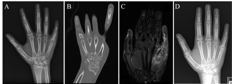
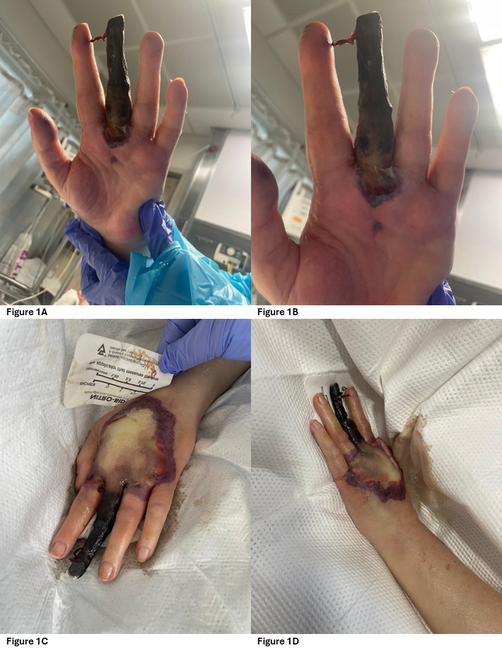
A 55-year-old, otherwise healthy, right-handed construction worker presented to the emergency department 2 days after an injury sustained while carrying a heavy piece of furniture. The furniture slipped in his hands, resulting in forced hyperextension of the metacarpophalangeal joints with flexion of the interphalangeal joints. There was an audible "pop" followed by immediate pain and swelling of the palm. On exam, he was unable to flex at the distal interphalangeal (DIP) joint. There were no wounds or lacerations, but there was a palpable mass over the ulnar aspect of the mid-palm. He had no nerve deficits. Radiographs were obtained, which demonstrated no fractures, dislocations, or arthrosis (Figure 1).
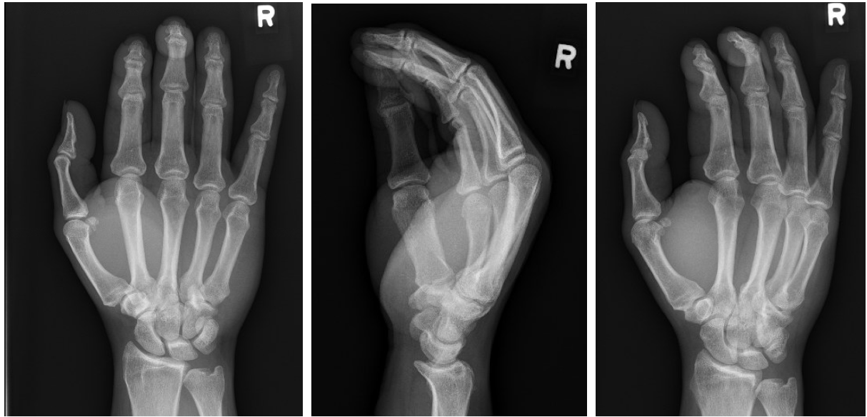
Figure 1. Three-view radiographs of the patient's right hand demonstrating no acute osseous abnormalities.
Given the clinical and radiographic findings, he was diagnosed with a type 1 Leddy and Packer FDP tendon avulsion. This type of tendon avulsion requires more urgent repair due to disruption of blood supply via the vincula.1
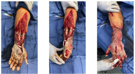
After a delay due to insurance issues, 9 days post-injury the patient underwent operative exploration and repair. Based on the assumption that this was a jersey finger, a Brunner incision was made over the small finger to expose the flexor tendon sheath. Surprisingly, both superficial and deep flexor tendons were intact. The A1 pulley was incised, and retraction of both tendons resulted in appropriate flexion of the finger. There was laxity of the FDP tendon proximally, indicating a proximal rupture. The incision was extended to the distal edge of the carpal tunnel where the distal stump was identified (Figure 2). Release of the transverse carpal ligament was performed, which revealed the small finger FDP tendon rupture at the level of the lumbrical origin (Figure 3). There was significant tenosynovitis involving the middle, ring, and small finger flexor tendons.
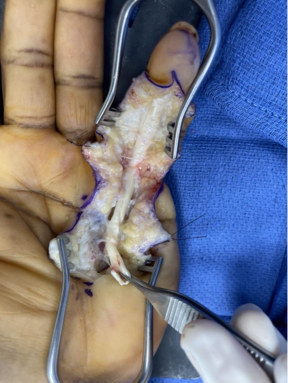
Figure 2. Distal stump of small finger FDP tendon.
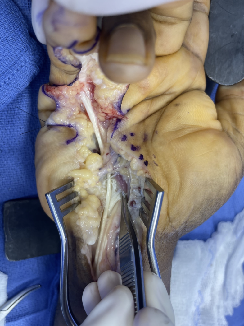
Figure 3. Forceps holds proximal stump of small finger FDP tendon within the carpal tunnel.
After tenolysis, the small finger FDP tendon was repaired primarily with a combination of a modified Kessler and figure-of-8 stitches using 3-0 Ethibond. He was placed into an ulnar gutter dorsal-blocking splint.
The patient was referred for early active motion therapy at 3 days but did not start until 3 weeks, which has resulted in tendon adhesions. He continues to improve with hand therapy, and we plan for tenolysis once he plateaus.
Discussion
The FDP tendon serves as the only flexor of the DIP joint and contributes to flexion at the wrist, metacarpophalangeal, and proximal interphalangeal joints of the index through small fingers. The muscle belly originates from the posterior and medial borders of the ulna and interosseous membrane with tendinous insertion on the volar base of the distal phalanx.2 The 2 ulnar FDP tendons are innervated by the ulnar nerve while the 2 radial FDP tendons are innervated by the anterior interosseous nerve. The FDP tendon is unique in that it serves as the origin of the lumbricals.
FDP tendon avulsion injuries, or jersey finger, was first described by Boyes et al in 1960.3 They were later classified by Leddy and Packer into 3 types. Type 1 is an avulsion without fracture with the tendon retracting into the palm with rupture of the vincula, type 2 involves a small fragment that retracts to the level of the proximal interphalangeal joint, and type 3 involves a large bony fragment which prevents retraction beyond the middle phalanx at the A4 pulley. This was subsequently modified with 2 additional types currently recognized; type 4 involves a simultaneous bony fragment with avulsion of the tendon from the bony fragment resulting in retraction into the palm, and type 5 involves avulsion with an osseous fragment with significant distal phalanx fracture extension.4
Type 1 and 4 injuries result in complete disruption of the extrinsic blood supply and result in tendon ischemia, necessitating repair within 10 days.
Most closed, traumatic FDP avulsions or ruptures involve sports like football or rugby in which the finger (most commonly the ring) is pulled in hyperflexion at the DIP joint, such as when an athlete is grabbing on another player's jersey – hence the term "jersey" finger.5 Conversely, nontraumatic, attritional rupture of the flexor pollicis longus occurs from intrinsic pathology such as rheumatoid arthritis, tumor, bony abnormality, or infection. Spontaneous rupture has been described but is very rare.
Masaki et al describe a unique case of spontaneous closed small finger FDP rupture in which the FDP tendon branched from the ring finger FDP in the mid-palm (zone III); magnetic resonance imaging (MRI) confirmed a symmetric contralateral variant of this anomaly.6 Wray described a case of spontaneous rupture of the FDP at the level of the lumbrical with no evidence of rheumatoid arthritis, gout, fractures, or carpal bone abnormalities, postulating an ischemic mechanism at the level of the lumbrical.7 Other attritional ruptures have occurred from sequelae of gouty infiltration of the tendon,8 hook of hamate fracture,9,10 pisotriquetral arthritis,10 and Kienbock disease.11 There are also cases of concomitant FDP and flexor digitorum superficialis avulsion,12,13 one of these occurring in both zones I and III of the small finger.14
A spontaneous attritional rupture of the small finger FDP has been described in a patient with acromegaly, attributed to acromegaly arthropathy of the hook of hamate.15 Cases of FDP rupture within the carpal tunnel have also been described.10,15,16 Lee et al published a review of spontaneous FDP ruptures of zones I, II, III, IV, and V in which there were 8, 2, 30, 59, and 5 cases in each of these zones, respectively. FDP ruptures labeled as "spontaneous" in this paper were predominantly in zone III. Most of the cases occurring in the other flexor zones had a component of antecedent trauma or associated pathology.
There are few reports of true spontaneous rupture without known or suspected morphologic etiologies. Yoon et al presented a case in which a closed rupture of the small finger FDP tendon occurred at the origin of the lumbrical while the patient was rotating a steering wheel while driving.18 The authors propose obtaining imaging via MRI or ultrasound (US) to determine the level of injury, which dictates surgical approach and timing.
All patients should have radiographs of the injured finger to identify associated avulsion fractures as these injuries define a Leddy and Packer type 2, 3, 4, and 5 and can be treated in a longer time frame as the vincular blood supply is intact and tendon retraction has not occurred. MRI and US can identify the level of tendon injury and location of the proximal stump,19,20 although the quality and accuracy of US is user dependent.19 MRI has a higher sensitivity but is costly and may take much longer to obtain.
In our case of closed FDP rupture with a convincing clinical picture indicating distal avulsion, our surgical approach included unnecessary incisions over the finger. Advanced imaging can be obtained to help the surgeon determine the site of injury, but in the current health care climate, it is hard to advocate for expensive imaging modalities that may also delay treatment.
In summation, closed FDP ruptures are commonly associated with distal avulsion injuries such as the jersey finger, which is categorized by Leddy and Packer into 4 stages; however, tendon ruptures can occur at more proximal levels, necessitating different incisions for exposure. We propose that when faced with a closed tendon rupture, the surgeon is best served by incising the A1 pulley first to determine if the tendon rupture is proximal or distal to this level. The surgeon can then appropriately plan for operative exposure of the tendon repair. MRI or US can aid in diagnosis, but one must weigh the financial burden and assess its impact on timing of surgery. Furthermore, this specific FDP injury pattern is not currently a part of the Leddy and Packer classification. We therefore recommend that a new Leddy and Packer type, type 6, be included in the modified classification.
Acknowledgements
Authors: Alexander M. Germann, MD; Sharon S. Stanley, MD
Affiliation: LSUHSC School of Medicine, New Orleans, Louisiana
Correspondence: Alexander M. Germann, MD; agerma@lsuhsc.edu
Ethics: Informed consent was obtained to use photos of the patients' surgery and imaging.
Disclosures: The authors disclose no relevant financial or nonfinancial interests.
References
1. Leddy JP, Packer JW. Avulsion of the profundus tendon insertion in athletes. J Hand Surg Am. 1977;2A:66–69.
2. Green DP, Hotchkiss RN, Pederson WC, et al. Flexor tendon injury, acute injuries. In Green's Operative Hand Surgery, 5th edition. Elsevier; 2005:454-487.
3. Boyes JH, Wilson JN, Smith JW. Flexor-tendon ruptures in the forearm and hand. J Bone Joint Surg Am. 1960;42-A:637-646.
4. Al-Qattan MM. Type 5 avulsion of the insertion of the flexor digitorum profundus tendon. J Hand Surg Br. 2001;26B:427-431.
5. dElzinga KE, Chung KC. Finger injuries in football and rugby. Hand Clin. 2017 Feb;33(1):149-160.
6. Masaki F, Isao T, Aya Y, et al. Spontaneous flexor tendon rupture of the flexor digitorum profundus secondary to an anatomic variant. J Hand Surg. 2007;32(8):1195-1199.
7. Wray RC Jr, Parlin LS. Spontaneous flexor tendon rupture in the palm. Ann Plast Surg. 1989 Oct;23(4):352-353.
8. Hankin F, Mayhew D, Compman R, et al. Gouty infiltration of a flexor tendon simulating rupture. Clin Orthop Relat Res. 1985;194:172-175.
9. Takami H, Takahashi S, Ando M. Rupture of flexor tendon associated with previous fracture of the hook of the hamate. Hand. 1983;15:73-76.
10. Grant I, Berger AC, Ireland DC. Rupture of the flexor digitorum profundus tendon to the small finger within the carpal tunnel. Hand Surg. 2005 Jul;10(1):109-114.
11. Masada K, Kawabata H, Ono K. Pathologic rupture of flexor tendons due to longstanding Kienbock's disease. J Hand Surg Am. 1987;12A:22-25.
12. de Villeneuve Bargemon JB, Jaloux C, Viaud-Ambrosino S, et al. A jersey finger diagnostic trap: rupture of the flexor digitorum profundus tendon and the flexor digitorum superficialis tendon. Trauma Case Rep. 2021 Apr 21;34:100476.
13. Johnson MA, Colville J. Closed traumatic avulsion of both ring finger flexors with successful primary repair more than 4 weeks after injury and a review of the literature. J Surg Case Rep. 2020 Jul 14;2020(7):rjaa160.
14. Soro MA, Christen T, Durand S. Unusual closed traumatic avulsion of both flexor tendons in zones 1 and 3 of the little finger. Case Rep Orthop. 2016;2016:6837298.
15. Lee MY, Jin YC. Attritional rupture of the little finger flexor digitorum profundus tendon in the carpal tunnel in a patient with acromegaly. J Hand Surg Asian Pac Vol. 2016 Feb;21(1):92-94.
16. Popov N, Escaré P, Allieu Y. A propos des ruptures primitives du fléchisseur du cinquième doigt au canal carpien. Essais de classification basée sur six cas cliniques et revue de la littérature [Primary flexor tendon ruptures of the little finger within the carpal tunnel. Proposed classification based on six clinical cases and review of the literature]. Chir Main. 2007 Feb;26(1):35-39. French.
17. Lee JS, McGrouth DA. Are flexor tendon ruptures ever spontaneous? A literature review on closed flexor tendon ruptures of the little finger. J Hand Surg Asian Pac Vol. 2019; 24(2):180-188.
18. Yoon, JH, Jung JS, Kim, H. Spontaneous zone III flexor tendon rupture of the little finger: a case report. J Wound Manag Res. 2022;18(1):53-57.
19. Drapé JL, Tardif-Chastenet de Gery S, Silbermann-Hoffman O, et al. Closed ruptures of the flexor digitorum tendons: MRI evaluation. Skeletal Radiol. 1998 Nov;27(11):617-624.
20. Lapegue F, Andre A, Brun C, et al. Traumatic flexor tendon injuries. Diagn Interv Imaging. 2015; 96(12):1279-1292.