Demographic Discrepancies of Vitamin D Deficiency in Craniofacial Fracture Patients
Abstract
Background. The relationship between craniofacial fracture and vitamin D status has not been studied. Given the important role vitamin D status plays in postfracture prognosis, a deep investigation into this relationship is due. The primary objective of this study was to assess the demographic discrepancies in the vitamin D status of patients with craniofacial fracture.
Methods. The Cerner Health Facts database was used to collect data on serum 25-hydroxyvitamin D, demographics, hospital admission and discharge, and fracture type of 76 craniofacial fracture patients with available vitamin D levels from October 2015 until May 2018. Pairwise Spearman Correlation was used for multivariate data analysis.
Results. Of the 76 craniofacial fracture patients identified, 55.3% were insufficient in vitamin D and 32.9% were deficient in vitamin D. Vitamin D deficiency and insufficiency were more common in women, African American individuals, and patients over 50 years of age.
Conclusions. The investigation revealed a high prevalence of vitamin D deficiency and insufficiency in craniofacial fracture patients. Despite the higher incidence of fractures in men and Caucasian individuals, women and African American individuals with craniofacial fractures had poorer vitamin D status. Clinicians should consider empiric vitamin D supplementation following craniofacial fracture in high-risk groups.
Introduction
Craniofacial fractures are a devastating source of soft tissue, bone, neurologic, and emotional disturbance. The danger of compromising the airway, brain, and an individual’s facial aesthetic justifies a thorough investigation into bone health and risk factors for craniofacial fracture. In 2017, the incidence of facial fracture worldwide was 98 per 100,000.1 A North American trauma database study demonstrated 34% of major traumas involved facial injuries, highlighting the importance of implementing prevention strategies and prompt medical and surgical care.2
Fracture healing is a complex process that can be broken down into 4 stages: inflammation, soft callus formation, hard callus formation, and bone remodeling. The steroid hormone vitamin D facilitates calcium and phosphorus absorption and bone mineralization. Mineralization is instrumental in hard callus formation and bone remodeling, 2 of the 4 key stages of fracture healing.3
The vitamin D precursor 25-hydroxyvitamin D (calcidiol) is measured routinely in the serum. Vitamin D deficiency is defined as calcidiol <20 ng/mL, and insufficiency is defined as <30 ng/mL. Calcidiol should be at least 30 ng/mL to effectively prevent fractures.4 Whereas the relationship between vitamin D levels and fractures of the vertebrae and hips has been studied extensively, vitamin D status of patients with craniofacial fracture has not been investigated. Given the progressively clearer role that vitamin D plays in bone healing, this study sought to examine vitamin D levels in a patient cohort diagnosed with facial fractures and hypothesizes that vitamin D status in this cohort mirrors that of the general population. In the long term, the study aims to elucidate the prevalence and discrepancies of vitamin D deficiency and insufficiency in craniofacial fracture patients to aid in optimal management of targeted patient populations.
Materials and Methods
Study Protocol
This is a retrospective study that utilized the Cerner Health Facts Database from October 2015 until May 2018. All patients with a facial fracture were identified using the International Classification of Diseases, Tenth Revision (ICD-10) code S02 (fracture of skull and facial bones). Inclusion criteria are patients 18 years or older with known serum 25-hydroxyvitamin D level. Variables collected for each patient included the following: race, gender, payer code and descriptions, census region and division, urban/rural status, teaching facility, bed size range, care setting description, admission time, discharge time, discharge disposition, age, diagnosis code and description, diagnosis priority and type, and calcidiol laboratory value. Variables pertaining to demographics, hospital admission and discharge, diagnosis, and calcidiol became the focus of the data analysis.
Statistical Analysis
The variables race, gender, age, hospital admission, hospital discharge, diagnosis, and calcidiol were considered in the data analysis. Correlations were considered statistically significant at P values < 0.05, and all analyses were performed using Microsoft Excel, Minitab, and RStudio. Excel was used to collect and organize the data. PivotTables were used to determine frequency of cases across variables. Excel was further used to produce histograms and other figures.
The data were coded into numerical values in Minitab. Pairwise Spearman Correlation was used to identify relationships among all variables of interest and associated P values. Two-sample t-tests were computed for variables with unequal sample sizes. Matrix Plots with a confidence interval (CI) of 95% were completed in Minitab as well. RStudio was used to calculate correlation coefficients and create a correlation matrix. Relationships among variables were visualized with a heatmap.
Results
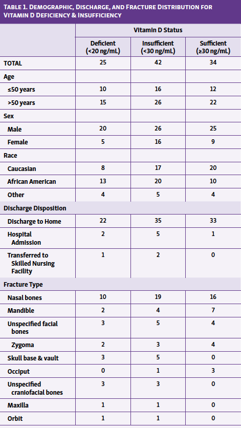
The database search yielded 76 incidents of craniofacial fracture from October 2015 until May 2018. Patients over 50 years of age accounted for 63% of fractures, whereas 36.8% of fractures occurred in patients 50 years old and younger. Male and female patients accounted for 67.1 and 32.9% of fractures, respectively. Caucasian individuals represented 48.7% of fractures, African American individuals 39.5%, and individuals of other races 11.8%. Of all patients presenting to the hospital with facial fractures, 89.5% were discharged to home, 7.9% were admitted to the hospital, and 2.6% were transferred to a skilled nursing facility. Fractures occurred mostly frequently in the nasal bones (46.1%), followed by mandible (14.5%), unspecified facial bones (11.8%), zygoma (9.2%), skull base and vault (6.6%), occiput (5.3%), unspecified craniofacial bones (3.9%), and maxilla (1.3%). All demographic, discharge, and fracture type data are listed in Table 1.
The range of vitamin D levels reported was 7 to 128 ng/mL. The rates of vitamin D insufficiency (<30 ng/mL) and deficiency (<20 ng/mL) were 55.3 and 32.9%, respectively. The mean vitamin D levels of several subgroups were considered insufficient (<30 ng/mL): individuals over 50 years old (27.72 ng/mL), women (26.80 ng/mL), Caucasian individuals (28.34 ng/mL), African American individuals (29.73 ng/mL), individuals of other races (29.86 ng/mL), individuals discharged to home (29.79 ng/mL), individuals admitted to the hospital (24.18 ng/mL), and individuals transferred to a skilled nursing facility (19.2 ng/mL). Of these subgroups, individuals transferred to a skilled nursing facility were also considered vitamin D deficient (<20 ng/mL) with a mean 25-hydroxyvitamin D of 19.2 ng/mL (Figure 1).
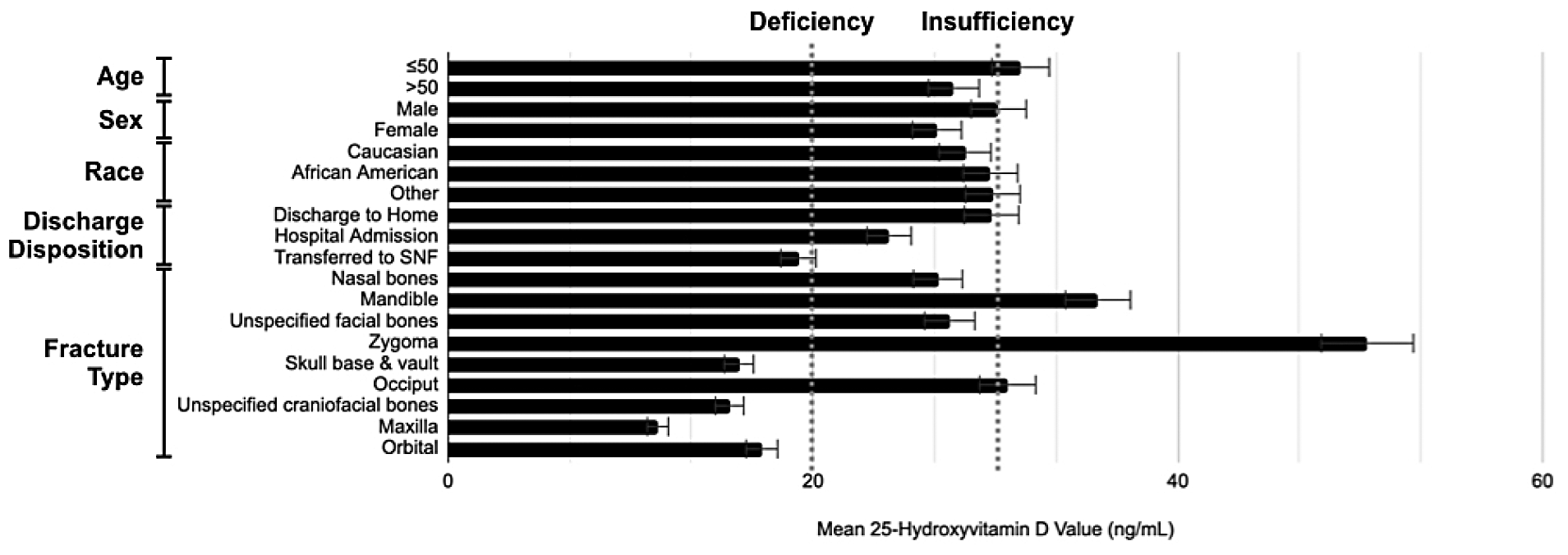
Certain fracture types were also associated with vitamin D insufficiency: nasal bones (26.85 ng/mL), unspecified facial bones (27.50 ng/mL), skull base and vault (15.94 ng/mL), unspecified craniofacial bones (15.43 ng/mL), maxilla (11.50 ng/mL), and orbital (17.20 ng/mL). Skull base and vault, unspecified craniofacial bones, maxilla, and orbital fractures also found 25-hydroxyvitamin D means that qualified as vitamin D deficiency (Figure 1).
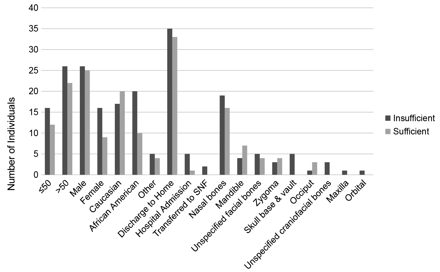
Across all ages and genders, more craniofacial fracture patients had vitamin D insufficiency than sufficiency. Men with facial fractures were just as likely to be vitamin D sufficient (51.0%) as insufficient (49.0%), whereas women with facial fractures were more likely to be vitamin D insufficient (64.0%) than sufficient (36.0%; Figure 2). Caucasian individuals with facial fractures were more likely to be vitamin D sufficient (54.1%) than insufficient (45.9%), whereas African American individuals with facial fractures were more likely to be vitamin D insufficient (66.7%) than sufficient (33.3%; Figure 2).
Discussion
Low levels of vitamin D are culpable in the pathophysiology of many chronic illnesses but are most notorious for their role in precipitating and exacerbating musculoskeletal diseases, namely, osteopenia, osteoporosis, osteomalacia, muscle weakness, and fractures.5 Though vitamin D is known to play an important role in fracture healing, further investigation is needed to fully delineate that role.3 Vitamin D deficiency is known to be common in the general population, but there are no studies to date specifically examining the vitamin D status of patients with facial fracture. The purpose of this study was to establish the prevalence and demographic discrepancy of vitamin D deficiency in a cohort of patients with facial fracture.
The overall prevalence of vitamin D deficiency in US adults is 24%.6 In this cohort of adult patients with craniofacial fractures from October 2015 until May 2018, the rate of vitamin D deficiency was 32.9%. The overall prevalence of vitamin D insufficiency in US adults has risen as high as 50 to 80% in the general population.7 This cohort had a rate of vitamin D insufficiency of 55.3%. Antifracture efficacy is achieved when 25-hydroxyvitamin D levels reach at least 30 ng/mL, putting all vitamin D deficient and insufficient patients at increased risk of fracture.5
Although recent systematic reviews still find the precise role of vitamin D in fracture healing to be incompletely investigated, experimental data demonstrate that vitamin D supplementation initiated after fracture and continued during the entire healing process prevents post-traumatic bone loss.8 Additionally, vitamin D has been shown to improve bone regeneration supporting the clinical potential of vitamin D supplementation in fracture healing.8 Given the higher prevalence of vitamin D insufficiency in this cohort compared with the general population, clinicians should consider empiric vitamin D supplementation in craniofacial fracture patients, even without well-established randomized controlled trials at this time.
Much of the research concerning bone health and fracture risk pertains to bones of the hip, vertebrae, and extremities. The mechanisms of these injuries, especially in the elderly, often relate to muscle weakness leading to increased fall risk and higher fracture rate. This leaves much to be wondered about the risk factors associated with craniofacial fractures. One study used osteomeatal unit computed tomography (CT) scans and dual-energy x-ray absorptiometry to analyze bone densities of facial bones in a group with osteoporosis and a control group. They found that facial bones deteriorate with age and severity of osteoporosis, just as bones in other locations in the body.9
Another study also identified racial and gender differences in facial fracture risk, with Caucasian and Asian individuals of either sex suffering higher incidence of facial fracture than African American individuals.10 Incidence of hip fractures maintains a racial component: controlling for age, Caucasian women are at twice the risk of hip fracture compared with African American women. Unlike facial fractures, the incidence of hip fractures has a strong sex component, with Caucasian women at 2.7 times the risk for hip fracture compared with Caucasian men.11
In this cohort, there were more male patients (67.1%) than female patients (32.9%) in all age groups. This might be explained by mechanisms of injury most common in craniofacial fractures, assault and motor vehicle crash, which men are more likely to be involved with than women.12 These results are also consistent with previous findings that showed a 3.6- to 5.4-fold higher rate of fracture in men than among women.13 Although men were more likely to suffer facial fractures overall, a relatively larger portion (64%) of women with facial fractures were vitamin D insufficient. This is consistent with studies that show women, across all age groups but especially those who are postmenopausal and osteoporotic, have a higher prevalence of vitamin D deficiency and insufficiency than men.14 Studies also demonstrate higher risk of fracture nonunion in women than in men.15 This might be explained in part by higher incidence of vitamin D insufficiency in women.
Caucasian patients were more likely to suffer facial fractures from this study population, but a relatively larger portion (67%) of African American people with facial fractures were vitamin D insufficient. Studies have shown that African American patients have a higher prevalence of calcidiol levels <30 ng/mL.16 Comparing the overall incidence of craniofacial fracture to the incidence of vitamin D insufficiency in specific groups (women and African American individuals) reveals significant discrepancies. Suffering facial fracture with underlying vitamin D deficiency is likely to delay healing, complicate surgical outcomes, and signal poor prognosis overall.17 Although not statistically significant in this study, these discrepancies might be concealing critical correlations and ought to be examined further.
Postmenopausal women are a group of particular interest in discussions about vitamin D deficiency and fracture risk. Physiologic changes to metabolism that occur during menopause, such as reduction in skin synthesis of vitamin D and changes in body composition, have implications for vitamin D status. Though patients’ menstrual status was not available, this study used the average age of menopause in the US—51 years—to estimate the relationship of menstrual status to vitamin D levels. In this study 60% (15 out of 25) of women were age 51 and above, of which 67% (10) were vitamin D insufficient and 20% (3) were vitamin D deficient. Low vitamin D levels could be associated with a number of physiologic and lifestyle factors associated with postmenopausal age. Studies show that empiric vitamin D supplementation for all postmenopausal women does not confer better clinical outcomes, but supplementing postmenopausal women who are vitamin D deficient does produce a significant reduction of fracture risk.18 Hence, menstrual status can be a useful characteristic to note when considering vitamin D supplementation in general but particularly in the setting of craniofacial fracture.
The laboratory measurement of 25-hydroxyvitamin D was vital to this study. However, this lab value is underutilized in facial fracture patients, usually drawn in clinical settings when a patient is suspected to have certain risk factors for vitamin D deficiency or if a patient reports a history of falls or fractures without significant trauma.19 Due to the availability of serum calcidiol in the patients found using the Cerner Health Facts database, we were able to conduct this study.
Future studies could expand on this research by performing a cost analysis of obtaining vitamin D levels for all patients. Given the high prevalence of vitamin D deficiency and insufficiency in the general population, assessing vitamin D status for more patients could help to elucidate significant relationships among vitamin D, fracture healing, and other chronic and acute diseases. Additionally, given the importance of mechanism of injury in the types and severity of craniofacial fractures, further investigation into vitamin D levels and mechanism of injury might prove to be useful. Furthermore, the natural history healing outcomes of fractures can be compared between vitamin D insufficient and sufficient cohorts. Randomized controlled trials should also be conducted to observe the outcomes of vitamin D supplementation after craniofacial fractures.
Limitations
One limitation of this study was its retrospective design. Another limitation was that calcidiol levels are not available in all patients with craniofacial fractures. Finally, the database utilized did not give access to clinical outcomes, mechanism of injury, preexisting medical conditions, menstrual status, or medications, which can be included in subsequent studies. The objective of this study was to focus on the prevalence and demographic discrepancies of vitamin D insufficiency and deficiency in craniofacial fractures
Conclusions
Fractures of the facial and skull bones can pose grave threats to physical and emotional wellbeing. Vitamin D plays a critical role in optimal bone health and fracture healing, with at least 30 ng/mL calcidiol needed to achieve antifracture efficacy. This is the first study investigating vitamin D status in adult craniofacial fracture patients. This study of craniofacial fractures found that more than half of patients had insufficient vitamin D levels and one-third were vitamin D deficient. Vitamin D insufficiency must be suspected in patients above 50 years of age, African American patients, and women. Serious consideration should be made to provide vitamin D supplementation following craniofacial fracture in these high-risk groups. Future studies can investigate the precise impact of vitamin D on healing and surgical outcomes in craniofacial fractures.
References
1. Lalloo R, Lucchesi LR, Bisignano C, et al. Epidemiology of facial fractures: incidence, prevalence and years lived with disability estimates from the Global Burden of Disease 2017 study. Inj Prev. 2020;26(Supp 1):i27-i35. doi:10.1136/injuryprev-2019-043297
2. Sastry SM, Sastry CM, Paul BK, Bain L, Champion HR. Leading causes of facial trauma in the major trauma outcome study. Plast Reconstr Surg. 1995;95(1):196-197. doi:10.1097/00006534-199501000-00039
3. Gorter EA, Hamdy NA, Appelman-Dijkstra NM, Schipper IB. The role of vitamin D in human fracture healing: a systematic review of the literature. Bone. 2014;64:288-297. doi:10.1016/j.bone.2014.04.026
4. Bischoff-Ferrari H. Vitamin D: what is an adequate vitamin D level and how much supplementation is necessary?. Best Pract Res Clin Rheumatol. 2009;23(6):789-795. doi:10.1016/j.berh.2009.09.005
5. Holick MF. Vitamin D deficiency. N Engl J Med. 2007;357(3):266-281. doi:10.1056/NEJMra070553
6. Amrein K, Scherkl M, Hoffmann M, et al. Vitamin D deficiency 2.0: an update on the current status worldwide. Eur J Clin Nutr. 2020;74(11):1498-1513. doi:10.1038/s41430-020-0558-y
7. Rosen CJ. Vitamin D insufficiency. N Engl J Med. 2011;364(3):248-254. doi:10.1056/NEJMcp1009570
8. Fischer V, Haffner-Luntzer M, Amling M, Ignatius A. Calcium and vitamin D in bone fracture healing and post-traumatic bone turnover. Eur Cell Mater. 2018;35:365-385. Published 2018 Jun 22. doi:10.22203/eCM.v035a25
9. Lee IJ, Lee JJ, Bae JH, et al. Significance of osteoporosis in facial bone density using computed tomography. J Craniofac Surg. 2013;24(2):428-431. doi:10.1097/SCS.0b013e3182801333
10. Hanba C, Svider PF, Chen FS, et al. Race and sex differences in adult facial fracture risk. JAMA Facial Plast Surg. 2016;18(6):441-448. doi:10.1001/jamafacial.2016.0714
11. Farmer ME, White LR, Brody JA, Bailey KR. Race and sex differences in hip fracture incidence. Am J Public Health. 1984;74(12):1374-1380. doi:10.2105/ajph.74.12.1374
12. Smith H, Peek-Asa C, Nesheim D, et al. Etiology, diagnosis, and characteristics of facial fracture at a midwestern level I trauma center. J Trauma Nurs. 2012;19(1):57-65.
13. Emodi O, Wolff A, Srouji H, et al. Trend and demographic characteristics of maxillofacial fractures in level I trauma center. J Craniofac Surg. 2018;29(2):471-475. doi:10.1097/SCS.0000000000004128
14. Muscogiuri G, Barrea L, Somma CD, et al. Sex differences of vitamin D status across BMI classes: an observational prospective cohort study. Nutrients. 2019;11(12):3034. Published 2019 Dec 12. doi:10.3390/nu11123034
15. Parker MJ, Raghavan R, Gurusamy K. Incidence of fracture-healing complications after femoral neck fractures. Clin Orthop Relat Res. 2007;458:175-179. doi:10.1097/BLO.0b013e3180325a42
16. Martins D, Wolf M, Pan D, et al. Prevalence of cardiovascular risk factors and the serum levels of 25-hydroxyvitamin D in the United States: data from the Third National Health and Nutrition Examination Survey. Arch Intern Med. 2007;167(11):1159-1165. doi:10.1001/archinte.167.11.1159
17. Bashutski JD, Eber RM, Kinney JS, et al. The impact of vitamin D status on periodontal surgery outcomes. J Dent Res. 2011;90(8):1007-1012. doi:10.1177/0022034511407771
18. Pérez-López FR, Chedraui P, Pilz S. Vitamin D supplementation after the menopause. Ther Adv Endocrinol Metab. 2020;11:2042018820931291. Published 2020 Jun 5. doi:10.1177/2042018820931291
19. Rockwell MS, Wu Y, Salamoun M, Hulver MW, Epling JW. Patterns of clinical care subsequent to nonindicated vitamin D testing in primary care. J Am Board Fam Med. 2020;33(4):569-579. doi:10.3122/jabfm.2020.04.200007