Evaluation of Tissue Apposition and Seroma Prevention in an Ovine Model of Surgical Dead Space Using a Novel Air-Purged Vacuum Closure System
Abstract
Background. Postoperative complications associated with seroma formation resulting from surgical dead space continue to present a challenge in modern surgery. There is an unmet need for new technologies that address surgical dead space as well as prevent seroma formation and associated downstream postoperative complications.
Methods. The novel implantable tissue apposition and drainage system ENIVO was developed and tested in a bilateral ovine external abdominal oblique (EAO) resection model of surgical dead space. The ENIVO system is a portable powered pump and wound interface featuring air-purged vacuum closure (APVC) that delivers a sustained level of vacuum pressure (80 and 100 mmHg) to the treatment site with an intermittent burst of sterile filtered air through the implanted wound interface. Seroma area, seroma volume, and drain migration were assessed at postoperative days 7 and 14, and all animals were euthanized at day 28 with gross assessment of treatment efficacy including the presence of residual seroma and tissue apposition.
Results. The bilateral model created relatively uniform defects of ~120 cm2 following excision of ~30 to 50 g of EAO muscle. Median seroma area of ENIVO-treated defects was statistically smaller than standard of care (SoC)-treated defects at days 7 and 14. Median seroma volume at 14 days was significantly reduced in ENIVO-treated defects relative to SoC-treated defects [1.3 (IQR 0.0-79.5) mL and 188.5 (IQR 27.6-342.9) mL, respectively]. At postoperative day 28, 40% (n = 4/10) of SoC defects showed a residual seroma, whereas in contrast, none of the ENIVO-treated defects showed signs of a residual seroma. Median tissue apposition scoring was higher in the ENIVO treatment group [3 (IQR 3-3)] compared with the SoC group [3 (IQR 0-3)].
Conclusions. The ENIVO system represents a new approach to dead space management and seroma prevention and was shown to outperform a SoC surgical drain in a challenging large defect model of surgical dead space management and seroma prevention.
Introduction
Surgical separation and/or excision of subcutaneous soft tissue as part of resections, reconstructions, or orthopaedic procedures often results in surgical dead space, where damaged vessels can fill the resultant void with plasma and lymph fluid.1-3 The resultant seroma is a collection of serous fluid that can increase patient discomfort, disrupt healing, and give rise to poor cosmetic outcomes.4 Seromas also increase the risk of more severe postoperative complications such as infection, wound dehiscence, lymphedema, and tissue necrosis.2,5 In some instances, the presence of seroma may delay downstream therapeutic interventions, eg, postmastectomy radiotherapy.6 Despite the risks associated with seroma formation, these are still relatively common surgical complications. For example, seromas have been reported as occurring in up to 20% of abdominoplasties,7 up to 52% of breast reconstructions,8 and between 20% to 79% of latissimus dorsi muscle harvest surgeries.9-11 Once established a seroma must be addressed surgically, often with repeated aspiration and compression. Where a seroma gives rise to infection, dehiscence, or tissue necrosis, additional surgeries are required to salvage the site, leading to increased surgical complexity, hospitalization, and overall cost while reducing the patient’s quality of life and potential to regain normal function.
The elimination of surgical dead space has received significant attention due to the negative patient outcomes that can occur in the event of seroma formation and the recognition that dead space is a key contributor to seroma formation.12 Several methods aim to directly mitigate dead space at the time of the surgical procedure via apposition of the tissue planes, whereas others seek to treat seroma prophylactically. Current standard of care (SoC) for mitigating surgical dead space includes meticulous surgical technique, tissue sealants such as fibrin adhesives,13 external compression dressings, surgical site immobilization, and quilting/progressive tension sutures.14,15 These approaches aim to mechanically affix the opposing tissue planes to reduce surgical dead space and therefore minimize seroma formation. Currently, the success of dead space management may be highly variable. For example, quilting sutures pexy the subcutaneous tissue to the deep fascia.16 However, the application of quilting sutures has been shown to reduce but not eliminate seroma from an incidence of 44.3% to 27.2% across a range of surgical procedures including abdominoplasty, breast reconstruction, and axillary dissection14 and in some instances leads to pain associated with nerve entrapment.17
The most common practice to prevent fluid accumulation and seroma formation is the prophylactic use of surgical drains.18 Surgical drains can be categorized as open or closed drains and both active or passive.19 Whereas open drains comprise corrugated or perforated tubes with an open outlet that drains fluid via gravity, closed drains provide a vacuum pressure (low or high negative pressure) to draw the serous and/or serosanguinous fluid from the surgical site to the collection vessel. There is now a wide range of closed surgical drains commercially available and in widespread use. The simplest and possibly most widely used consist of a thin silicone or PVC tube coupled to a mechanical vacuum source, such as a squeeze bulb or spring-loaded reservoir, that when depressed delivers negative pressure via the tube to the surgical site. These simple closed systems include the Jackson-Pratt (Cardinal Health) and the Hemovac systems (Zimmer Biomet). The vacuum pressure delivered by these simple systems is variable and can change based on the fluid volume contained within the reservoir and the method of mechanically priming the device.20,21 Though the application of surgical drains may decrease the incident of seroma, seroma and associated complications (eg, infection, hematoma, dehiscence, and necrosis) are still commonplace with existing technologies. The accumulation of fibrinous debris within the drain occurs universally and requires regular “stripping” or “milking” to force fluid, tissue, and clots through the tubing to return function.22 The accumulation of debris reduces drain efficacy and, when left unchecked, may result in complete drain blockage.23 Additionally, drain removal may result in significant patient discomfort, and drains are known to migrate postoperatively, potentially leading to reduced function or complication.24,25
The clinical need for technologies to effectively manage postsurgical dead space and prevent seroma formation led to the development of a new class of powered surgical closure device, termed air-purged vacuum closure (APVC). The first of these APVC systems (ENIVO) includes a portable battery-powered vacuum device and flexible fluid collection reservoir: a dual lumen silicone drain catheter fitted with an extracellular matrix (ECM) biomaterial sleeve interface. The ENIVO system has been designed to overcome the limitations of current systems by improving tissue apposition and fluid management as well as negating drain migration at the defect site. To validate the ENIVO system, a large animal model of dead space management was developed. Surgical dead space was surgically treated in a novel ovine bilateral external abdominal oblique (EAO) excision model, and surgical defects were treated with either the ENIVO system or a SoC drain.
Methods and Materials
General
Descriptive statistics (median, IQR, mean, SEM) were calculated using GraphPad Prism (version 9.0.0, Graphpad Software LLC). The normality of distribution of continuous variables was tested by Kolmogorov-Smirnov test. Continuous variables with normal distribution were presented as mean (SEM); non-normal variables were reported as median (IQR). For continuous non-normal variables, significance between treatment groups (SoC vs ENIVO) was determined using paired nonparametric (Wilcoxon) t tests, whereas normal variables were compared via paired t test. Differences within a treatment group (effect of time) were compared using a paired nonparametric Friedman test. For categorical variables P values were determined via a Chi-squared test. A P value less than .05 was considered statistically significant.
Investigational Devices
The SoC device (Cardinal Health Jackson Pratt 3-Spring Reservoir, with Cardinal Health Jackson Pratt 15Fr PVC round drain) was used according to the product instructions for use without modification. The SoC device is a mechanical closed vacuum drain system and comprised a (15Fr) Ø5-mm OD PVC surgical drain tethered to a 3-spring mechanical vacuum canister with 400-mL capacity.
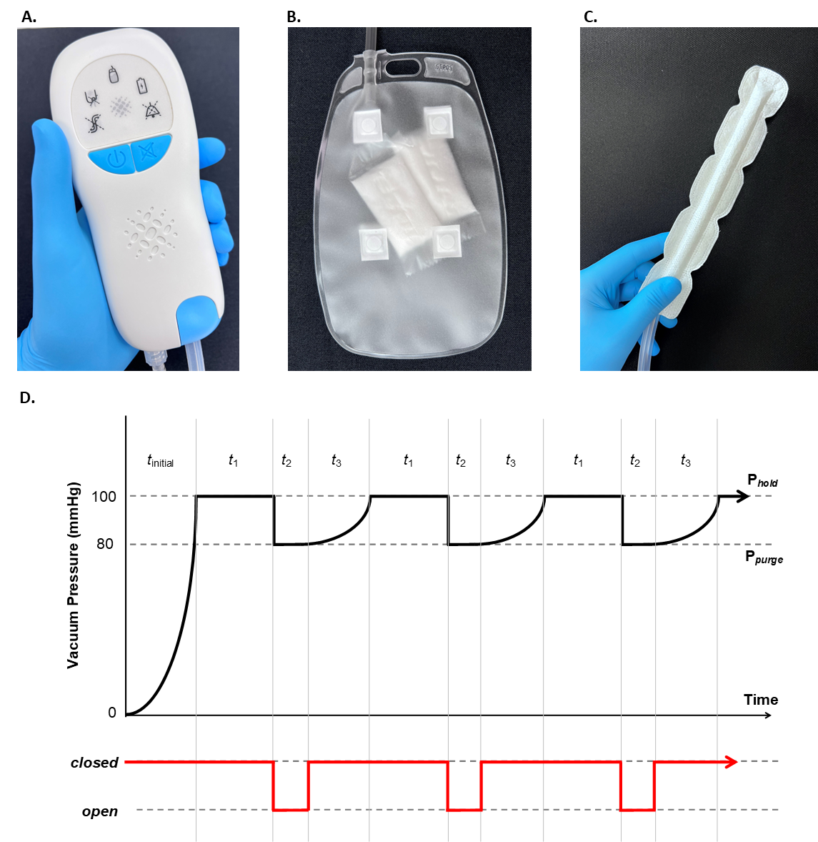
The ENIVO device (Aroa Biosurgery Limited) comprised 2 main components: a silicone dual lumen drain catheter, fitted with an ovine forestomach matrix (OFM) sleeve interface, tethered to a proprietary vacuum pump system preprogrammed to deliver APVC (Figure 1). The OFM catheter sleeve comprised 2 layers of OFM, fabricated and reinforced with braided (multifilament) PGA resorbable suture, to create a linear opening [~Ø1-cm inner diameter (ID )] to accommodate the silicone drain catheter (Figure 1c). The OFM sleeve was perforated [~Ø0.5-mm outer diameter (OD )] on both sides with the perforations spaced ~2 mm apart across the length to enable fluid egress at the treatment site. A dual lumen catheter (~Ø10-mm OD) was preassembled into the OFM sleeve prior to surgical placement. The drain catheter had 2 adjacent internal pathways of Ø1.6-mm ID and Ø4.8-mm ID. The larger Ø4.8-mm ID tube was connected to the vacuum outlet, whereas the smaller Ø1.6-mm ID tube connected to the air purge inlet. The drain catheter had Ø2-mm and Ø3-mm OD perforations across the larger Ø4.8-mm ID lumen at the portion interfacing with the OFM sleeve to enable fluid egress at the treatment site.
The drain catheter connected directly to a proprietary vacuum device system (Figure 1a) fitted with a 700-mL fluid collection bag, which included solidifier (sodium polyacrylate powder contained within a polyvinyl acylate film sachet; Figure 1b). The vacuum pump and associated air purge valve delivered the novel proprietary APVC treatment algorithm that oscillates the vacuum pressure between a first target pressure Phold and second target pressure Ppurge (Figure 1d). The system initially depressurised to a first target vacuum pressure of ~100 mmHg (Phold) over ~30 seconds (tinitial). Once the target Phold had been reached, a constant vacuum pressure (~100 mmHg) was maintained for a further 2 minutes (t1), then the air purge valve opened while the vacuum pump was operating to introduce sterile filtered (0.22 µm) air into the system, resulting in a vacuum pressure drop to a second target vacuum pressure ~80 mmHg (Ppurge) over ~14 secs (t2). At this point, the air purge valve automatically closes causing the system to increase vacuum pressure (t3 < 30 sec) and return to the first target vacuum pressure Phold. Oscillations in the vacuum pressure continue for ~30 hours, at which point the pump delivers a continuous ~150 mmHg. The OFM sleeve interface and silicone drain catheter were terminally sterilized prior to use.
Ovine Bilateral External Abdominal Oblique Model of Seroma
Ten female, nonpregnant Ovis aries (Romney or Romney-Texel Cross; <2 years, ~60-90 kg) were acclimatized to the indoor enclosure for a minimum of 7 days prior to surgery with daily monitoring, with feed and water ad libitum. During the acclimatization period all animals were fitted with an equipment harness that was worn continuously for >5 days prior to surgery. The custom harness, constructed of polyether polyurethane foam (H35-200, Nexus Foams) and thermoplastic (ORFIT ECO 3.2 mm perforated, ORFIT Industries), was fitted to straddle the length of the animal’s spine and secured via hook and loop fasteners (Velcro) at 3 locations (hindlegs, legs, and collar). The harnesses were designed to enable free range of motion and not to occlude the bilateral surgical sites. Animals were housed during the acclimatization period and postoperatively up to day 14 in an indoor pen (6 x 6 m).
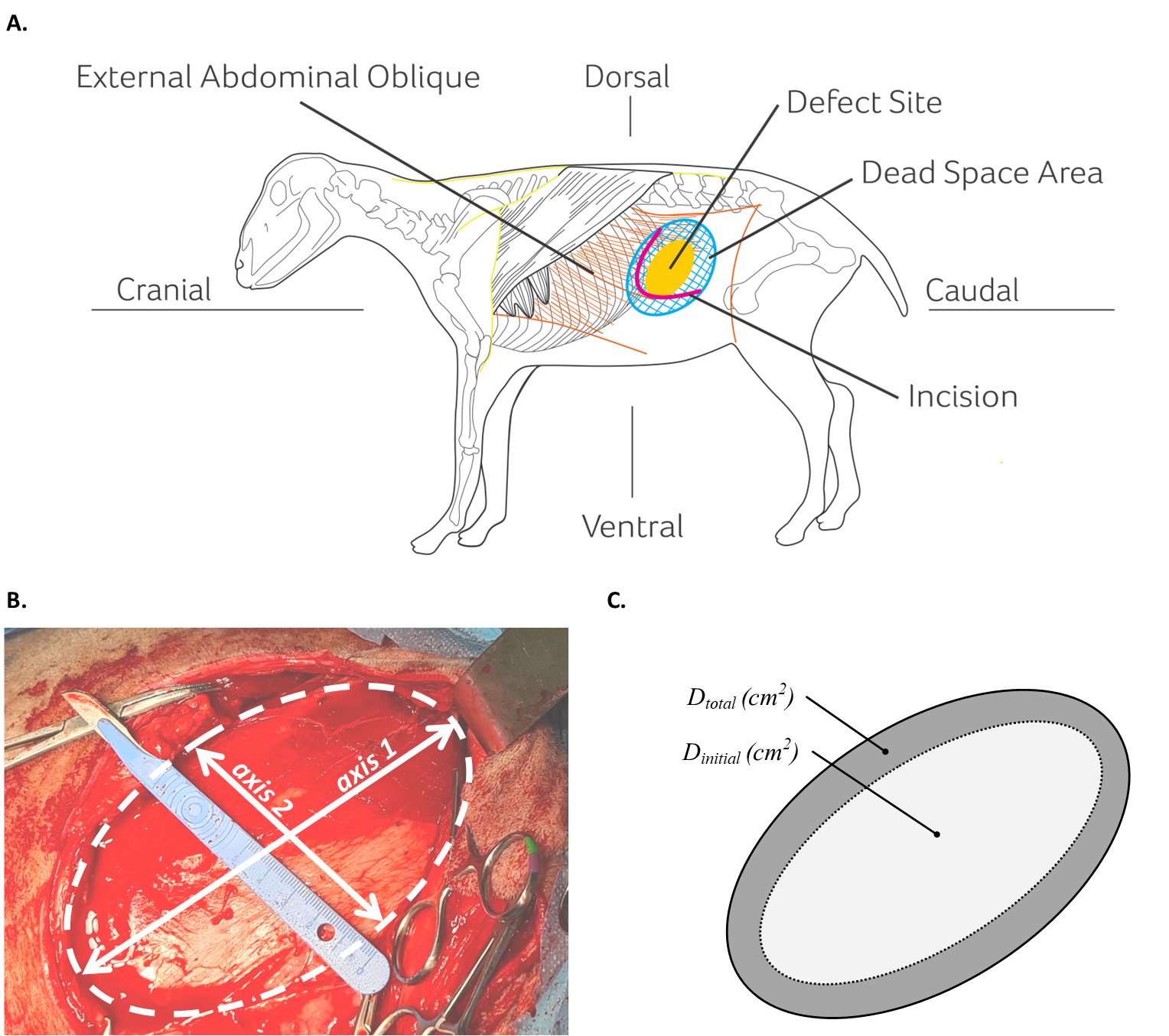
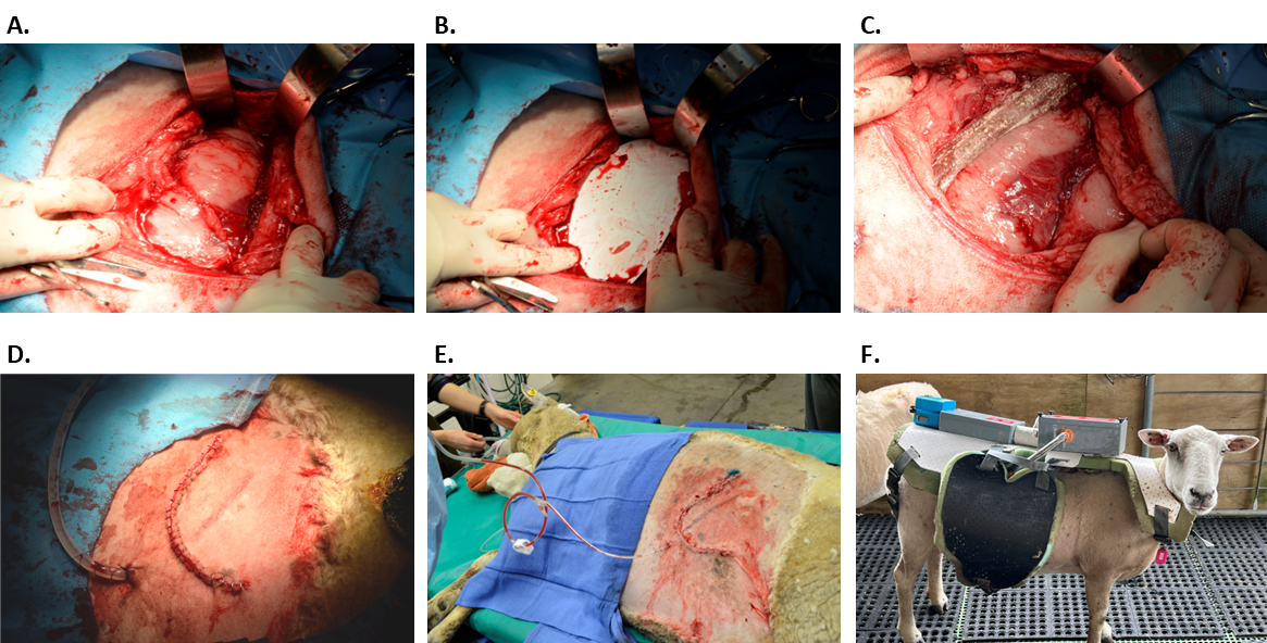
Animals were induced with intravenous diazepam (0.2-0.3 mg/kg, 6 mL) then intravenous ketamine (2-5 mg/kg, 6 mL). A rumen tube was passed to empty ruminal contents and stayed in place throughout the duration of the surgery. An endotracheal tube was inserted aided by a laryngoscope and secured in place with the cuff inflated to allow administration of isoflurane (3%–5%) to maintain anaesthesia. Lignocaine hydrochloride (2-4 mg/kg, 2.5 mL) was administered topically onto the larynx as required to aid in the insertion of the endotracheal tube. Animals were placed in the lateral recumbency, the wool was removed with electric clippers, and the surgical site was prepared with chlorhexidine/ethanol (5%/95% w/v) and rinsed with sterile saline. Animals were administered prophylactic antibiotics (Bivatop 200, oxytetracycline, 20 mg/kg, 6 mL) and analgesic (Meloxicam, Loxicom LA, 1 mg/kg, 3 mL) prior to incision. On the animal’s left or right abdomen, the caudal edge of the last rib was located, and from this point an arcing skin incision (approximately 20 cm in length) was made. The external abdominal oblique muscle was identified and subcutaneous tissue dissected away using a scalpel from the external abdominal oblique muscle in a caudal direction to expose the entire muscle. Vessels were isolated and clamped using hemostats as required. The muscle above the rib was excised in an arcing shape extending caudally and ventrally to create the ~90 x 100 x 10-mm defect area (Figure 2a and Figure 3a). The excised tissue was full thickness through the EAO muscle bundle. Following initial excision, the defect was further enlarged by selective undermining of tissues extending ~3 to 4 cm around the perimeter of the initial excisional defect. A sterile template was used to standardize the area of the excision (Figure 3b).
Prior to placement of the test devices the initial defect area (Dinitial) and total defect area (Dtotal) were estimated by the length (cm) of the 2 perpendicular axes that define the elliptical defect (Figure 2b and c).
The 2 defect sites (left and right) on each animal were randomized to receive either treatment, such that each animal received both an ENIVO and an SoC treatment. A single small cutaneous incision (~1 cm) was made approximately 3 cm distal from the primary incision to port the device tubes through the cutaneous tissue (Figure 3d). The drains were passed through the cutaneous tissue and laid into the defect vertically (Figure 3c). The OFM sleeve of the ENIVO device was tack sutured to the underlying defect tissues at 8 points spaced ~2 cm apart using polypropylene suture and rehydrated in sterile saline (1-2 minutes). The tubes of both devices were secured at the skin portal using a poliglecaprone 25 suture on the outside of the porting incision. Care was taken to ensure the drains were not pierced. The subcutaneous tissues were closed using poliglecaprone 25 suture and the skin incision closed using staples. The approximate location of both drain devices was marked for reference on the skin with a surgical pen (Figure 3e). Oxytetracycline hydrochloride (Tetravet Blue) was applied to the closed incisions as well as intramuscular bupivacaine (1-2 mg/kg, 40 mL). Equipment harnesses were fitted to each animal ensuring that no load was being applied to the to the surgical sites (Figure 3f).
Animals were extubated when actively swallowing and monitored during recovery. A further subcutaneous injection of meloxicam (Loxicom LA, R&D Labs, 1 mg/kg, 3 mL) and intramuscular oxytetracycline (Bivatop 200, Boehringer Ingelheim, 20 mg/kg, 6 mL) were given at 48 hours post surgery, then as needed. Food and water were available ad libitum, and animals were moved to open pasture at postoperative day 14. Animals were monitored twice daily for 5 days then daily for signs of pain, central nervous system complications, inappetence, comparably decreased activity, or weight loss. Additionally, the surgical defects were assessed for hemorrhage, edema, swelling, and infection. The test drains were not milked or stripped postoperatively.
Ultrasound Assessment
Ultrasound assessment of the defect was performed on postoperative days 7 and 14 to quantify the seroma volume via Xaxis, Yaxis, and Zaxis measurements (cm) of the defect. Animals were secured in a pinch gate during the examination. Ultrasound (Sonosite Edge II, Fujifilm) was performed using a transducer (HFL50, Fujifilm) set to operating mode “M.” Methylated spirits was applied to the skin to reduce acoustic impedance during the ultrasound. To define the Xaxis and Yaxis, the scanner was passed over the skin of the surgical defect and the boundary of any seroma identified and marked with a surgical pen. The distances Xaxis and Yaxis were then measured using a ruler, and seroma area (cm2) that occupies the original defect area (Dtotal) was calculated using the formula:
The seroma depth( Zaxis ) was determined by moving the scanner across the skin of the defect to identify the maximum depth, then Zaxis was measured using the ultrasound measurement function. Seroma volume (mL) was calculated using the ellipsoid formula:
Seroma area at postoperative days 7 and 14 was also expressed as the percentage change relative to the total defect area (Dtotal) using the formula:
Drain Migration Assessment
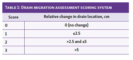
A drain migration assessment was performed on postoperative days 7 and 14 to determine the retention or migration of the treatment drains within the defect site over the course of the study. The position of each implanted drain within the defect was located using a combination of ultrasound and gentle palpation of the skin, and the position was assessed relative to the original location, as indicated by the skin markers applied at the time of surgery. The relative change in the drain location versus baseline position was quantitatively assessed according to Table 1.
Drain Fluid Output Volume
Drain output volume was collected once daily following the surgery until the volume of fluid collected was less than 15 mL following 2 consecutive days. Cumulative fluid volume output was expressed as the sum of the total drain fluid outputs.
Drain Removal
At postoperative day 14, animals were restrained in a pinch gate with the drain stay sutures removed prior to drain removal. A handheld 50N force gauge (FL 50, Sauter) was secured to the distal end of each drain, ~ 5 cm above the skin, and force applied to remove the drain from the defect. Maximum force (N) was recorded for each treatment drain.
Euthanization and Gross Assessment of Tissue Apposition
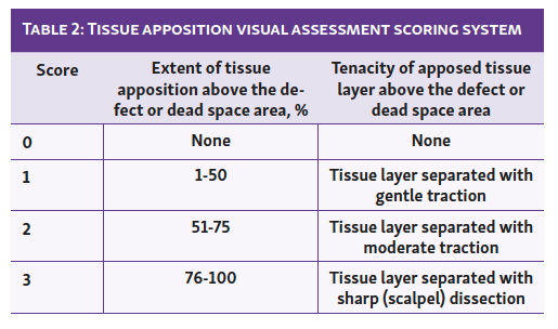
At postoperative day 28 all animals were euthanized via intramuscular administration of xylazine (1.5-2.0 mL, PhoenixPharm), followed by intravenous pentobarbitone (20 mL, ProVet). A gross visual assessment of each defect site was conducted immediately following euthanasia to observe the presence of any residual seroma and assess the extent and tenacity of apposed tissue. Qualitative scoring was assessed according to Table 2.
Results
Using the bilateral EAO model, relatively uniform soft tissue defects could be created. The mean tissue mass excised were equivalent in the ENIVO and SoC treatment groups (36.8 ± 2.9 g and 36.3 ± 2.2 g, respectively; Table 3), and there were no differences in excised tissue mass between the left- and right-side defects (data not shown). The resulting defect area following the initial excision (Dinitial) was ~65 cm2, whereas the final defect area (Dtotal) was nearly double in size (~120 cm2) once tissue undermining was taken into consideration (Table 3). There was no significant difference in defect size between the 2 treatment groups (Table 3). All animals recovered well post surgery and returned to typical daily function within ~4 days. A single ENIVO drain catheter was dislodged prior to day 7 and excluded from analysis, along with the contralateral SoC drain. A SoC drain was dislodged following ultrasound and prior to drain pull out and drain migration assessments.
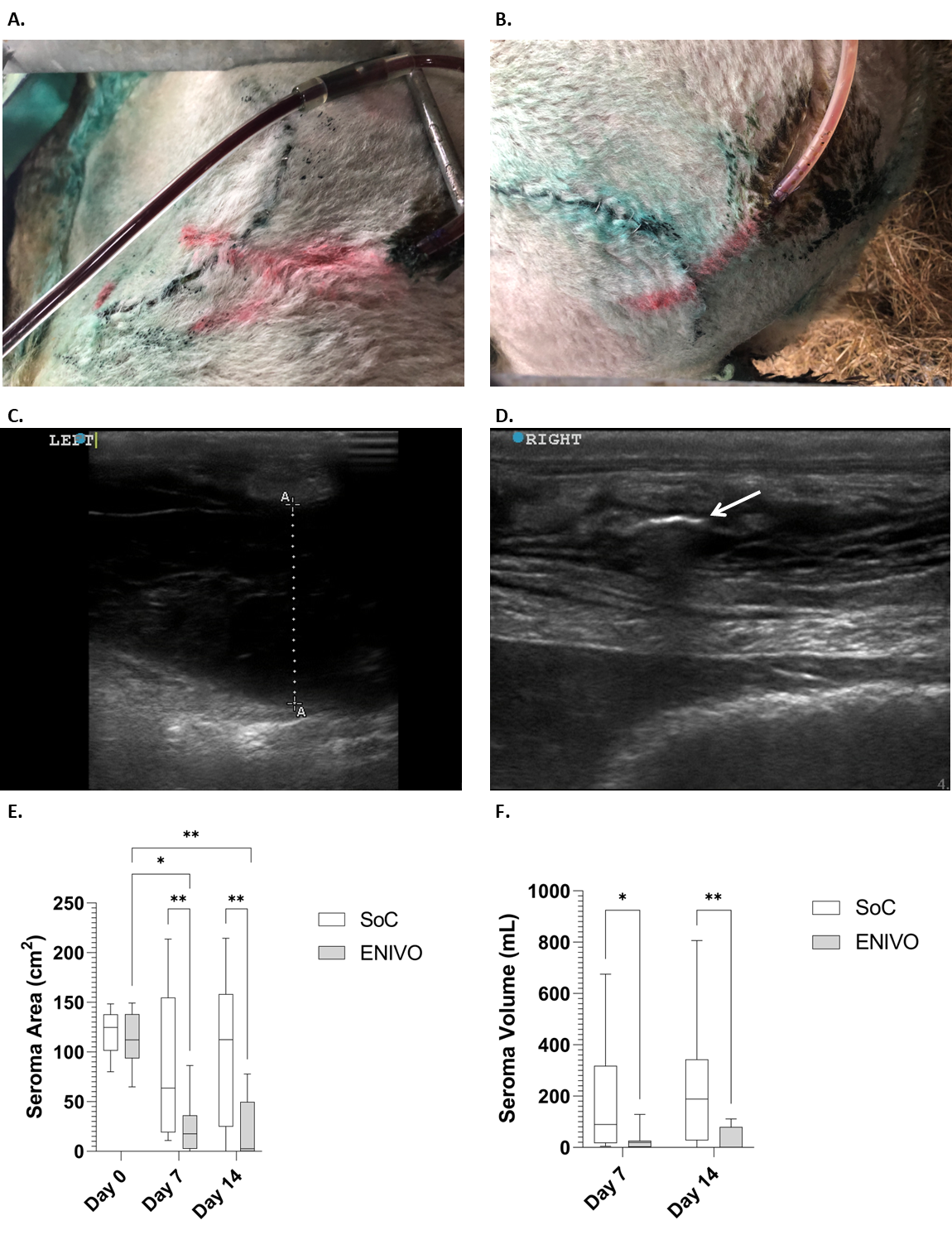


Fibrous material accumulated in the SoC drains from postoperative day ~1 to 2, whereas the extent of fibrinous material in the ENIVO drain catheter was less apparent visually (Figure 4a and 4b). Seromas were clinically observable by postoperative days 3 to 7 and were clearly visible via ultrasound (Figure 4c). Seroma area and seroma volume as determined from ultrasound are presented in Table 4 and Figure 4. By day 7, defects treated with SoC had a median seroma area and volume of 63.6 (IQR 19.1-154.7) cm2 and 89.9 (IQR 17.3-318.3) mL, respectively (Table 4). In comparison ENIVO treatment led to a significantly reduced median seroma area [17.7 (IQR 2.7-36.2) cm2], and seroma volume [19.6 (IQR 2.4-26.3) mL], compared with SoC treatment. At day 14 the differences were more pronounced with ENIVO-treated defects having significantly reduced median seroma area and seroma volume [2.5 (IQR 0.0-49.6) cm2 and 1.3 (IQR 0.0-79.5) mL] compared with that of the SoC group. Seroma area of ENIVO-treated defects at day 7 and day 14 were statistically smaller relative to the day 0 total defect area (Dtotal). The seroma areas at day 7 and day 14 were also expressed as a percentage change relative to the total defect areas (Dtotal;Table 4). Percentage seroma area change at day 7 and day 14 for the SoC-treated defects were 79.0% (IQR 15.5%-142.0%) and 98.0% (IQR 19.5%-128.5%), respectively, with 4 of the 10 defects increasing in area relative to the measured area at day 0 (Dtotal). In contrast, ENIVO-treated defects at days 7 and 14 were 13.0% (IQR 2.5%-42.5%) and 2.0% (IQR 0.0%-55.0%) of the original total defect area (Dtotal), respectively. The cumulative drain fluid output was determined over the course of the study (Table 5 and Figure 5). At all timepoints ENIVO-treated defects resulted in an increase in the cumulative drain fluid volume output (Figure 5a). At day 14 the mean cumulative volume collected by the ENIVO devices was approximately triple that of the SoC devices, 178.4 ± 23.2 mL versus 64.1 ± 14.8, P = .0026 (Figure 5b). In both treatment groups the majority of the fluid output was collected in the first 3 days (Figure 5a). At day 14 treatment in both groups was ceased. Following the postoperative day 14 ultrasound assessment, ENIVO and SoC drains were removed and the maximum force required to remove each drain measured. The mean maximum drain pull force required to remove an ENIVO drain catheter was statistically higher than that for the SoC drains, 11.68 ± 2.11 N and 3.66 ± 1.12 N, respectively (Table 4 and Figure 6a). Drain migration was assessed at postoperative days 7 and 14 and scored according to Table 1. At days 7 and 14, 8 of 9 (89%) ENIVO drains were judged as having no migration from the original position within the defect (Figure 6b). In contrast, SoC drains were relatively mobile, with over half the drains being assessed as having migrated from their original position by postoperative day 7.
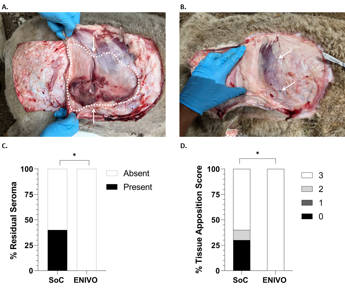
All animals were euthanized at postoperative day 28, and treatment success was assessed by visual examination of the defect site. Where treatment had been unsuccessful and a seroma had formed, evidence of the residual seroma was apparent, as characterized by fibrinous deposits and/or a seroma capsule (Figure 7a). In contrast, where treatment was successful and no residual seroma present, the defect contained only dense connective tissue (Figure 7b). Animals treated with the ENIVO system showed no evidence of residual seroma at the 28-day euthanization (Table 4 and Figure 7c), whereas 40% (n = 4/10) of the SoC group showed evidence of residual seroma within the defect. Visual assessment of residual seroma was complemented by tissue apposition scoring, a qualitative assessment based on the tenacity of the adjacent tissue planes (Table 2). Where treatment had been unsuccessful and a residual seroma was present at necropsy, the strength and tenacity of the opposing tissue planes was relatively low (eg, score = 0, Table 2). There was a statistically significant (P = .0348) difference between the 2 treatment groups, where ENIVO-treated defects had a median tissue apposition score of 3 (IQR 3-3) compared with the SoC group, 3 (IQR 0-3; Table 4 and Figure 7d). At necropsy the OFM drain sleeve was not apparent in the defect, indicating early incorporation of the biomaterial into the regenerating tissues of the opposing tissue planes.
Discussion
There are few established models of dead space management and seroma formation with which to meaningfully evaluate device-based interventions. Ex vivo models of seroma are available; however, these are less physiologically relevant than animal models and do not necessarily recapitulate the clinical problem.26,27 Rodent models of seroma have been published, mostly focused on excision of the major pectoralis muscle28-30 or latissimus dorsi muscle.31-33 The use of small animals has some advantage, being relatively inexpensive and easy to execute; however, due to the small size of the animals and the resultant surgical defect, these models are generally more suited to validation of pharmacotherapeutic interventions. Previously, several groups have provided more physiologically relevant large animal models of seroma.34-37 The bilateral porcine model of dead space creates a 25-cm incision on the backs of animals, with partial removal of the back muscle.38 Klima et al35 published a porcine bilateral axillary lymphadenectomy model using an incision made over the axilla and then blunt, sharp, and cautery dissection of the axillary lymph node. However, the model employed a relatively small defect area (8-9–cm incision) that may not recapitulate features of human surgeries. Gilbert et al.36,37 developed a canine model of abdominoplasty, whereby cutaneous tissues were lifted to created 2 adjacent subcutaneous pockets. The model results in a large surface-area defect (15 x 10 cm) that mirrors human abdominoplasty, and seroma was shown to develop within the subcutaneous pockets. However, this model does not include volumetric tissue excision to create dead space as is common in various human surgeries, eg, mastectomy. The ovine bilateral EAO dead space seroma model was developed to address these shortcomings. The EAO muscle of an adult sheep spans a large area, and following partial excision the resulting dead space area is positioned between the ribs and rear limb at the dorsal-caudal quadrant of the muscle. Partial excision of the abdominal oblique muscle and subsequent undermining gave a dead space area of ~120 cm2 at a depth of ~1 cm, with the mass of excised tissue ~30 to 50 g. All steps were taken to ensure consistency across the defects created, including the use of a surgical template to mark the area of excision. The size of the defect and mass of tissue excised was consistent with excisions made during mastectomy (tissue mass 30 g, tissue volume of 210-1588 cm3)39 but is less than the excised tissue mass reported for latissimus dorsi musculocutaneous flap for breast reconstruction (180-610 g).40 In this model ineffective treatment resulted in a visible seroma by day 3 to 7, and by day 14 the seroma volume could reach up to ~800 mL. In this respect the model represents a clinically relevant model of surgical dead space and an especially challenging scenario for intervention. This is the first instance of high-resolution ultrasound being used as part of a large animal in vivo model of seroma formation, though this technique is common practice clinically in the diagnosis and management of seroma. The use of ultrasound to assess seroma area and seroma volume negated the need to terminate animals at interim timepoints (days 7 and 14) and meant aspirations of the seromas were not required to assess seroma volume.
The ENIVO system has been designed to address shortcomings with existing technologies for dead space management and seroma prophylaxis. Treatment with the ENIVO system resulted in reduced seroma area and seroma volume at days 7 and 14 (Figure 4) and over the course of 14 days drew more fluid from the defect versus SoC (Figure 5a). At the 28-day euthanization, 9 of 9 (100%) defect treatments were judged clinically successful with use of the ENIVO system versus only 6 out of 10 (60%) for the SoC treatment. The absence of any residual seroma correlated to strength and tenacity of the tissue apposition, with ENIVO-treated sites having a statistically higher tissue apposition score versus SoC-treated sites.
One of the main limitations of existing closed surgical drains is ineffective fluid removal due to the accumulation of fibrinous material within the drain, blockage, and loss of vacuum pressure. This is especially apparent ~1 to 3 days postoperative when fluid production from tissue trauma is greatest. As a result, it is best practice with existing drains to milk the drain tube to prevent accumulation of fibrinous material and blockage. To overcome these challenges the ENIVO system has been designed to deliver a novel oscillating negative pressure that we have defined as “air-purged vacuum closure” to the defect site (Figure 1d). To achieve this, the system utilizes an air purge valve that sequentially opens and closes during typical operation. When the air purge valve opens while the system is under vacuum, a short (t2~14 secs ) bolus of higher-pressure sterile air is introduced into the system, forcing fluids through the vacuum line and into the collection bag. This approach has a similar effect as manual milking of surgical drain tubes that occurs with traditional systems. As a result of this functionality the risk of complete blockage of the ENIVO drain catheter in the early and critical 1 to 3–day postoperative period is greatly reduced, and experimentally the build-up of fibrinous materials and cellular debris in ENIVO catheters was less apparent than that of SoC drains (Figure 4a and 4b). It should be noted that this approach to the delivery of negative pressure is quite distinct from intermittent negative pressure wound therapy, where oscillations in vacuum pressure result from the vacuum pump cycling between on (at a set vacuum pressure level) and off (ambient or near-ambient pressure level), not from the introduction of air into the vacuum system.
Typically, surgical drains have been used prophylactically to reduce the risk of seroma formation. However, available technologies to address the underlying cause, namely surgical dead space, have been limited. This is addressed in part by a more effective delivery of consistent vacuum pressure to the surgical defect in the immediate postoperative period to draw and hold the 2 opposing tissue planes together. Additionally, the ENIVO system included a tissue interface comprising OFM to enable early tissue ingrowth and adhesion between opposing tissue planes. OFM is a resorbable decellularized ECM biomaterial prepared from ovine forestomach tissue41 that retains the native structure of tissue ECM42,43 and a large number of naturally occurring matrisomal proteins.44 When implanted OFM is infiltrated with host cells, scaffolds soft tissue regeneration, and over time is remodelled to give highly vascular and functional tissue.45,46 OFM has seen widespread use in wound healing, burns, and complex soft tissue reconstruction.47-51 The inclusion of the OFM sleeve interface in the design of the ENIVO system was based off similar biological interfaces that have been developed for hardware implants, including cardiac implantable electronic devices and neurostimulators. For example, in the clinical use of cardiac implantable electronic devices, ECM biologic interfaces are now commonplace (eg, CanGaroo ECM, CorMatrix Cardiovascular) and have been shown to reduce erosion of the surrounding soft tissue, dehiscence, and infection.52-54 The rationale for these ECM sleeves is to buffer any host tissue inflammatory response to the implanted foreign material while also serving to scaffold new tissue formation at the interface as the ECM is infiltrated with host cells and ultimately remodelled into healthy well-vascularized tissue. From a practical standpoint, ECM sleeves, like that included in the ENIVO system, enable fixation of the implantable hardware or drain to the surrounding tissues.55 Thus, we considered that the OFM sleeve and drain catheter interface could improve the efficacy of the ENIVO system by reducing drain migration, host tissue inflammatory response, and importantly enabling more effective tissue apposition via integration of the tissue planes into the OFM sleeve during the early postoperative healing period. Other researchers have studied the addition of decellularized ECM28 and collagen-based devices56 to surgical defects to reduce seroma formation and shown this approach to be effective in vivo. For example, the addition of bovine dermal ECM reduced seroma formation in a dose-dependent manner in a rodent model of mastectomy and axillary dissection.28 The current study found that tissue apposition was significantly improved in ENIVO-treated surgical defects.
Limitations
The current study represents the first description of the ENIVO system and the APVC algorithm in a clinically relevant animal model of surgical dead space management. The bilateral ovine EAO model presented a challenging environment for the evaluation of the test devices, but further clinical evaluations across a range of surgical procedures will be required to prove the efficacy of the system. In the current study we did not histologically assess the local inflammation or remodelling of the OFM catheter sleeve, but based on existing published data from in vivo and human studies,45,46,57 we don’t expect any untoward local tissue reaction as a result of using OFM in combination with a surgical drain. For ethical reasons an untreated control group was not included in the current study. However, based on outcomes for the SoC group we would expect spontaneous seroma formation in untreated animals using the bilateral EAO model.
Conclusions
Effective surgical dead space management and seroma prevention remains a challenge in modern surgery. The novel ENIVO system aims to reduce the risk of seroma formation by addressing dead space management via the effective delivery of local vacuum pressure to the defect to draw opposing tissue planes together and via use of a biomaterial catheter interface to biologically adhere tissue planes through tissue integration. Initial findings in a challenging surgical model of dead space management demonstrated that the ENIVO system outperformed current SoC device in fluid removal, seroma prevention, and tissue apposition.
Acknowledgments
The authors acknowledge the assistance of staff at Aroa Biosurgery Limited (Auckland, New Zealand), AgResearch (Ruakura, New Zealand), and Vet Associates Limited (Auckland, New Zealand) for execution of the in vivo study.
Affiliations: 1Aroa Biosurgery Limited, Auckland, New Zealand; 2Vet Associates Equine, Auckland, New Zealand
Funding: This work was supported by Aroa Biosurgery Limited and Callaghan Innovation Limited (Wellington, New Zealand) Growth Grant (MSMA1402).
Ethics: Ethical approval for animal manipulations and surgery was obtained from the AgResearch Animal Ethics Committee (AE15327, Hamilton, New Zealand) and executed in accordance with the NZ Animal Welfare Act 1999.
Disclosures: ITTM, HJR, SFW, ATJ, SJG, HDC, CCHW, AJD, SBT, YEP and BCHM are employees and shareholders of Aroa Biosurgery Limited.
References
1. Ousey K, Djohan R, Dowsett C, Ferreira F, Hurd T, Romanelli M et al. Surgical wound dehiscence : Improving prevention and outcomes. World Union of Wound Healing Societies (WUWHS); 2018:1-48.
2. Bullocks J, Basu CB, Hsu P, Singer R. Prevention of hematomas and seromas. Semin Plast Surg. 2006;20(4):233-240. doi:10.1055/s-2006-951581
3. Klima DA, Brintzenhoff RA, Tsirline VB, et al. Application of subcutaneous talc in hernia repair and wide subcutaneous dissection dramatically reduces seroma formation and postoperative wound complications [published correction appears in Am Surg. 2014 Apr;80(4):422]. Am Surg. 2011;77(7):888-894.
4. van Bemmel AJ, van de Velde CJ, Schmitz RF, Liefers GJ. Prevention of seroma formation after axillary dissection in breast cancer: a systematic review. Eur J Surg Oncol. 2011;37(10):829-835. doi:10.1016/j.ejso.2011.04.012
5. Toyserkani NM, Jørgensen MG, Haugaard K, Sørensen JA. Seroma indicates increased risk of lymphedema following breast cancer treatment: A retrospective cohort study. Breast. 2017;32:102-104. doi:10.1016/j.breast.2017.01.009
6. Kumar S, Lal B, Misra MC. Post-mastectomy seroma: a new look into the aetiology of an old problem. J R Coll Surg Edinb. 1995;40(5):292-294.
7. Seretis K, Goulis D, Demiri EC, Lykoudis EG. Prevention of seroma formation following abdominoplasty: a systematic review and meta-analysis. Aesthet Surg J. 2017;37(3):316-323. doi:10.1093/asj/sjw192
8. Barwell J, Campbell L, Watkins RM, Teasdale C. How long should suction drains stay in after breast surgery with axillary dissection? Ann R Coll Surg Engl. 1997;79(6):435-437.
9. Clough KB, Louis-Sylvestre C, Fitoussi A, Couturaud B, Nos C. Donor site sequelae after autologous breast reconstruction with an extended latissimus dorsi flap. Plast Reconstr Surg. 2002;109(6):1904-1911. doi:10.1097/00006534-200205000-00020
10. Kat CC, Darcy CM, O'Donoghue JM, Taylor AR, Regan PJ. The use of the latissimus dorsi musculocutaneous flap for immediate correction of the deformity resulting from breast conservation surgery. Br J Plast Surg. 1999;52(2):99-103. doi:10.1054/bjps.1997.3035
11. Delay E, Gounot N, Bouillot A, Zlatoff P, Rivoire M. Autologous latissimus breast reconstruction: a 3-year clinical experience with 100 patients. Plast Reconstr Surg. 1998;102(5):1461-1478. doi:10.1097/00006534-199810000-00020
12. Pollock TA, Pollock H. No-drain abdominoplasty with progressive tension sutures. Clin Plast Surg. 2010;37(3):515-524. doi:10.1016/j.cps.2010.03.004
13. Grossman JA, Capraro PA. Long-term experience with the use of fibrin sealant in aesthetic surgery. Aesthet Surg J. 2007;27(5):558-562. doi:10.1016/j.asj.2007.07.004
14. Janis JE, Khansa L, Khansa I. Strategies for postoperative seroma prevention: a systematic review. Plast Reconstr Surg. 2016;138(1):240-252. doi:10.1097/PRS.0000000000002245
15. Massey LH, Pathak S, Bhargava A, Smart NJ, Daniels IR. The use of adjuncts to reduce seroma in open incisional hernia repair: a systematic review. Hernia. 2018;22(2):273-283. doi:10.1007/s10029-017-1690-z
16. Aho JM, Nickerson TP, Thiels CA, Saint-Cyr M, Farley DR. Prevention of postoperative seromas with dead space obliteration: A case-control study. Int J Surg. 2016;29:70-73. doi:10.1016/j.ijsu.2016.03.004
17. Pollock TA, Pollock H. Progressive tension sutures in abdominoplasty: a review of 597 consecutive cases [published correction appears in Aesthet Surg J. 2012 Sep;32(7):910]. Aesthet Surg J. 2012;32(6):729-742. doi:10.1177/1090820X12452294
18. Bercial ME, Sabino Neto M, Calil JA, Rossetto LA, Ferreira LM. Suction drains, quilting sutures, and fibrin sealant in the prevention of seroma formation in abdominoplasty: which is the best strategy? Aesthetic Plast Surg. 2012;36(2):370-373. doi:10.1007/s00266-011-9807-8
19. Mamuyac EM, Pappa AK, Thorp BD, et al. How much blood could a jp suck if a jp could suck blood? Laryngoscope. 2019;129(8):1806-1809. doi:10.1002/lary.27710
20. Whitson BA, Richardson E, Iaizzo PA, Hess DJ. Not every bulb is a rose: a functional comparison of bulb suction devices. J Surg Res. 2009;156(2):270-273. doi:10.1016/j.jss.2009.03.096
21. Khansa I, Khansa L, Meyerson J, Janis JE. Optimal use of surgical drains: evidence-based strategies. Plast Reconstr Surg. 2018;141(6):1542-1549. doi:10.1097/PRS.0000000000004413
22. Kinney MR, Kirchhoff KT, Puntillo KA. Chest tube removal practices in critical care units in the United States. Am J Crit Care. 1995;4(6):419-424.
23. Shalli S, Boyle EM, Saeed D, Fukamachi K, Cohn WE, Gillinov AM. The active tube clearance system: a novel bedside chest-tube clearance device. Innovations (Phila). 2010;5(1):42-47. doi:10.1097/IMI.0b013e3181cf7ce3
24. Radhakrishna V, Tanga SM. Lesson learnt from a migrated drain: A case report. Ann Med Surg (Lond). 2017;20:80-83. Published 2017 Jul 5. doi:10.1016/j.amsu.2017.07.015
25. Karabulut M, Gönenç M, Bozkurt MA, Kalayci MU, Alış H. A rare complication of abdominal drain: transmigration of drainage tube into the diverting loop ileostomy. Dis Colon Rectum. 2011;21(4):187-190.
26. Mehdorn M, Jansen-Winkeln B. Modified incisional negative pressure wound therapy increases seroma evacuation: an ex vivo model. Biomed Res Int. 2021;2021:5846724. Published 2021 Oct 21. doi:10.1155/2021/5846724
27. Wang W, Li J, Li J, Bi H. Promoting Wound Healing Using Bilayer Negative Pressure Wound Therapy to Minimize Subcutaneous Dead Space: A Porcine Model Investigation and Retrospective Clinical Review. Wounds. 2021;33(11):277-284.
28. Ağalar C, Sevinç Aİ, Aysal A, Egeli T, Aksoy ÖS, Koçdor MA. Porcine dermal collagen prevents seroma formation after mastectomy and axillary dissection in rats. Eur J Breast Health. 2017;13(4):200-205. Published 2017 Oct 1. doi:10.5152/ejbh.2017.3616
29. Zawaneh PN, Singh SP, Padera RF, Henderson PW, Spector JA, Putnam D. Design of an injectable synthetic and biodegradable surgical biomaterial. Proc Natl Acad Sci U S A. 2010;107(24):11014-11019. doi:10.1073/pnas.0811529107
30. Lindsey WH, Masterson TM, Spotnitz WD, Wilhelm MC, Morgan RF. Seroma prevention using fibrin glue in a rat mastectomy model. Arch Surg. 1990;125(3):305-307. doi:10.1001/archsurg.1990.01410150027005
31. Bacilious N, Kulber DA, Peters ED, et al. Harvesting of the latissimus dorsi muscle: a small animal model for seroma formation. Microsurgery. 1995;16(9):646-649. doi:10.1002/micr.1920160911
32. Chen XK, Walters TJ. Muscle-derived decellularised extracellular matrix improves functional recovery in a rat latissimus dorsi muscle defect model. J Plast Reconstr Aesthet Surg. 2013;66(12):1750-1758. doi:10.1016/j.bjps.2013.07.037
33. Lese I, Tsai C, Matter M, et al. Mixed metal oxide nanoparticle formulations for the treatment of seroma. ACS Biomater Sci Eng. 2021;7(6):2676-2686. doi:10.1021/acsbiomaterials.1c00283
34. Suh H, Lee AY, Park EJ, Hong JP. Negative pressure wound therapy on closed surgical wounds with dead space: animal study using a swine model. Ann Plast Surg. 2016;76(6):717-722. doi:10.1097/SAP.0000000000000231
35. Klima DA, Belyansky I, Tsirline VB, et al. Application of subcutaneous talc after axillary dissection in a porcine model safely reduces drain duration and prevents seromas. J Am Coll Surg. 2012;214(3):338-347. doi:10.1016/j.jamcollsurg.2011.11.004
36. Gilbert TW, Badylak SF, Beckman EJ, Clower DM, Rubin JP. Prevention of seroma formation with TissuGlu® surgical adhesive in a canine abdominoplasty model: long term clinical and histologic studies. J Plast Reconstr Aesthet Surg. 2013;66(3):414-422. doi:10.1016/j.bjps.2012.09.029
37. Gilbert TW, Badylak SF, Gusenoff J, et al. Lysine-derived urethane surgical adhesive prevents seroma formation in a canine abdominoplasty model. Plast Reconstr Surg. 2008;122(1):95-102. doi:10.1097/PRS.0b013e31817743b8
38. Kim JJ, Hou L, Huang NF. Vascularization of three-dimensional engineered tissues for regenerative medicine applications. Acta Biomater. 2016;41:17-26. doi:10.1016/j.actbio.2016.06.001
39. Chan SW, Cheung PS, Lam SH. Cosmetic outcome and percentage of breast volume excision in oncoplastic breast conserving surgery [published correction appears in World J Surg. 2010 Jul;34(7):1446. Chueng, Polly S Y [corrected to Cheung, Polly S Y]]. World J Surg. 2010;34(7):1447-1452. doi:10.1007/s00268-009-0278-x
40. Lee JW, Chang TW. Extended latissimus dorsi musculocutaneous flap for breast reconstruction: experience in Oriental patients. Br J Plast Surg. 1999;52(5):365-372. doi:10.1054/bjps.1998.3045
41. Lun S, Irvine SM, Johnson KD, et al. A functional extracellular matrix biomaterial derived from ovine forestomach. Biomaterials. 2010;31(16):4517-4529. doi:10.1016/j.biomaterials.2010.02.025
42. Sizeland KH, Wells HC, Kelly SJR, et al. Collagen fibril response to strain in scaffolds from ovine forestomach for tissue engineering. ACS Biomater Sci Eng. 2017;3(10):2550-2558. doi:10.1021/acsbiomaterials.7b00588
43. Smith MJ, Dempsey SG, Veale RW, et al. Further structural characterization of ovine forestomach matrix and multi-layered extracellular matrix composites for soft tissue repair. J Biomater Appl. 2022;36(6):996-1010. doi:10.1177/08853282211045770
44. Dempsey SG, Miller CH, Hill RC, Hansen KC, May BCH. Functional insights from the proteomic inventory of ovine forestomach matrix. J Proteome Res. 2019;18(4):1657-1668. doi:10.1021/acs.jproteome.8b00908
45. Irvine SM, Cayzer J, Todd EM, et al. Quantification of in vitro and in vivo angiogenesis stimulated by ovine forestomach matrix biomaterial. Biomaterials. 2011;32(27):6351-6361. doi:10.1016/j.biomaterials.2011.05.040
46. Overbeck N, Nagvajara GM, Ferzoco S, May BCH, Beierschmitt A, Qi S. In-vivo evaluation of a reinforced ovine biologic: a comparative study to available hernia mesh repair materials. Hernia. 2020;24(6):1293-1306. doi:10.1007/s10029-019-02119-z
47. Bosque BA, et al. Retrospective real world comparative effectiveness of ovine forestomach matrix and collagen/oxidized regenerated cellulose in the management of diabetic foot ulcers. Presented at: Symposium on Advanced Wound Care - Fall, 2021; October 29-31, 2021; Las Vegas, NV.
48. Desvigne MN, Bauer K, Holifield K, Day K, Gilmore D, Wardman AL. Case report: Surgical closure of chronic soft tissue defects using extracellular matrix graft augmented tissue flaps. Front Surg. 2021;7:559450. Published 2021 Jan 26. doi:10.3389/fsurg.2020.559450
49. Bohn GA, Chaffin AE. Extracellular matrix graft for reconstruction over exposed structures: a pilot case series. J Wound Care. 2020;29(12):742-749. doi:10.12968/jowc.2020.29.12.742
50. DeNoto G 3rd, Ceppa EP, Pacella SJ, et al. A prospective, single arm, multi-center study evaluating the clinical outcomes of ventral hernias treated with OviTex® 1S permanent reinforced tissue matrix: The BRAVO study 12-month analysis. J Clin Med. 2021;10(21):4998. Published 2021 Oct 27. doi:10.3390/jcm10214998
51. Ankney C, Banaschak C, Sowers B, Szotek P. Minimizing retained foreign body in hernia repair using a novel technique: reinforced biologic augmented repair (ReBAR). J Clin Med Res. 2021;3(4): 1-11. doi: 10.37191/Mapsci-2582-4333-3(4)-073
52. Buchanan E, Yoo D. Use of biologic extracellular matrix in two ways to reduce cardiac electronic device infection. Cureus. 2021;13(1):e13037. Published 2021 Jan 31. doi:10.7759/cureus.13037
53. Nayak H, Beaser AD, Aziz ZA. Patient profiles in the utilization of the CanGaroo® envelope. Cureus. 2021;13(1):e12702. Published 2021 Jan 14. doi:10.7759/cureus.12702
54. Pothineni NVK, Kumareswaran R, Schaller RD. Pacemaker pocket stabilization utilizing a novel envelope and a three-point anchoring technique. Cureus. 2021;13(2):e13108. Published 2021 Feb 3. doi:10.7759/cureus.13108
55. Kloosterman EM, Rosman J, Rosenbaum M. The axillary fossa: an uncovered hidden site as a new alternative for cardiac pacemaker and defibrillator implantation. J Innov Card Rhythm Manag. 2019;10(4):3593-3599. Published 2019 Apr 15. doi:10.19102/icrm.2019.100404
56. Bakır H, Uysal E, Kurt AH, Kirdak T. Analysis of the Effect of Locally Applied Bovine Collagen Sponge and Adipose-Derived Mesenchymal Stem Cells on Seroma Development in Rats Undergone Mastectomy and Axillary Dissection. J Invest Surg. 2017;30(4):252-259. doi:10.1080/08941939.2016.1236856
57. Simcock J, May BC. Ovine forestomach matrix as a substrate for single-stage split-thickness graft reconstruction. Eplasty. 2013;13:e58. Published 2013 Nov 7.