Soft Tissue Support as an Adjunct to Implant-Based Cosmetic Breast Surgery: A 500+ Case Experience
Julia A. Chiemi, BSc; S. Sean Kelishadi, MD, FACS
© 2024 HMP Global. All Rights Reserved.
Any views and opinions expressed are those of the author(s) and/or participants and do not necessarily reflect the views, policy, or position of ePlasty or HMP Global, their employees, and affiliates.
Abstract
Background. Intraoperative techniques to maximize control are crucial to achieving an aesthetic result in cosmetic breast surgery with implants. The authors describe their experience with the use of polydioxanone (PDO) internal support matrix in a high volume of primary augmentation, primary mastopexy augmentation, and revision augmentation cases.
Methods. A high-volume (n = 522) single-surgeon experience followed patient outcomes in consecutive cases from September 2020 to June 2023. All patients received smooth-shelled silicone gel breast implant augmentation surgeries with PDO mesh as an adjunct. Each case used at least 1 sheet of PDO mesh, with a small set (n = 9) receiving 2 sheets. Patients were followed (range 6-37 months), with 6 months minimum follow-up to assess outcomes.
Results. PDO mesh is associated with decreased malposition in primary augmentation with smooth implants and improved scarring in primary and revision augmentations involving a mastopexy. Augmentations performed with PDO mesh as an adjunct allowed for the use of larger implant volumes with less concern over poor soft tissue stores and compromised skin quality to hold the weight of the implant.
Conclusions. PDO mesh is a safe and effective adjunct to smooth silicone gel implants to obtain greater pocket control and optimal aesthetic results in cosmetic breast surgeries.
Introduction
Internal support matrices, hereinafter referred to as "mesh," have been used in both reconstructive and cosmetic breast surgery to provide soft tissue reinforcement and prevent complications such as implant malposition and wound dehiscence.1 Various matrices and scaffolds currently exist in the plastic surgery armamentarium, including acellular dermal matrices, permanent synthetic polymer meshes, and increased numbers of absorbable synthetic polymer meshes.2 Durasorb (Integra Life Sciences), a synthetic bioabsorbable polydioxanone (PDO) mesh, was cleared by the FDA in 2018 for soft tissue reinforcement3 and has been used widely at the senior author's practice since 2020 in primary breast augmentations and mastopexy-augmentations as well as implant-based breast revisions. The authors have previously shown benefits of the use of Durasorb mesh in these surgeries: improved pocket control and a decrease in scar malposition rates with PDO mesh in breast augmentation cases4 as well as improved scar aesthetics with PDO mesh reinforcement in mastopexy-augmentations.5 Recently, the highest volume case series of over 100 revision-augmentation surgeries using PDO mesh was published , where improved pocket control, greater support of high-volume implants, and safety of PDO mesh as an adjunct to smooth implants were demonstrated.6
With these 3 studies taken together, as well as additional surgeries performed at the senior author's practice between March 2022 and June 2023, a total of 522 cosmetic breast surgeries using PDO soft tissue support as an adjunct to smooth silicone gel implants have been performed. The authors seek to describe their experience from over 2 years developing the world's largest experience using PDO mesh uniformly in aesthetic breast cases.
Methods and Materials
A retrospective cohort analysis was performed of 522 cosmetic breast surgeries performed from September 2020 to June 2023 utilizing smooth silicone gel breast implants plus PDO mesh: 162 primary augmentation mammaplasty cases; 133 primary mastopexy-augmentations utilizing a wise-pattern or vertical mastopexy scar; 181 revision-augmentations with a wise-pattern or vertical mastopexy, and 46 revision-augmentations without mastopexy. All patients were women, ages 18 to 70 years, with an average age of 34.4 years. The vast majority of patients (n = 505) had no preexisting medical conditions, and 17 had non-acute medical conditions managed by medication (7 with hypothyroidism; 4 with hypertension; 2 with autoimmune diseases; 2 patients with prior cancer history; 1 patient with a congenital adrenal disorder; and 1 patient with epilepsy). For the purposes of this study, all patients were considered similarly healthy, with patient body mass indexes (BMIs) ranging from 18.3 to 32.4 and an average patient BMI of 21.8. Patients received smooth, round silicone gel breast implants of all volumes ranging from 240 to 800 mL. Written consent was provided at the preoperative appointment, by which the patients agreed to the retrospective and prospective review of their case data. All surgeries were performed by the senior author (S.S.K.) in Newport Beach, California.
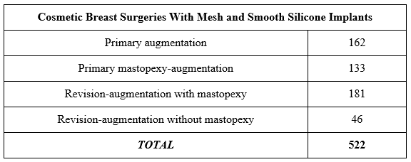
Table 1. Case type breakdown of the breast surgeries performed in this study.
Primary Augmentation
Primary breast augmentations proceeded as previously described by the authors (antibiotics, inframammary fold incision, dual-plane technique, atraumatic dissection with electrocautery, nipple shields, pocket irrigation, etc).4 Smooth silicone gel implants were inserted using an introduction sleeve. After removal from sterile packaging, the 10 × 25-cm mesh was soaked in the same preferred pocket irrigation solution as described above and was cut in half. Each half was used to stabilize the respective breast pocket from the entire medial to lateral border of the inframammary fold (IMF) along its most inferior edge and covered at least half the height of the breast implant along its superior border. The mesh was inset with deep stitches to the Scarpa fascia and sometimes the periosteum of the rib using 2-0 Vicryl suture along its inferior edge spanning as far medial and lateral as could be reached along the IMF incision; most of the patients received 3 or 4 interrupted sutures approximately 1 cm apart (Figure 1). The mesh was subsequently unfurled, making sure that its smooth surface faced the implant and its rough surface was against the breast tissue. Three-layered suture closure was then performed per the usual routine: breast fascia to Scarpa fascia (2-0 Vicryl), deep dermis (3-0 PDS), and subcuticular layer (4-0 Monocryl).
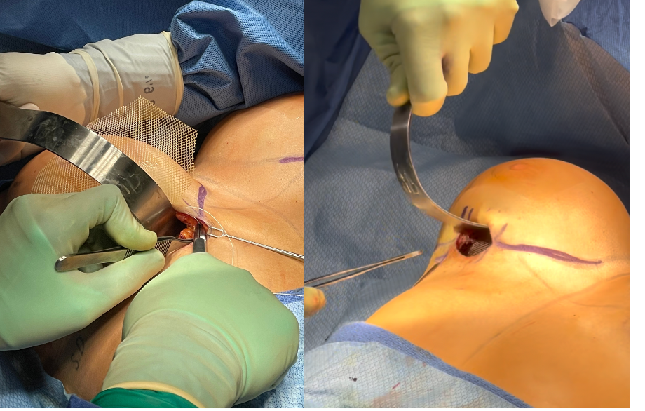
Figure 1. Mesh implantation technique inside of the pocket in a primary breast augmentation. This placement technique was also utilized for revision-augmentations without mastopexy.
Primary Mastopexy-Augmentation
Primary mastopexy-augmentations proceeded as follows5: In a pure vertical mastopexy-augmentation, breast implants were placed and the mesh was cut in half and soaked in the irrigation solution as described above, identical to the technique for breast augmentation. After that, total pocket closure of the breast implants and mesh occurred, and mastopexy followed.
In cases where a wise-pattern mastopexy-augmentation was performed, the implants were placed, and the breast fascia was then closed to have total implant coverage. Then the mastopexy was carried out, flaps were elevated, the breast tissue debulked as necessary, and the mesh was sewn outside the breast implant pocket, but still with its most inferior border securing the inframammary fold border. While the mesh was still oriented with the smooth surface toward the breast implant (but outside the implant pocket) and its rough surface toward the mastopexy flaps, the main difference was that its superior border and any dead space were quilted with light 2-0 Vicryl interrupted sutures tacked down to the underlying soft tissue, being sure not to go too deep and inadvertently puncture the underlying implant (Figure 2). If the patient's soft tissue was very thin, the mesh would sometimes be placed inside of the breast implant pocket, similar to what is done in a vertical mastopexy, in order to have more tissue coverage over the mesh.
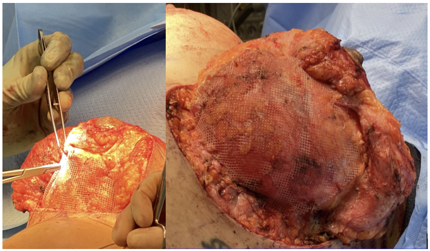
Figure 2. Mesh implantation technique outside of the pocket when a wise-pattern mastopexy is performed as a part of the breast surgery.
Revision Augmentation
For revision-augmentation cases, all surgeries were performed in the dual plane unless patients had a preexisting subglandular capsule/pocket deemed healthy enough to reuse. A small subset of cases (n = 15) required pocket exchange from subglandular to dual-plane. The technique for implantation of the mesh was done as described above for primary breast enhancement surgeries, and based on the incisions used: IMF, vertical, or wise pattern mastopexy. Cases requiring a mastopexy used a superior or superomedial dermal pedicle blood supply or random blood supply (if prior pedicle was unknown) and with a wise-pattern or vertical mastopexy scar.
In situations where increased internal pocket lateral support was needed, the mesh was parachuted inside the breast implant pocket, creating a "retaining wall" from near the axillary dome superolaterally down to the IMF, and then across the IMF medially to also give some lower pole support. In a subset of more complex revision cases that also required a wise pattern mastopexy (n = 9), 2 pieces of mesh were used to construct a "fortress," with mesh inset inside and outside of the pocket using the strategies described above.
Other Details
Drains were used only in cases requiring a total capsulectomy due to ruptured silicone implants (n = 7). Drains were never used in any primary surgeries. Postoperative antibiotic prophylaxis was employed, with all patients receiving a 7-day course of cephalosporin antibiotic (Cephalexin 500 mg, PO TID) except in the case of a known allergy.
A minimum of 6 months of postoperative evaluation to assess patient outcome was required for this study, with follow-up time ranging from 3 to 32 months. The outcome factors that the authors measured were scar malposition, reoperation rates, recurrent ptosis in mastopexy patients, scar quality in patients who received a mastopexy, the change in implant volume in revision patients, and postoperative complications including but not limited to implant extrusion, wound dehiscence, tissue necrosis, seroma, hematoma, infection, capsular contracture, and implant malposition.
Scar malposition was classified into 3 categories according to a previously published scale utilized by the authors (Figure 3)7: minor (1 < x ≤ 2 mm, single-side or bilateral), moderate (2 < x ≤ 3 mm, single-side or bilateral), and major (x > 3 mm, single-side or bilateral). The authors used the scar position gathered at the time of latest follow-up for this study.
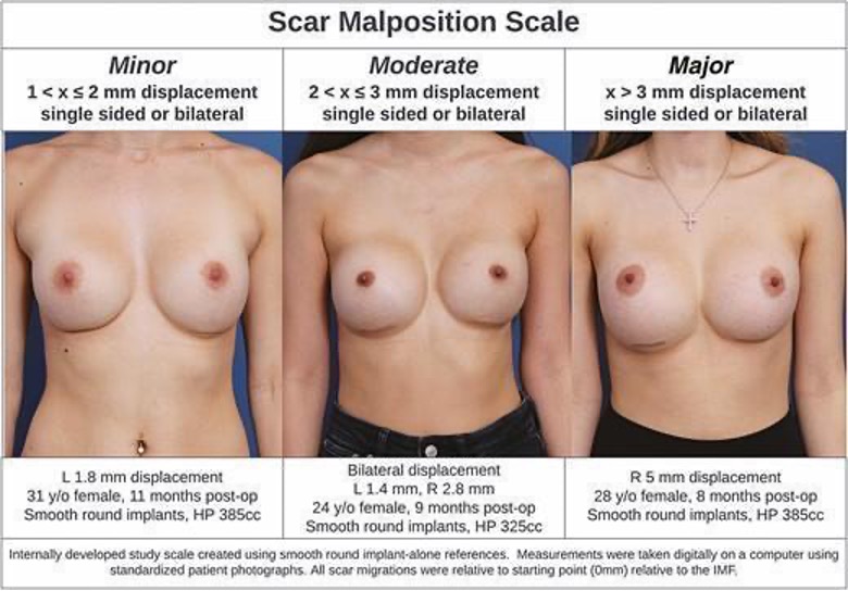
Figure 3. Scar malposition classification scale, used with permission from Chiemi & Kelishadi (2022).7
Scar quality in patients who received a mastopexy was evaluated by photography and then scored by an independent observer according to a previously described 5-point scar scale, where 1 corresponds to a hypertrophic/keloid scar and 5 denotes a thin, well-toned scar that blends with the native breast tissue (Figure 4).5 The authors used the scar quality score assessed at the time of latest follow-up for this study.
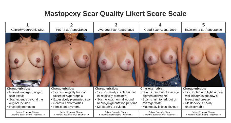
Figure 4. Mastopexy Scar Scale, used with permission from Chiemi & Kelishadi (2022).5
Results
Cohort-Wide
The average follow-up length was 13.7 months, ranging from 6 to 37 months. The average implant volume utilized was 474 mL (range 240-800 mL); however, the average implant size varied largely across surgery type. Patients undergoing a primary augmentation received an average implant volume of 396 mL (range, 240-800 mL), while patients undergoing a primary mastopexy-augmentation received an average implant volume of 494 mL (range, 350-800 mL). Patients undergoing a revision augmentation without mastopexy received an average new implant volume of 563 mL (range, 325-800 mL), and patients undergoing a revision augmentation with mastopexy received the largest average implant volume for their new prostheses of 577 mL (range, 350-800 mL; Table 2).
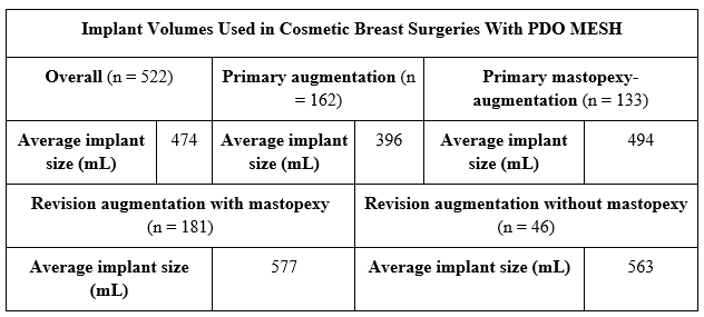
Table 2. Implant size data in the cohort, including overall trends and differences among patients receiving different surgeries.
Primary Augmentation
Scar malposition rates. In this study, 97.5% of patients (n = 158) developed no scar malposition (151, 93.2%) or minor malposition (7, 4.3%) over the duration of follow-up. There were 3 incidences of moderate scar malposition (1.8%) and 1 incident of major scar malposition (0.6%), with an overall scar malposition rate of 6.8% in the primary augmentation group (Table 3).
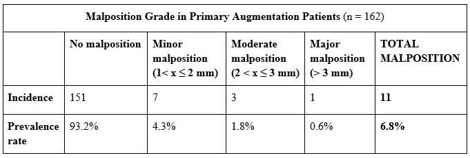
Table 3. Incidence, scaled prevalence rates, and total prevalence of scar malposition recorded in primary augmentation patients with PDO mesh.
Complications
The authors recorded a total of 2 complications (1.2%) comprising 2 single-sided implant infections. There were no mesh-related complications. The infected implants were removed, and the breasts were re-augmented 3 months post-surgery. Both patients had positive outcomes post-revision.
Reoperation Rates
Twelve patients (8.0%) in the primary augmentation group went on to have a later breast revision surgery: 7 electively sought a desired size change; 2 patients were brought back surgically to treat infections as mentioned above, and 3 patients sought revision due to malposition.
Primary Mastopexy-Augmentation
Scar quality. The mean scar quality scale score across the primary mastopexy-augmentation group was 4.3 (good-excellent; Table 4). No patients scored below 3 on the scar quality index (Figure 4). The mean Fitzpatrick phototype7 across the cohort was 3.8.
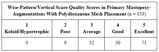
Table 4. Distribution of mastopexy scar quality Likert scores across primary mastopexy-augmentation patients. Final scar scores were coded by an independent observer at uniform follow-ups post-surgery.
Recurrent Ptosis
The authors recorded a total of 8 (6.0%) cases of recurrent ptosis within this cohort. The average implant volume of the patients who developed recurrent ptosis was 549 mL.
Complications
The authors recorded a total of 17 complications (11.7%) within the primary mastopexy-augmentation group: 9 superficial wound dehiscences; 5 deep wound dehiscences; 1 seroma, and 2 single-sided implant infections. There were no mesh-related complications. Of these complications, 10 were resolved nonsurgically; all 9 superficial dehiscences and 1 superficial-to-deep dehiscence (included in the 5 deep dehiscences for consistency) were resolved by secondary intention in-office. The seroma was drained in the operating room (OR) with no further complications. Three of the deep dehiscences were brought back to the OR for debridement and revision of wound closure, and the other was treated in-office but will likely request scar revision in the OR in the near future. In the cases of both infections, the infected implants were removed, and the breasts were re-augmented without revision to the original mastopexy after 3 months post-surgery. All patients with complications who underwent a reoperation had no further complications. Of note, patients who developed dehiscence on one side were noted to develop wider scars on the incision that dehisced, while the opposite breast obtained a better scar score. Breasts that developed asymmetrical scar quality scores were placed in the lower of the two scar score categories for consistency in the cohort (for example, a patient who scored a 3 on the left breast's scar score and a 4 on the right breast was placed in the 3 category).
Reoperation Rates
Thirteen patients had a later breast revision surgery, yielding a 9.7% rate of reoperation for any reason: 4 underwent reoperation for a desired size change (3.0%); 6 patients were brought back to the OR to resolve a complication (4.5%), and 3 received a revision mastopexy without implant exchange to resolve recurrent ptosis (2.3%).
Breast Revision
Prior breast surgeries. Patients in the breast revision group had received an average of 1.55 prior breast surgeries, ranging from 1 to 7. The majority (n = 158, 68.0%) had 1 prior breast surgery; most others had undergone 2 or 3 prior surgeries (n = 59, 26.0%), and 10 patients (4.4%) had a surgical history of greater than 3 breast surgeries. Twenty-one patients (10.3%) had undergone their most recent prior breast surgery at our practice (Table 5).
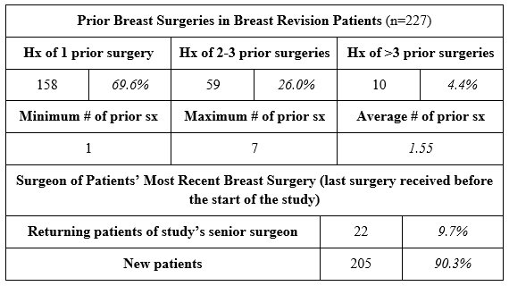
Table 5. Breast surgical histories of patients in the breast revision group.
Implant Size Changes
The average prior implant volume in all revision patients was 428 mL (range, 165-800 mL), while the average new implant volume was 574 mL (range, 325-800 mL). The average implant volume change was +146 mL. At the time of consultation, 178 patients (87.7%) stated a desire to receive larger implants than their previous size as a goal for their revision surgery. For such patients seeking a size increase, the average implant volume change was +189 mL, with achieved size increases ranging from 10 to 510 mL. Patients receiving a mastopexy had an average implant volume change of +143 mL, and patients not receiving a mastopexy (excluding the 7 patients undergoing pocket revision without new implants) had an average implant volume change of +154 mL (Table 6).
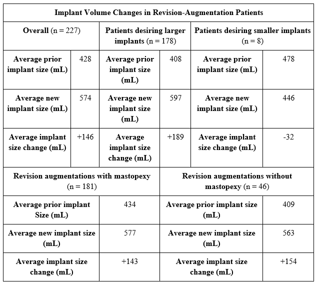
Table 6. Implant size change data in the breast revision group including overall trends, differences between patients who stated their intent to go larger or smaller at pre-operation evaluation, and patients receiving or not receiving mastopexies.
Scar Quality in Mastopexy Patients
There were 181 patients who received a mastopexy as part of their breast revision surgery. The mean scar quality scale score across the breast revision with mastopexy group was 4.4 (good-excellent). No patients scored below 3 on the scar quality index (Figure 4). The mean Fitzpatrick phototype8 across the breast revision with mastopexy group was 3.2.
Reoperation Rates
In the revision-augmentation group, 26 patients had reoperations (11.5%): 4 desired size changes (1.8%); 13 complications requiring surgical intervention (5.7%), and 9 pocket revisions to resolve breast shape/malposition/recurrent ptosis (3.9%). The average implant volume of patients who had a complication or sought revision for malposition or ptosis (n = 22) was 583 mL; these patients had undergone an average of 2.1 prior breast surgeries (range, 1-6). The authors noted that a higher proportion of revision patients who sought reoperation had undergone a mastopexy as part of their revision (Table 7).
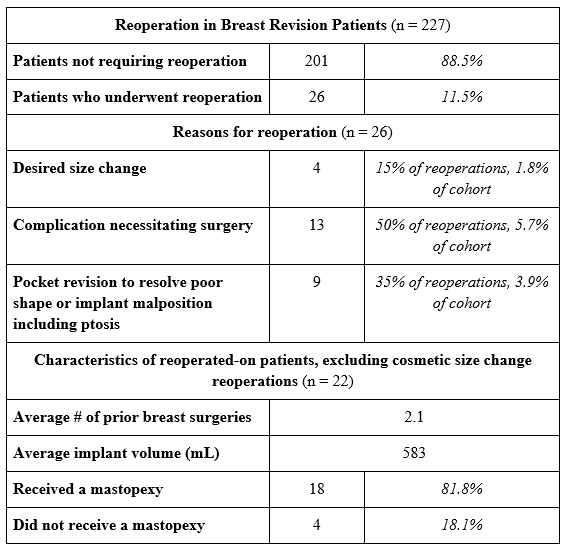
Table 7. Reoperation data for patients in the breast revision group.
Complications
The authors recorded a total of 23 complications (10.1%) within the breast revision group; 19 occurred in patients who received mastopexy while 4 were in non-mastopexy patients. These included 4 infections, 4 seromas, 3 partial nipple-areola complex necroses, 6 deep wound dehiscences, 1 capsular contracture, 1 nonfatal pulmonary embolism, and 4 superficial wound dehiscences. There were no mesh-related complications. Of these complications, 10 were resolved nonsurgically: the superficial wound dehiscences and partial nipple necroses were healed in office by secondary intention; 1 infection was treated with oral antibiotics; 1 seroma was drained in office; and the pulmonary embolism was treated in the emergency room with anticoagulants. The other 3 infections were treated with implant removal and staged re-augmentation. Three seromas were drained in the OR. The deep wound dehiscences were debrided and reclosed in the OR.
In the cohort, 13 patients (5.7%) had a prior history of capsular contracture; all had prior smooth-shelled breast implants. The 1 patient who developed new capsular contracture had undergone 4 prior breast surgeries with 2 previous instances of capsular contracture; of note, in this particular patient, her mesh was placed outside of the breast implant pocket. Unless a total capsulectomy was performed at the time of breast revision, the majority of the cases in this study had preservation of prior capsule, which was repaired during the surgery. In all of the revision patients not requiring mastopexy, the mesh was placed against the implant, and this was also the case in roughly one third of the cases where a mastopexy was performed. Therefore, there were no cases of capsular contracture noted in this cohort of breast revision cases when the mesh was placed against the implant.
Discussion
After 3 years of experience using PDO mesh uniformly in all types of cosmetic breast implant surgeries, the authors have discovered manifold benefits in obtaining greater pocket control, long-term stability, and overall breast aesthetics. Due to the lack of observed mesh-related complications in this large case series (where all primary augmentation, vertical mastopexy, and revision augmentation patients received mesh placed inside the pocket against the implant), the authors judge PDO mesh to be a safe adjunct to silicone gel implants. Infection would be exceedingly rare with this material. Polydioxanone is the same inert synthetic polymer that composes PDS suture (Ethicon), which is generally well-tolerated by patients. The literature documents polydioxanone's low rates of adverse site reactions and surgical site complications.8 Some inflammatory response and foreign body reaction are expected following the implantation of any biomaterials, with the reaction's duration and strength being an intersection between material-dependent and material-independent processes. Synthetic materials such as PDO are not known to elicit a specific biological reaction.9 This mesh's porosity and short-term integration profile make it a biologically compatible (and thus, feasible) option for the vast majority of patients.
In various other surgical disciplines such as orthopedics and otolaryngology, soft tissue splints are commonly used in order to gain greater control in the repair. However, due to the lack of bony repair in aesthetic breast cases, the fact that breast tissue is being stressed and a device is being placed that may move within the soft tissue is often neglected. A breast implant's pocket is vastly different from devices such as pacemakers, which are implanted underneath the skin on top of a bony structure. Additionally, pregnancy and hormonal changes in female patients lead to unpredictable changes in the soft tissue. The authors believe that prioritizing pocket control and maximizing soft tissue support in breast implant cases is extremely important in determining the outcome.
Mesh has been used in aesthetic breast surgery to reinforce soft tissue and prevent complications like implant malposition, but the cost constraints associated with traditional meshes such as acellular dermal matrices have restricted their use in aesthetic cases to essential soft tissue adjuncts, primarily in very complex revisions.2 The market influx of various synthetic mesh options with diverse compositions and absorption profiles provides plastic surgeons with more cost-effective and inert alternatives than biologic products. Notably, synthetic absorbable meshes, particularly those with an absorption profile conducive to optimal support during initial soft-tissue healing, have gained attention in aesthetic breast surgeries. Recent studies supporting the safety and aesthetic benefits of resorbable matrices in cosmetic breast revisions, specifically those using poly-4-hydroxybutyrate matrices, call for further investigation.10 The selection of PDO mesh for aesthetic revision cases is based on its shorter absorption profile (3-12 months), combined with its thin, nonpalpable nature and robust strength. This matrix integrates into tissue within 4 weeks of implantation, gradually absorbing over 1 year, leaving behind 1 to 2 mm of neo-collagenous vascular tissue. While concerns about mesh infections in breast reconstruction have deterred some surgeons, the elective nature of cosmetic cases, coupled with the biologically compatible nature of PDO mesh minimizes the risk of infection.
Benefits in Primary Augmentation
In a 2021 study,11 the authors previously showed that mesh in conjunction with smooth implants yielded a significantly lower rate of scar malposition compared with smooth implants alone in primary augmentation patients. The previous study also found that mesh with smooth implants performed statistically similarly to microtextured implants in preventing malposition; mesh with smooth shells even trended towards a lower prevalence of scar malposition, approaching yet not reaching significance (Figure 5).
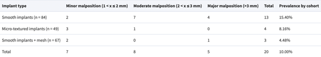
Figure 5. Scar malposition recorded between smooth implant, microtextured implant, and smooth implant + mesh groups, used with permission from Chiemi & Kelishadi (2022).4
In this study, the authors once again observed a low rate of scar malposition in primary augmentation cases (Figures 6-8). Though a 0% malposition rate and mesh's total protection against scar movement would have been desirable, the authors believe that the uniform use of PDO mesh with smooth implants offered a similar level of control to that which a microtextured device offers without the risks associated with texture such as breast implant-associated anaplastic large cell lymphoma or double capsule. This is especially attractive for patients with certain anatomical features necessitating additional pocket control such as rib asymmetries, an indiscrete or mobile inframmamary fold, or limited soft tissue stores; however, the uniform employment of mesh even in "straightforward" primary augmentation cases without these anatomical challenges served as a pull factor for aesthetic patients wishing for greater long-term stability and a chance at avoiding subsequent revision surgeries in the future. The senior author still uses microtextured shaped implants at their practice when an anatomical prosthesis is indicated, but mesh in conjunction with smooth implants has completely replaced textured round implants in this application. Additionally, one of the unique characteristics and advantages of Durasorb mesh is that it allows "shaping" of the breast pocket due to its strength yet at the same time malleability – this advantage is one that cannot be achieved with an implant alone and has not been seen by the senior author when using other meshes.
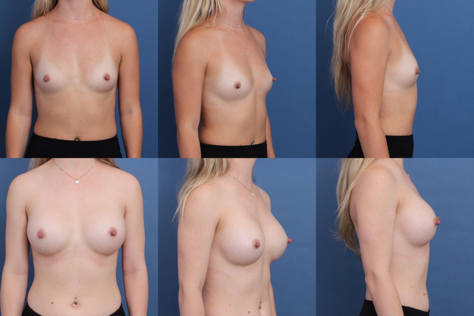
Figure 6. Patient (28 y/o female) shown 25 months after primary augmentation using Sientra HSC + High Profile Round Smooth Implants, 415 cc, plus DuraSorb mesh.
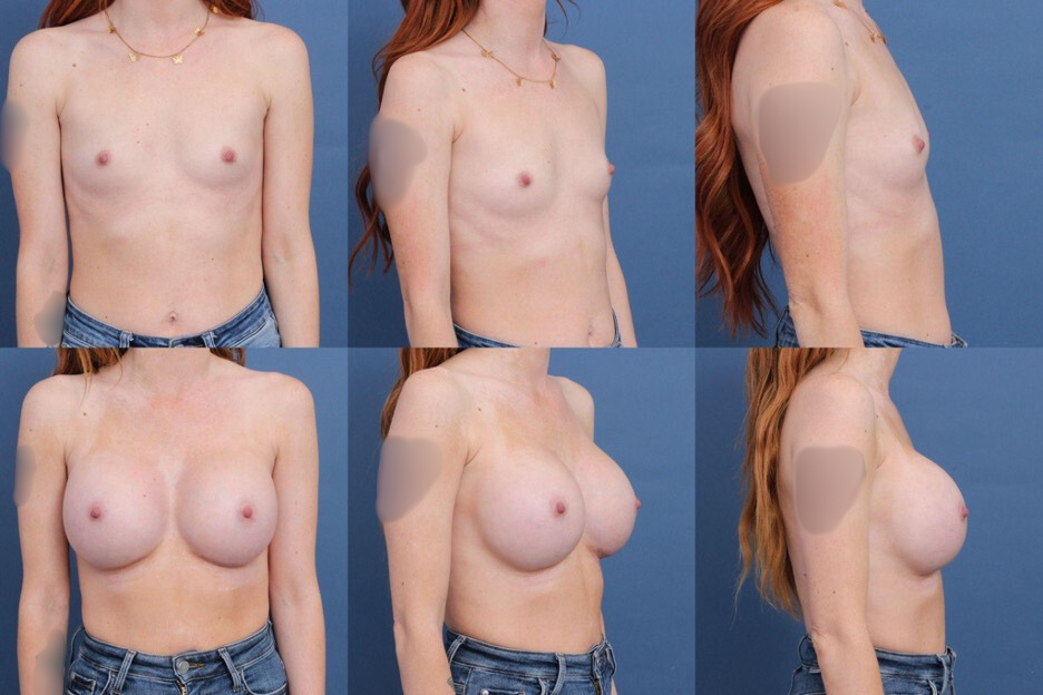
Figure 7. Patient (29 y/o female) shown 7 months after primary augmentation using Mentor MemoryGel Boost High Profile Round Smooth Implants, 545-mL implant on the right and 480-mL implant on the left, plus DuraSorb mesh.
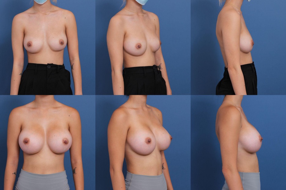
Figure 8. Patient (22 y/o female) shown 9 months after primary augmentation using Allergan Natrelle Inspira SoftTouch SSF Smooth Round Implants, 365 mL, plus DuraSorb mesh.
Benefits in Primary Mastopexy Augmentation
A mastopexy is a powerful and often necessary part of many cosmetic breast surgeries to obtain an aesthetic shape, but the vertical scar is a potent deterrent for many patients wishing to avoid unsightly visible scarring on the breast mound. Unfortunately, when these patients refuse a mastopexy and opt for implants alone, they often report high rates of dissatisfaction with a ptotic appearance that has only been "augmented" by the presence of an implant. Clearly, scarring is incredibly important to aesthetic patients; the vertical mastopexy is unavoidable for certain patients to obtain the shape they desire, and the modern plastic surgeon must seek out intraoperative and postoperative measures in their tool belt to provide patients with optimal scarring outcomes, barring individual genetics and wound healing factors.
Both tension forces from a tight mastopexy closure and the weight of the implant may contribute to poor scarring outcomes. The rationale for using mesh uniformly in mastopexy-augmentations is that the additional soft tissue support can take pressure off the wise pattern or vertical incisions from the inside, especially over the critical first 3 months of soft tissue healing as the mesh absorbs and deposits neocollagen in its place.
In consistency with the author's previously described results,5 this study showed largely positive aesthetic outcomes for scarring with uniform placement of PDO mesh in the lower pole of the breast pocket in mastopexy-augmentations (Figures 9-11). Not only did mesh mastopexy-augmentation patients score above average on the scar quality scale, but the authors noticed thinner, better-toned mastopexy scars earlier on in the healing process. A diverse range of skin tones and ethnicities were represented in this cohort, and the scar aesthetics also held up across the Fitzpatrick phototype scale, with positive scarring outcomes observed in both the lightest- and darkest-skinned patient groups of the study. This is important given that genetics do contribute to scarring outcomes, with Asian skin being more prone to hyperpigmented scars13 and more melanated skin being more susceptible to keloid scarring.13 Mesh may have promise as an effective prophylaxis that delivers a protective and more predictable effect against unfavorable scarring in all demographics. In addition to patient demographics, we recognize that patient behavior and economic status play crucial roles in scar prevention and wound healing – wealthier patients may have access to higher-quality postoperative care that can play a role in scar management outcomes. They also may be more able to pursue scar-reducing treatments aimed at improving scar aesthetics. A more in-depth exploration of how these aspects interplay with the use of PDO mesh could offer valuable insights.
Lowering the tension forces at play on the internal aspect of the incision was also hypothesized to lead to an improvement in wound healing outcomes through a lower dehiscence rate. Dehiscences at the T-junction are a common sequela in mastopexy-augmentations; although most superficial dehiscences heal by secondary intention, some persistent superficial or deep dehiscences present a challenge to early wound healing that can later result in poor scarring aesthetics or breast aesthetics. Though this study lacked a formal control group to compare with the wound-healing outcomes of the mastopexy-augmentation group, the authors believe that mesh greatly contributed to the overall low wound healing complication rate in high–implant volume augmentations with aggressive mastopexies performed and all risk factors taken into account.
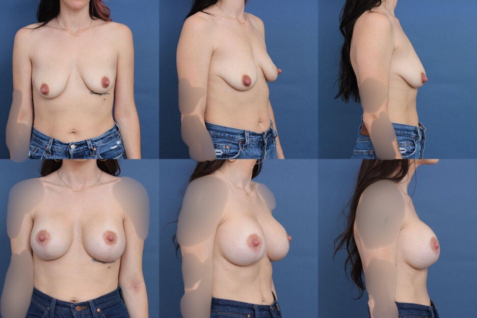
Figure 9. Patient (32 y/o female) shown 14 months after primary mastopexy-augmentation using Allergan Natrelle Inspira SoftTouch SSF Smooth Round Implants, 415 mL, plus DuraSorb mesh. The patient scored a 5 on the Scar Quality Likert scale and is a Fitzpatrick I.
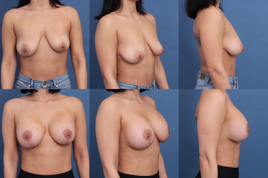
Figure 10. Patient (32 y/o female) shown 6 months after primary mastopexy-augmentation using Allergan Natrelle Inspira SoftTouch SSM Smooth Round Implants, 275 mL, plus DuraSorb mesh. The patient scored a 4 on the Scar Quality Likert scale and is a Fitzpatrick III.
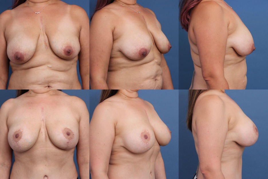
Figure 11. Patient (41 y/o female) shown 12 months after primary mastopexy-augmentation using Allergan Natrelle Inspira SoftTouch SSM Smooth Round Implants, 405-mL implant on the right, 445-mL implant on the left, plus DuraSorb mesh. The patient scored a 4 on the Scar Quality Likert scale and is a Fitzpatrick II. The patient had a prior history of cardiac surgery; of note, the 1-year-old vertical mastopexy scars show comparable tone to the 15-year-old sternotomy scar.
Benefits in Revision Augmentation
Using mesh in revision-augmentations allowed the authors to utilize larger implant volumes in patients, better meeting their aesthetic goals, regardless of their soft tissue stores and/or skin quality (Figures 12-15). The average implant used in this cohort was 571 mL, considered to be a high-volume breast implant surgery, which poses challenges to wound healing in patients with limited soft tissue and previous capsule.14 The authors acknowledge that large-volume implants were utilized in this study, with each patient group receiving an average volume well above the national average size used in the US.15 The main selection criteria for implant size in this study were patients' stated preferences during consultations (with many patients seeking out the senior surgeon's practice specifically to obtain a certain aesthetic result delivered to prior practice patients), as well as the senior surgeon's technical preference to perform an aggressive mastopexy with debulking of ptotic native tissue, relying largely on the implant to shape the breast.
Substantial size increases averaging 189 mL were achieved for the revision patients who wished to go larger, and the authors believe that this would not have been feasible without the provision of additional soft tissue support in the average lean patient. Though patients will still contend with the effects of gravity and normal aging, and there is no technique that is bulletproof against these, mesh helped to deliver greater stability and pocket integrity in these complex, high-risk aesthetic breast surgeries, a precaution that can certainly contribute to fewer later unsatisfactory aesthetic outcomes. We find that patients who have undergone many prior breast surgeries often demand great precision in their results and want to push the limits with greater implant volumes, and using mesh as a uniform adjunct in revision patients allowed us to deliver such results with greater confidence in their wound healing outcomes and safety.
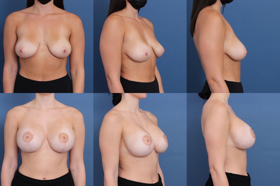
Figure 12. Patient (22 y/o female) shown 14 months after revision mastopexy-augmentation using Mentor MemoryGel Boost High Profile Round Smooth Implants, 740 mL, plus DuraSorb mesh. The patient scored a 5 on the Scar Quality Likert scale and is a Fitzpatrick II. This patient had a history of 3 prior breast surgeries.

Figure 13. Patient (32 y/o female) shown 8 months after revision augmentation. Allergan Natrelle Inspira Responsive SRF Smooth Round Implants, 560 mL, were exchanged for Allergan Natrelle Inspira SoftTouch SSX Smooth Round Implants, 560 mL plus DuraSorb mesh. This patient had a history of 2 prior breast surgeries.
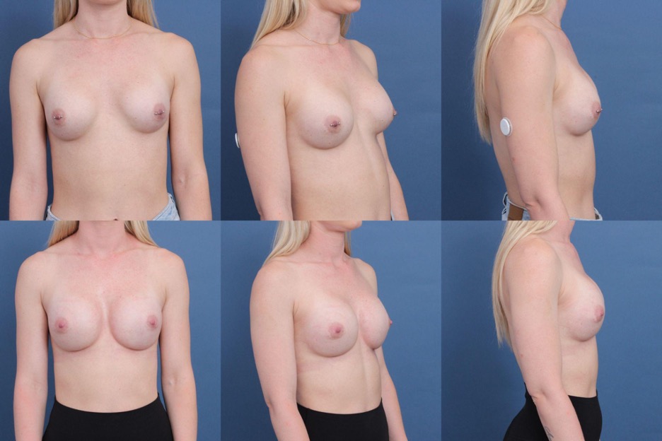
Figure 14. Patient (31 y/o female) shown 5 months after revision augmentation. Mentor Saline Smooth Round Implants, 225 mL, were exchanged for Allergan Natrelle Inspira SoftTouch SSM Smooth Round Implants, 360 mL plus DuraSorb mesh. This patient had a history of 1 prior breast surgery.
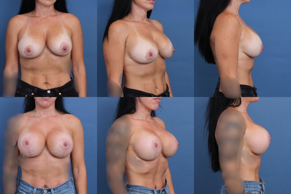
Figure 15. Patient (32 y/o female) shown 11 months after revision mastopexy-augmentation. Allergan Natrelle Inspira Responsive SRLP Smooth Round Implants, 440 mL, were exchanged for Allergan Natrelle Inspira SoftTouch SSX Smooth Round Implants, 650 mL plus DuraSorb mesh. This patient had a history of 2 prior breast surgeries.
Pearls Using PDO Mesh in Implant-Based Cosmetic Breast Surgery
The authors discovered a variety of pearls using the mesh during the course of the case series. The PDO mesh utilized has preferential stretch and is more pliable in 1 orientation, with the orientation 90 degrees to this having less give to it. Patients who are at risk for inferior malposition generally received the mesh oriented vertically in its least stretchy/pliable orientation in order to reduce inferior malposition over time. This technique was utilized in both primary and revision patients. The orientation of the mesh was factored into the surgical decision-making when the properties of preferential stretch were deemed advantageous; unfortunately, some early cases with scar malposition in primary breast augmentation or recurrent ptosis in mastopexy-augmentation could have been avoided with this insight.
It was also found that, when large implants were utilized or a patient had a looser pocket, using a whole sheet of mesh on each side was more helpful than using a single 10 × 25-cm sheet bisected. Over the duration of this study, the senior surgeon gradually used a lower threshold for making the decision to employ a whole sheet of mesh on each side. This clinical decision-making generally took place intraoperatively, where 2 sheets of mesh would be ordered for the patient and the final decision on whether to open and soak both sheets would be made in the OR once the breast was open.
Limitations
A longer period of follow-up certainly could have captured additional complications, including longer-term sequelae such as capsular contracture and ptosis, and would have naturally yielded higher complication and reoperation rates for the study. The period of follow-up used in this study was partly chosen due to author convenience as it can be difficult to maintain extended follow-up with cosmetic patients.16 Additionally, the complications that the authors sought to capture in this study were based in surgical techniques and control, which tend to be shorter-term sequelae, whereas longer-term complications are usually related to the unique biologic phenomena and wound healing circumstances in each person.
The authors also acknowledge that there was not a formal control group (except in a previously published primary breast augmentation paper4) to compare with in this study, as mesh was used uniformly in these cosmetic breast surgery patients. Thus, comparing the senior surgeon's outcomes with their previous cosmetic breast patients prior to the uniform use of mesh may be impacted by natural technical improvement as time-in-practice elapsed.
Conclusions
The use of round, smooth silicone gel breast implants with the adjunct placement of PDO internal support matrix is a safe and effective method of increasing pocket control and improving outcomes in cosmetic breast implant surgery.
Acknowledgments
Authors: Julia A. Chiemi, BSc1; S. Sean Kelishadi, MD, FACS2
Affiliations: 1Yale School of Medicine, New Haven, Connecticut; 2SSK Plastic and Reconstructive Surgery, Newport Beach, California
Correspondence: S. Sean Kelishadi, MD, FACS; drssk@sskplasticsurgery.com
Ethics: Patients provided written consent for the use of images.
Disclosures: S.S.K. was formerly a SIA consultant shareholder from June 2021 to November 2023. The authors disclose no other relevant financial or nonfinancial interests.
References
1. Jacobs JM, Salzberg CA. Implant-based breast reconstruction with meshes and matrices: biological vs synthetic. Br J Hosp Med (Lond). 2015;76(4):211-216. doi:10.12968/hmed.2015.76.4.211
2. Becker H. Update on the use of synthetic mesh in reconstructive and cosmetic breast surgery. Aesthetic Plast Surg. 2020;44(4):1128-1129. doi:10.1007/s00266-020-01767-2
3. Gutowski KA. DuraSorb Monofilament Mesh (Polydioxanone, PDO) for Tissue Support. 2019. Plastic Surgery The Meeting, San Diego.
4. Chiemi JA, Kelishadi SS. Polydioxanone internal support matrix: a rationale for prophylactic internal bra support in breast augmentation. Aesthet Surg J Open Forum. 2022 Mar 30:4:ojac021. doi:10.1093/asjof/ojac021
5. Chiemi JA, Kelishadi SS. "Never Trust the Skin": a rationale for using polydioxanone internal support matrix to minimize scarring in primary mastopexy–augmentation–an observational study. Aesthet Surg J Open Forum. 2022 May 19;4:ojac048. doi:10.1093/asjof/ojac048
6. Chiemi JA, Kelishadi SS. Polydioxanone monofilament mesh: a safety net for complex breast implant revision surgery, Aesthet Surg J. 2023 Feb 21;43(3):NP155-NP166.
7. Chiemi JA, Kelishadi SS. Polydioxanone internal support matrix: A rationale for prophylactic internal bra support in breast augmentation. Aesthet Surg J Open Forum. 2022;4:ojac021. Published 2022 Mar 30. doi:10.1093/asjof/ojac021
8. Martins JA, Lach AA, Morris HL, Carr AJ, Mouthuy PA. Polydioxanone implants: a systematic review on safety and performance in patients. J Biomater Appl. 2020;34(7):902-916. doi:10.1177/0885328219888841
9. Anderson JM, Cook G, Costerton B, et al. Host reactions to biomaterials and their evaluation. In: Ratner B, Hoffman A, Schoen F, Lemons J, eds. Biomaterials Science – An Introduction to Materials in Medicine. Academic Press; 1997:293-296.
10. Nair NM, Mills DC. Poly-4-Hydroxybutyrate (P4HB) scaffold internal support: preliminary experience with direct implant opposition during complex breast revisions. Aesthet Surg J. 2019;39(11):1203-1213. doi:10.1093/asj/sjy276
11. Chiemi JA, Kelishadi SS. A rationale for micro-textured breast implant augmentation. Aesthet Surg J Open Forum. 2022 Mar 30;4:ojac020.
12. Kim S, Choi TH, Liu W, Ogawa R, Suh JS, Mustoe TA. Update on scar management: guidelines for treating Asian patients. Plast Reconstr Surg. 2013;132(6):1580-1589.
13. Gao FL, Jin R, Zhang L, Zhang YG. The contribution of melanocytes to pathological scar formation during wound healing. Int J Clin Exp Med. 2013;6(7):609-613.
14. Pacifico MD. Commentary on: Polydioxanone monofilament mesh: a safety net for complex breast implant revision surgery, Aesthet Surg J. 2023 Feb 21;43(3):NP167-NP169.
15. Swanson E. Underestimating implant volumes in cosmetic breast augmentation. Plast Reconstr Surg Glob Open. 2017;5(9):e1483. doi:10.1097/GOX.0000000000001483
16. Honigman RJ, Phillips KA, Castle DJ. A review of psychosocial outcomes for patients seeking cosmetic surgery. Plast Reconstr Surg. 2004;113(4):1229-1237.