Closure of a Very Large Patent Ductus Arteriosus Using the Amplatzer Duct Occluder
September 2002
Closure of the small to moderate sized patent ductus arteriosus (PDA) is accomplished at most large pediatric heart centers by transcatheter techniques, most often by inserting thrombogenic fabric-stranded Gianturco coils. However, closure of the large PDA is more challenging, especially when the morphology provides little substrate to anchor the initial coil. Even if the coils do remain stable, many are required to effectively plug the defect. As a result, the Amplatzer Duct Occluder (AGA Medical Corporation, Golden Valley, Minnesota) was manufactured in different sizes to allow implantation of a single device to completely close a PDA. Significant experience worldwide has been obtained regarding the effectiveness of the device,1–3 and a United States multicenter phase II clinical trial is currently underway. Nonetheless, there has been very little experience closing very large PDAs with the largest Amplatzer device (smallest diameter, 14 mm). This case describes complete closure of a very large PDA using the largest Amplatzer Duct Occluder size and represents closure of the largest PDA using this device in the US.
Case Report. A 4.5-year-old girl from the Philippines presented to a Florida pediatrician shortly after her arrival in the US. A heart murmur prompted referral to our institution, where the diagnosis of a large PDA was made. A chest x-ray showed significant cardiomegaly and prominent central pulmonary vascularity (Figure 1). Anticipating availability of the Amplatzer Duct Occluder device, we performed standard right and left heart catheterization under general anesthesia to determine the feasibility of nonsurgical PDA closure. Angiography demonstrated a very large PDA (type C) measuring about the same size as the descending aorta (Figure 2). In room air oxygen, the pulmonary artery (PA) pressure was 60/30/45 mmHg (70% systemic). Temporary balloon occlusion of the PDA with a Tyshak II 12 x 2 valvuloplasty catheter decreased the PA pressure to 38/12/24 mmHg, while administration of 100% oxygen with the PDA open resulted in a PA pressure of 50/26/36 mmHg. The pulmonary to systemic flow ratio was 2.75, and the resistance ratio was 0.19. Hemodynamic data are given in Table 1.
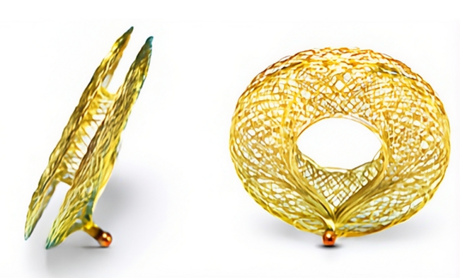
Three weeks later, the patient underwent repeat cardiac catheterization. Because she had mild rhinorrhea, the anesthesiologist was reluctant to provide complete general anesthesia. Instead, the procedure was performed under intravenous sedation with fentanyl and ketamine. This time the PA pressure was 95/50/70 mmHg (86% systemic), and there were no gradients from main to left branch pulmonary artery or from ascending to descending aorta. Angiographic measurements of the PDA were: length, 20.5 mm; diameter at aortic end, 18.1 mm; and diameter at pulmonary end, 11.2 mm. PDA closure proceeded as previously described1 using the largest available Amplatzer Duct Occluder (a 16-14 device). Because of the length of the PDA, the entire device was delivered within the PDA (Figure 3). Rounding of the retention disk produced by compression within the PDA created a “fish”-shaped appearance of the implanted device. Final PA pressure was 48/24/33 mmHg (40% systemic) and no PA or aortic gradients were detected. A small residual shunt persisted acutely, primarily from foaming through the device. Procedure and fluoroscopy times were 33 minutes and 9.1 minutes, respectively.
The following day, the chest x-ray showed unchanged position of the device, and a repeat echocardiogram showed only a small residual ductal shunt at the edge of the device. The patient subsequently moved to another state, so follow-up was delayed until she returned for follow-up at 1 year. A chest x-ray confirmed a stable device position and shape, as well as a significant decrease in heart size and pulmonary vascularity (Figure 4). Follow-up echocardiography showed no residual shunt with normal flow velocities in the left PA and descending aorta.
Discussion. To our knowledge, this case represents the largest PDA to undergo closure with the Amplatzer Duct Occluder in the US multicenter clinical trial. Although the ductal diameters at the aortic and pulmonary ends differ significantly, the aortic measurement from superior to inferior along its inner curvature is oblique. In fact, the PDA was tubular in shape, tapering little throughout its length from aorta to pulmonary artery. Surgical closure of such a large PDA would likely involve more than simple ligation with or without division. Instead, patch closure using either homograft or synthetic material may be necessary to repair the resulting defects in the walls of the great vessels.4
Given the reactivity of this patient’s pulmonary vascular resistance, nonsurgical closure was entertained. Protocol guidelines for implanting the Amplatzer Duct Occluder are to select a device size at least 2 mm greater than the smallest ductal diameter, considering the smallest diameter of the device when making this decision. Consequently, a 14-12 (12 mm) device may have been too small for this 11.2 mm PDA, so a 16-14 device was chosen. Because the retention disk is 4 mm larger than the larger end of the device (i.e., 20 mm diameter for the 16-14 device), we had concerns about aortic obstruction related to leaving this portion of the device in the aortic lumen. Because the retention disk rounds its shape when the midportion of the device is compressed (Figure 5), a decision was made to intentionally place the device completely within the PDA rather than allow the retention disk to be anchored against the aortic end. As a result, unique placement of the device was achieved within the PDA, and potential compromise of the aortic lumen by the large retention disk was avoided. With the diagnostic information already completed 3 weeks prior, the delivery was straightforward and the procedure time was short. The higher PA pressures measured during the second procedure likely reflected the difference between mechanical ventilation and spontaneous breathing with sedation.
Implantation of an Amplatzer Duct Occluder acutely decreased this child’s pulmonary blood flow. Even if the permanent result was a small residual PDA, as seen on the echocardiogram 1 day later, this would constitute clinical success in improving patient symptoms and long-term outcome. The only remaining cardiac risk would be infective endarteritis. However, it is more reassuring to learn that the small residual ductal flow remaining acutely after implantation of this large device disappeared completely at follow-up, obviating the need for endarteritis prophylaxis. In an effort to maximize applicability of the device to all PDA sizes and shapes during the U.S. Multicenter Trial, the device was deployed somewhat atypically in our patient and created a vascular plug within the ductus arteriosus. Although the retention disk is designed to lay flush against the aortic lumen, its shape rounds somewhat when the midportion of the device is compressed. This characteristic made unacceptable protrusion into the aortic lumen very likely. The fact that the largest device did not compromise blood flow at either end of the PDA makes the clinical utility of the Amplatzer Duct Occluder appealing for patients with a wide range of ductal sizes. For our patient, transcatheter implantation of the device resulted in a complete cure, leaving no residual abnormalities that might lead one to consider a surgical approach more desirable.
Even very large PDAs can now be closed with endovascular techniques, once the pulmonary vascular resistance is determined to be acceptably low. This was possible in our patient because of her older age and larger size. The small infant with a proportionately large PDA still presents a challenge to the interventional cardiologist because the smaller surrounding vascular structures may not provide adequate space to accommodate a device without becoming at least partially obstructed. Further advances in device technology will only expand the ranges of size and age for patients who will benefit from these techniques in the future.
Acknowledgment. I wish to thank Ziyad Hijazi, MD, for his assistance and sharing of technical considerations that helped produce a successful result.
1. Masura J, Walsh KP, Thanopoulos B, et al. Catheter closure of moderate- to large-sized patent ductus arteriosus using the new Amplatzer Duct Occluder: Immediate and short-term results. J Am Coll Cardiol 1998;31:878–882.
2. Thanopoulos BD, Hakim FA, Hiari A, et al. Further experience with transcatheter closure of the patent ductus arteriosus using the Amplatzer Duct Occluder. J Am Coll Cardiol 2000;35:1016–1021.
3. Bilkis AA, Alwi M, Hasri S, et al. The Amplatzer Duct Occluder: Experience in 209 patients. J Am Coll Cardiol 2001;37:258–261.
4. Toda R, Moriyama Y, Yamashita M, et al. Operation for adult patent ductus arteriosus using cardiopulmonary bypass. Ann Thorac Surg 2000;70:1935–1937.