Coronary Thrombosis From Superficial Calcific Sheet
Ariana Gonzálvez-García, MD; Harold Hernández-Matamoros, MD; Santiago Jiménez-Valero, MD; Alfonso Jurado-Román, MD, PhD; Guillermo Galeote, MD, PhD; Raúl Moreno, MD, PhD; José Luis López-Sendón, MD, PhD
Key words: acute coronary syndrome, calcified plaque, superficial calcific sheet
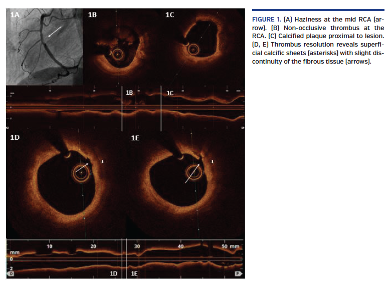
We present the case of a 63-year-old woman, former smoker, transferred to the catheterization laboratory for primary percutaneous coronary intervention in the midst of anterior ST-segment elevation myocardial infarction. Coronary angiography showed acute total occlusion of the proximal left anterior descending (LAD) (Video 1). After the guidewire passed through the distal LAD, TIMI-3 flow revealed severe proximal stenosis, which was treated with an everolimus-eluting stent with good outcome (Video 2). Afterward, angiographic haziness was recognized at the mid right coronary artery (RCA) (Figure 1A). Optical coherence tomography (OCT) exhibited non-occlusive thrombus (Figure 1B) and calcified plaques were observed proximal to the lesion (Figure 1C). Tirofiban perfusion was administered. After 10 days under dual-antiplatelet therapy (DAPT) with aspirin and prasugrel, OCT was repeated, displaying thrombus resolution with superficial calcific sheets (SCS) (Figures 1D and 1E) and minimal disruption of overlying fibrous tissue (Figure 1D-E: arrows) without compromise of the luminal area. Prolonged DAPT was recommended.
SCS has been implicated recently in the pathogenesis of acute coronary syndromes, and could have caused the acute RCA thrombosis in our patient. The underlying mechanism seems to be different from eruptive calcified nodules, and different theories have been speculated. OCT was useful to characterize in vivo SCS and exclude other causes of acute coronary syndromes, such as plaque erosion. Nevertheless, the management and potential prognostic and clinical implications of this type of calcified plaque are not clear, so further investigation needs to be done.
View the Supplemental Video Series Here
From the Interventional Cardiology Unit. Cardiology Department. La Paz University Hospital, Madrid, Spain.
Disclosure: The authors have completed and returned the ICMJE Form for Disclosure of Potential Conflicts of Interest. The authors report no conflicts of interest regarding the content herein.
The authors report that patient consent was provided for publication of the images used herein.
Manuscript accepted January 6, 2020.
Address for correspondence: Ariana Gonzálvez-García, MD, La Paz University Hospital, Paseo de la Castellana, 261, 28046 Madrid, Spain. Email: arianagonzalvez@gmail.com