

Impact of Proximal Cap Ambiguity on Outcomes of Chronic Total Occlusion Percutaneous Coronary Intervention: Insights From a Multicenter US Registry
Abstract: Objectives. We sought to determine the impact of proximal cap ambiguity on procedural techniques and outcomes for coronary chronic total occlusion (CTO) percutaneous coronary intervention (PCI). Methods. We examined the clinical and angiographic characteristics and outcomes of 1021 CTO-PCIs performed between 2012 and 2015 at 11 United States centers. Results. Proximal cap ambiguity was present in 31% of target lesions and was associated with increased clinical and angiographic complexity (prior coronary artery bypass graft surgery: 43% vs 33%; P=.01; moderate/severe calcification 66% vs 51%; P<.001) and lower technical success (85% vs 93%; P<.001) and procedural success (84% vs 91%; P=.01), but similar incidence of major adverse cardiac events (3.2% vs 2.9%; P=.77). A retrograde approach was more commonly utilized among cases with proximal cap ambiguity (68% vs 33%; P<.001), and was more likely to be the initial (39% vs 13%; P<.001) and successful approach (42% vs 20%; P<.001). Proximal cap ambiguity was associated with increased use of intravascular ultrasound (49% vs 36%; P=.01) and contrast (281 mL vs 250 mL; P<.001), higher air kerma radiation dose (4.0 Gy vs 3.0 Gy; P<.001), and longer procedure time (161 min vs 119 min; P<.001). Conclusions. Proximal cap ambiguity is present in one-third of CTO-PCI target lesions and is associated with lower success rates, higher utilization of the retrograde approach, and lower procedural efficiency, but no significant difference in the incidence of major adverse cardiac events.
J INVASIVE CARDIOL 2016;28(10):391-396
Key words: chronic total occlusion, percutaneous coronary intervention, techniques, outcomes
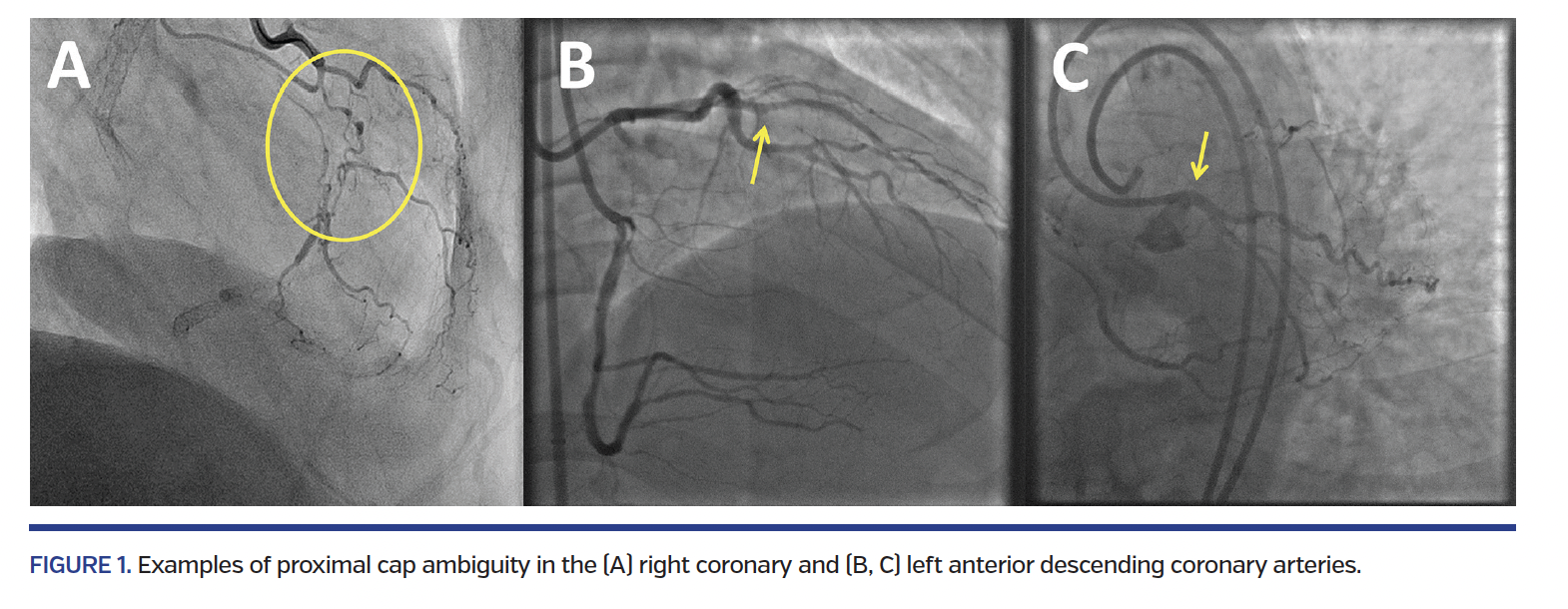
Proximal cap ambiguity is defined as inability to unequivocally determine the proximal entry point into a chronic total occlusion (CTO) (Figure 1). This can be due to the presence of side branches, vessel tortuosity, or flush ostial coronary artery occlusion preventing engagement of the guide catheter. Proximal cap ambiguity is one of the four major angiographic characteristics utilized to determine the preferred initial crossing strategy for coronary CTOs according to the hybrid crossing algorithm (the other three being occlusion length, quality of distal vessel, and presence of interventional collaterals).1 The difficulties posed by proximal cap ambiguity can be overcome via the use of multiple angiographic views, intravascular ultrasound (IVUS) or coronary computed tomography angiography (CCTA), use of subintimal dissection/reentry strategies,2 or use of a primary retrograde approach. We analyzed a multicenter CTO percutaneous coronary intervention (PCI) registry to determine the prevalence of proximal cap ambiguity and its impact on procedural techniques and outcomes.
Methods
We examined the clinical and angiographic records of patients who underwent CTO-PCI between May 2012 and December 2015 by experienced, high-volume operators at 11 centers in the United States: Appleton Cardiology, Appleton, Wisconsin; Columbia University, New York, New York; Henry Ford Hospital, Detroit, Michigan; Massachusetts General Hospital, Boston, Massachusetts; Medical Center of the Rockies, Loveland, Colorado; Piedmont Heart Institute, Atlanta, Georgia; PeaceHealth St. Joseph Medical Center, Bellingham, Washington; St. Luke’s Health System’s Mid-America Heart Institute, Kansas City, Missouri; Torrance Memorial Center, Torrance, California; VA North Texas Health Care System, Dallas, Texas; and VA San Diego Healthcare System, San Diego, California. Data collection was performed prospectively and retrospectively and recorded in a database (PROGRESS CTO, clinicaltrials.gov identifier: NCT02061436).3-11 Some centers only enrolled patients during part of the study period due to participation in other studies. The study was approved by the institutional review board of each site.
Definitions. Coronary CTOs were defined as coronary lesions with Thrombolysis in Myocardial Infarction (TIMI) grade 0 flow of at least 3-month duration. Estimation of the occlusion duration was based on first onset of anginal symptoms, prior history of myocardial infarction in the target vessel territory, or comparison with a prior angiogram. Proximal cap ambiguity was defined as inability to adequately define the location and shape of the proximal cap, as determined by the operator. Calcification was assessed by angiography as mild (spots), moderate (involving ≤50% of the reference lesion diameter), or severe (involving >50% of the reference lesion diameter). Moderate proximal vessel tortuosity was defined as the presence of at least 2 bends >70° or 1 bend >90° and severe tortuosity as 2 bends >90° or 1 bend >120° in the CTO vessel. Interventional collaterals were defined as collaterals deemed amenable to crossing by a guidewire and a microcatheter by the operator. Technical success of CTO-PCI was defined as successful CTO revascularization with achievement of <30% residual diameter stenosis within the treated segment and restoration of TIMI grade 3 antegrade flow. Procedural success was defined as achievement of technical success with no in-hospital major adverse cardiac event (MACE). In-hospital MACE included any of the following adverse events prior to hospital discharge: death, myocardial infarction (MI), recurrent symptoms requiring urgent repeat target-vessel revascularization with PCI or coronary artery bypass graft (CABG) surgery, tamponade requiring either pericardiocentesis or surgery, and stroke. Periprocedural and late in-hospital MI were defined according to the Third Universal Definition of Myocardial Infarction.12 The J-CTO score was calculated as described by Morino et al.13 and the Progress-CTO score as described by Christopoulos et al.4
Statistical analysis. Categorical variables were expressed as percentages and compared using Pearson’s c2 test or Fisher’s exact test. Continuous variables were presented as mean ± standard deviation or median with interquartile range (IQR) and were compared using the t-test or Wilcoxon rank-sum test, as appropriate. Logistic regression analysis was performed to identify predictors of technical failure for CTO-PCI: variables with P≤.10 in univariable analysis (patient age, hypertension, history of MI, prior CABG, smoking status, CTO length, proximal cap ambiguity, degree of calcification and tortuosity, bifurcation at the distal cap, and presence of interventional collaterals) were included in a multivariable model. Two-sided P-values of .05 were considered statistically significant. All statistical analyses were performed with JMP 12.0 (SAS Institute).
Results
The present analysis included 1021 CTO-PCIs performed in 1002 patients. Proximal cap ambiguity was present in 317 lesions (31%). Patients whose lesions had proximal cap ambiguity were more likely to have had prior MI (49% vs 42% with no cap ambiguity; P=.049), PCI (70% vs 62%; P=.02), CABG (43% vs 33%; P=.01), and peripheral arterial disease (20% vs 14%; P=.03), and to have a lower ejection fraction (48 ± 15% vs 51 ± 14%; P=.02) (Table 1).
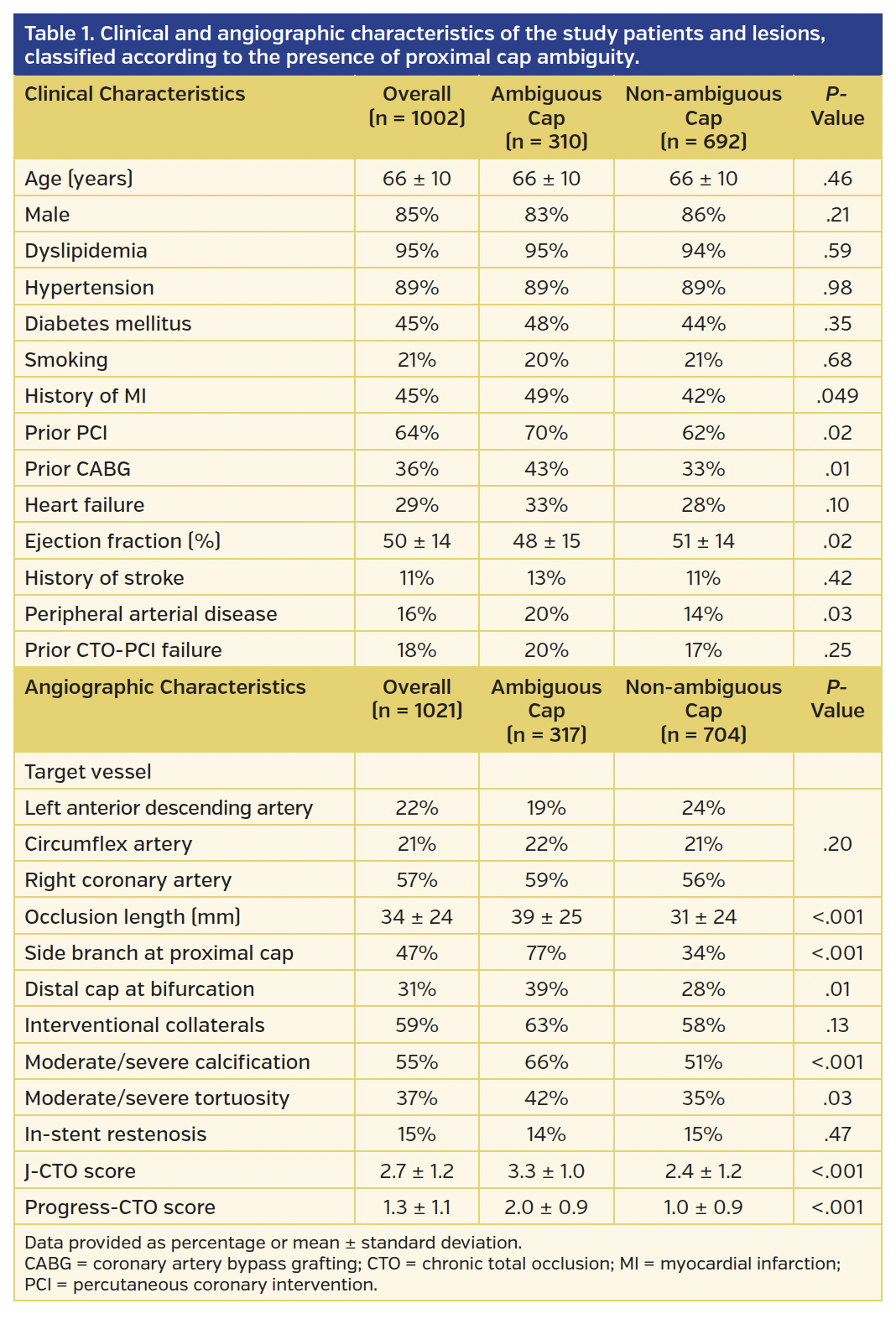
The right coronary artery was the most common target vessel (57%), followed by the left anterior descending (22%) and circumflex (21%) arteries. Lesions with proximal cap ambiguity were more likely to have a longer occlusion length (39 ± 25 mm vs 31 ± 24 mm; P<.001), moderate to severe proximal vessel tortuosity (42% vs 35%; P=.03) and calcification (66% vs 51%; P<.001), a side branch in the vicinity of the proximal cap (77% vs 34%; P<.001) and a bifurcation at the distal cap (39% vs 28%; P=.01).
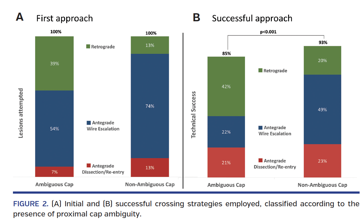
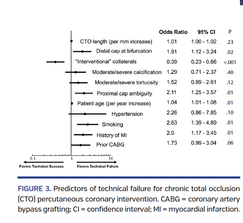
Overall, a larger number of approaches were utilized for recanalizing lesions with proximal cap ambiguity (1.7 ± 0.7 vs 1.5 ± 0.7; P<.001) (Table 2). When compared with lesions with non-ambiguous caps, lesions with ambiguous proximal caps were more frequently attempted using a retrograde approach (68% vs 33%; P<.001) and less frequently with antegrade wire escalation (64% vs 80%; P<.001); they also had lower overall technical success (85% vs 93%; P<.001) and procedural success (84% vs 91%; P=.01), and the final successful crossing strategy was more frequently a retrograde approach (42% vs 20%; P<.001) (Figure 2). In multivariable regression analysis, proximal cap ambiguity was found to be an independent predictor for technical failure, along with older patient age, smoking, history of MI, vessel bifurcation at the location of the distal cap, and lack of interventional collaterals (Figure 3).
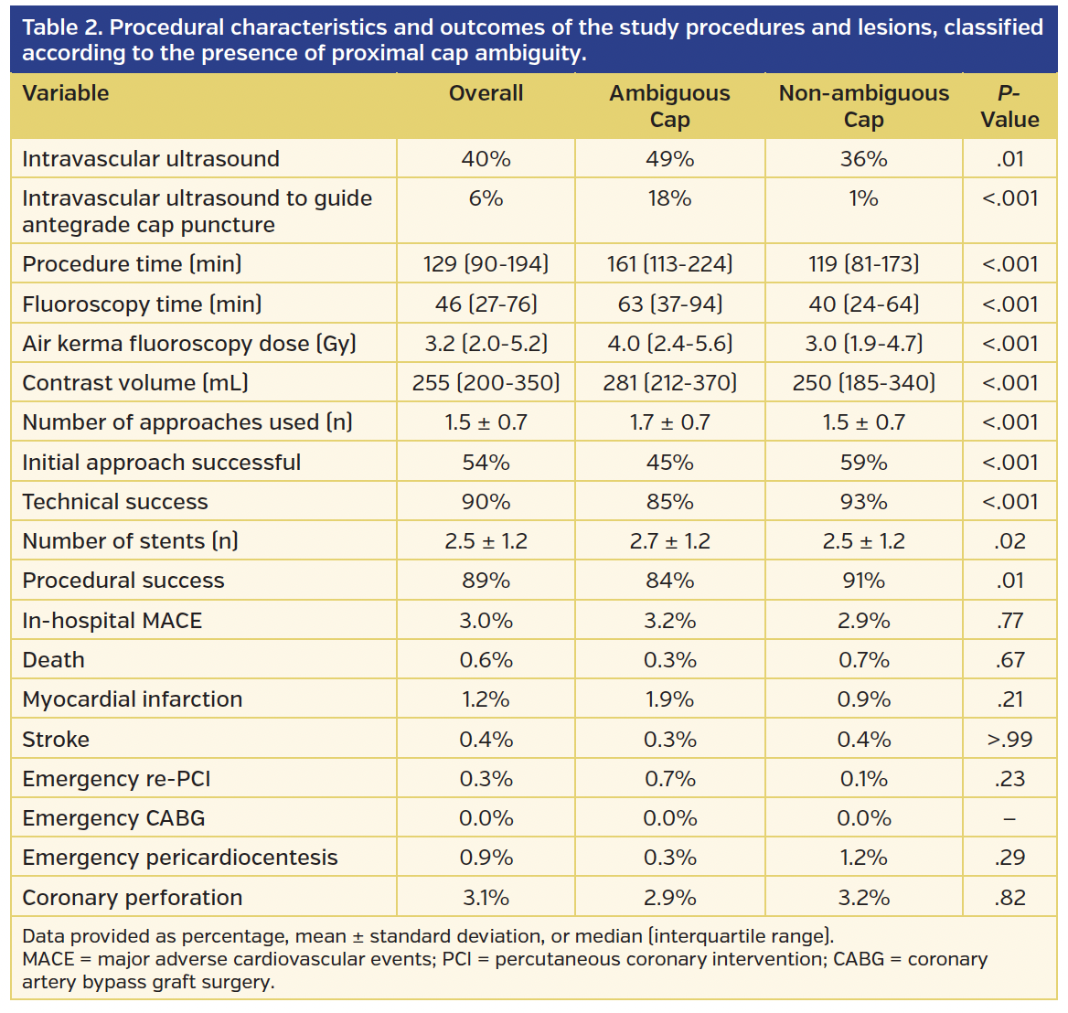
Treating CTOs with proximal cap ambiguity required more contrast (281 mL [IQR, 212-370 mL] vs 250 mL [IQR, 185-340 mL]; P<.001), radiation (air kerma dose, 4.0 Gy [IQR, 2.4-5.6 Gy] vs 3.0 Gy [IQR, 1.9-4.7 Gy]; P<.001), fluoroscopy time (63 min [IQR, 37-94 min] vs 40 min [IQR, 24-64 min]; P<.001), and procedural time (161 min [IQR, 113-224 min] vs 119 min [IQR, 81-173 min]; P<.001) to complete (Table 2). In addition, such lesions more frequently required the use of IVUS (49% vs 36% [P=.01], with IVUS used to  guide antegrade cap puncture in 18% vs 1% [P<.001]), and involved implantation of a larger number of stents per lesion (2.7 ± 1.2 vs 2.5 ± 1.2; P=.02).
Thirty patients (3.0%) experienced in-hospital MACE. There was no significant difference in the incidence of MACE (3.2% vs 2.9%; P=.77), any of the components of MACE, or coronary perforation (2.9% vs 3.2%; P=.82) between lesions with and without proximal cap ambiguity (Table 2).
Discussion
The main findings of our study are that proximal cap ambiguity: (1) is common in CTO-PCI, being encountered in approximately one-third of lesions; (2) is associated with other indicators of clinical and angiographic complexity; (3) requires more frequent use of the retrograde approach; and (4) is associated with lower technical and procedural success and efficiency, but similar incidence of MACE.
In our study, proximal cap ambiguity was present in 31% of CTOs. Moreover, lesions with an ambiguous cap displayed additional markers of angiographic complexity including longer occlusion length, more extensive tortuosity and calcification, and more frequent origin of side branches at the proximal cap. Resolving proximal cap ambiguity is important to improve the likelihood of success and decrease the risk during antegrade wiring of lesions with ambiguous caps, and can sometimes be achieved by dual coronary injection, use of additional angiographic projections (such as the lateral projection for right coronary artery lesions), and possibly identification of additional markers of the vessel course, such as calcification. If cap ambiguity cannot be resolved by angiography, it can sometimes be resolved by intravascular imaging. IVUS can be useful to identify the location of the occluded vessel, but may not always be able to be delivered to the occlusion. IVUS can be particularly useful if a side branch is present next to the occlusion, and can be facilitated by use of short-tip catheters (such as the short-tip Eagle Eye; Volcano Corporation). Coronary CTA can also help determine the vessel course, especially when co-registration with angiographic imaging can be performed.
Should imaging fail to resolve proximal cap ambiguity, treatment options include use of a primary retrograde approach or antegrade dissection/reentry. If the retrograde approach is feasible, a retrograde wire can either cross the occlusion, or help clarify the ambiguity, thereby enabling antegrade guidewire crossing. Antegrade dissection and reentry can be used to cross ambiguous proximal caps (the “move the cap technique”), by using a stiff guidewire (the “scratch and go” technique), or by using a slightly oversized balloon (the “balloon-assisted subintimal entry” technique) to create a proximal dissection though which a knuckled guidewire is advanced around the occluded segment, followed by reentry into the distal true lumen.2
As expected, in our study, lesions with proximal cap ambiguity were approached more frequently with the retrograde approach (68% vs 33%; P<.001), which was the initial crossing strategy in 39% and the final successful crossing strategy in 42% of such lesions. Despite increased utilization of the retrograde approach, we found no significant increase in MACE or coronary perforation in cases with cap ambiguity, which may be explained by careful lesion selection and utilization of the retrograde approach as a primary strategy, as opposed to bail-out after failed (and potentially dangerous) antegrade attempts.6,14 Of note, lesions with ambiguous proximal caps that exhibited interventional collaterals had significantly better outcomes than ambiguous lesions with no such collaterals (technical success, 88% vs 79%; P=.04); the retrograde approach accounted for 66% of the technical success in the former group. Thus, the presence of unresolvable proximal cap ambiguity in combination with the presence of good collaterals remains, in our opinion, a strong indication for selection of a primary retrograde strategy. The use of IVUS in the cap ambiguity subgroup was also significantly increased, demonstrating the usefulness of this technique in characterizing the proximal cap, as long as an adequate side branch allows deployment of the IVUS catheter. Despite the availability of the aforementioned techniques to resolve cap ambiguity, technical and procedural success remained lower for lesions with ambiguous proximal caps.
Proximal cap ambiguity was also associated with lower procedural efficiency in our study; procedure and fluoroscopy times were longer, there was a higher utilization of contrast and radiation, and more stents were implanted for lesions with ambiguous caps, with the latter likely due to longer occlusion length and more frequent entry into the subintimal space (planned or inadvertent).
Study limitations. Our study has important limitations. It is observational, subject to selection bias, and includes procedures performed by experienced CTO operators; thus, our results may not apply to procedures performed by less experienced operators.15 The presence or absence of proximal cap ambiguity was at the discretion of each operator, and it is possible that different operators may have different abilities to clarify the location of the proximal cap based on angiographic analysis. There was no core laboratory adjudication of the angiograms or centralized clinical event adjudication.
Conclusion
Proximal cap ambiguity is a frequently encountered indicator of angiographic complexity during CTO-PCI and is associated with lower procedural efficiency and success rates, but no difference in MACE rates. IVUS and the retrograde approach are tools frequently used to assist recanalization of these complex lesions.
Acknowledgment. We would like to thank Aya J. Alame, BA, Jose R. Martinez-Parachini, MD, Erica Resendes, MS, and Michele Roesle, RN for their assistance with the present study.
Study data were collected and managed using REDCap electronic data capture tools hosted at University of Texas Southwestern Medical Center.1 REDCap (Research Electronic Data Capture) is a secure, web-based application designed to support data capture for research studies, providing: (1) an intuitive interface for validated data entry; (2) audit trails for tracking data manipulation and export procedures; (3) automated export procedures for seamless data downloads to common statistical packages; and (4) procedures for importing data from external sources.16
References
1. Brilakis ES, Grantham JA, Rinfret S, et al. A percutaneous treatment algorithm for crossing coronary chronic total occlusions. JACC Cardiovasc Interv. 2012;5:367-379.
2. Vo MN, Karmpaliotis D, Brilakis ES. “Move the cap” technique for ambiguous or impenetrable proximal cap of coronary total occlusion. Catheter Cardiovasc Interv. 2016;87:742-748. Epub 2015 Sep 2.
3. Alaswad K, Menon RV, Christopoulos G, et al. Transradial approach for coronary chronic total occlusion interventions: insights from a contemporary multicenter registry. Catheter Cardiovasc Interv. 2015;85:1123-1129.
4. Christopoulos G, Kandzari DE, Yeh RW, et al. Development and validation of a novel scoring system for predicting technical success of chronic total occlusion percutaneous coronary interventions: the PROGRESS CTO (Prospective Global Registry for the Study of Chronic Total Occlusion Intervention) score. JACC Cardiovasc Interv. 2016;9:1-9.
5. Christopoulos G, Karmpaliotis D, Alaswad K, et al. The efficacy of “hybrid” percutaneous coronary intervention in chronic total occlusions caused by in-stent restenosis: insights from a US multicenter registry. Catheter Cardiovasc Interv. 2014;84:646-651.
6. Christopoulos G, Karmpaliotis D, Alaswad K, et al. Application and outcomes of a hybrid approach to chronic total occlusion percutaneous coronary intervention in a contemporary multicenter US registry. Int J Cardiol. 2015;198:222-228.
7. Christopoulos G, Karmpaliotis D, Wyman MR, et al. Percutaneous intervention of circumflex chronic total occlusions is associated with worse procedural outcomes: insights from a multicentre US registry. Can J Cardiol. 2014;30:1588-1594.
8. Christopoulos G, Menon RV, Karmpaliotis D, et al. The efficacy and safety of the “hybrid” approach to coronary chronic total occlusions: insights from a contemporary multicenter US registry and comparison with prior studies. J Invasive Cardiol. 2014;26:427-432.
9. Christopoulos G, Menon RV, Karmpaliotis D, et al. Application of the “hybrid approach” to chronic total occlusions in patients with previous coronary artery bypass graft surgery (from a contemporary multicenter US registry). Am J Cardiol. 2014;113:1990-1994.
10. Christopoulos G, Wyman RM, Alaswad K, et al. Clinical utility of the Japan-chronic total occlusion score in coronary chronic total occlusion interventions: results from a multicenter registry. Circ Cardiovasc Interv. 2015;8:e002171.
11. Sapontis J, Christopoulos G, Grantham JA, et al. Procedural failure of chronic total occlusion percutaneous coronary intervention: Insights from a multicenter US registry. Catheter Cardiovasc Interv. 2015;85:1115-1122.
12. Thygesen K, Alpert JS, Jaffe AS, et al. Third universal definition of myocardial infarction. Circulation. 2012;126:2020-2035.
13. Morino Y, Abe M, Morimoto T, et al. Predicting successful guidewire crossing through chronic total occlusion of native coronary lesions within 30 minutes: the J-CTO (multicenter CTO registry in Japan) score as a difficulty grading and time assessment tool. JACC Cardiovasc Interv. 2011;4:213-221.
14. Michael TT, Mogabgab O, Fuh E, et al. Application of the “hybrid approach” to chronic total occlusion interventions: a detailed procedural analysis. J Interv Cardiol. 2014;27:36-43.
15. Brilakis ES, Banerjee S, Karmpaliotis D, et al. Procedural outcomes of chronic total occlusion percutaneous coronary intervention: a report from the NCDR (National Cardiovascular Data Registry). JACC Cardiovasc Interv. 2015;8:245-253.
16. Harris PA, Taylor R, Thielke R, Payne J, Gonzalez N, Conde JG. Research electronic data capture (REDCap) – a metadata-driven methodology and workflow process for providing translational research informatics support. J Biomed Inform. 2009;42:377-381.
From the 1VA North Texas Health Care System and University of Texas Southwestern Medical Center, Dallas, Texas; 2Columbia University, New York, New York; 3Henry Ford Hospital, Detroit, Michigan; 4Massachusetts General Hospital, Boston, Massachusetts; 5Beth Israel Deaconess Medical Center, Boston, Massachusetts; 6VA San Diego Healthcare System and University of California San Diego, La Jolla, California; 7Torrance Memorial Medical Center, Torrance, California; 8PeaceHealth St. Joseph Medical Center, Bellingham, Washington; 9Mid America Heart Institute, Kansas City, Missouri; 10Piedmont Heart Institute, Atlanta, Georgia; 11Medical Center of the Rockies, Loveland, Colorado; 12Minneapolis VA Health Care System and University of Minnesota, Minneapolis, Minnesota; and 13Boston Scientific, Natick, Massachusetts.
Funding: Research reported in this publication was supported by the Clinical and Translational Science Awards Program of the National Institutes of Health, (Bethesda, Maryland, USA) under grant number UL1-RR024982. The content is solely the responsibility of the authors and does not necessarily represent the official views of the NIH.
Disclosures: The authors have completed and returned the ICMJE Form for Disclosure of Potential Conflicts of Interest. Dr Karmpaliotis reports speaker’s bureau fees from Abbott Vascular, Medtronic, and Boston Scientific; consultant fees from BridgePoint Medical. Dr Alaswad reports consulting fees from Terumo and Boston Scientific; consultant (non-financial support) for Abbott Laboratories. Dr Jaffer is a consultant for Boston Scientific, Siemens, and Merck; non-financial research support from Abbott Vascular; research grant from National Institutes of Health (HL-R01-108229), Siemens, and Kowa. Dr Yeh has received a Career Development Award (1K23HL118138) from the National Heart, Lung, and Blood Institute; consultant for Abbott Vascular, Gilead Sciences, and Boston Scientific; advisory board for Abbott Vascular; salary support from Harvard Clinical Research Institute. Dr Patel is on the speaker’s bureau for Astra Zeneca. Dr Wyman reports honoraria/consulting/speaking fees from Boston Scientific, Abbott Vascular, and Asahi Intecc. Dr Lombardi reports equity with BridgePoint Medical; consultant for Boston Scientific, Abiomed, and Abbott Vascular. Dr Grantham reports speaking fees, consulting fees, and honoraria from Boston Scientific and Asahi Intecc; research grants from Boston Scientific, Asahi Intecc, Abbott Vascular, and Medtronic. Dr Kandzari reports research/grant support and consulting honoraria from Boston Scientific and Medtronic Cardiovascular; research/grant support from Abbott Vascular. Dr Lembo reports speaker’s bureau fees from Medtronic; advisory board fees from Abbott Vascular and Medtronic. Dr Moses is a consultant to Boston Scientific and Abiomed. Dr Kirtane reports institutional research grants to Columbia University from Boston Scientific, Medtronic, Abbott Vascular, Abiomed, St. Jude Medical, Vascular Dynamics, Glaxo SmithKline, and Eli Lilly. Dr Garcia reports consulting fees from Medtronic and Surmodics. Dr Parikh reports personal fees (speaker’s bureau and/or advisory board) from Abbott Vascular, Medtronic, CSI, Boston Scientific, and Philips. Dr Thompson reports that he is an employee of Boston Scientific. Dr Banerjee reports research grants from Gilead and the Medicines Company; consultant/speaker honoraria from Covidien and Medtronic; ownership in MDCare Global (spouse); intellectual property in HygeiaTel. Dr Brilakis reports consulting/speaker honoraria from Abbott Vascular, Asahi Intecc, Cardinal Health, Elsevier, GE HealthCare, and St. Jude Medical; research support from Boston Scientific and InfraRedx; spouse is employee of Medtronic. The remaining authors report no disclosures regarding the content herein.
Manuscript submitted April 26, 2016, provisional acceptance given May 3, 2016, final version accepted May 13, 2016.
Address for correspondence: Emmanouil S. Brilakis, MD, PhD, Dallas VA Medical Center (111A), 4500 South Lancaster Road, Dallas, TX 75216. Email: esbrilakis@gmail.com


