Percutaneous Closure of a Traumatic Fistula Between the Right Coronary Artery and the Right Atrium (Full title below)
Percutaneous Closure of a Traumatic Fistula Between the Right Coronary Artery and the Right Atrium in a Young Adult: A New Covered Stent Strategy
_______________________________________________
From the Liverpool Heart and Chest Hospital, Liverpool, United Kingdom. The authors report no conflicts of interest regarding the content herein. Manuscript submitted February 3, 2009, provisional acceptance given February 16, 2009, and final version accepted February 17, 2009. Address for correspondence: Telal O. Mudawi, MRCP (UK), Liverpool Heart and Chest Hospital, Thomas Drive, Liverpool, L14 3PE, United Kingdom. E-mail: telalmudawwi@gmail.com
_______________________________________________
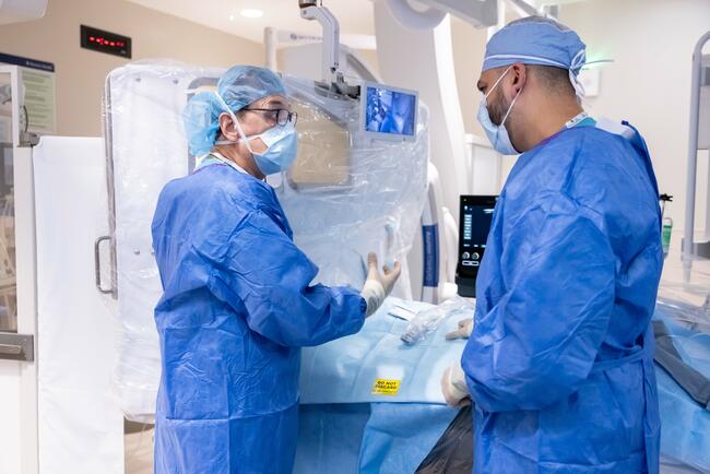
J INVASIVE CARDIOL 2009;21:E161-E163 We present a case of a young adult who suffered penetrating chest trauma. Coronary angiographic evaluation revealed the development of a hemodynamically significant fistula between the right coronary artery and the right atrium. Effective long-term fistula closure was achieved using percutaneous intervention with a covered stent strategy. Case Presentation. A previously fit 20 year-old male sustained a stab wound injury to the right anterior parasternal chest wall. The assault weapon was a 3-inch blade knife. He was admitted to the nearest cardiothoracic unit and was treated for right hemothorax with a chest drain. He also required the transfusion of 4 units of blood. Serial chest X-rays revealed acceptable continuing regression of the hemothorax. Serial echocardiograms only showed a very tiny rim of fluid at the right ventricular (RV) wall, with normal structure and function of all the chambers and valves. He made a seemingly uneventful recovery and was subsequently discharged home after having spent 2 days in the hospital. Six weeks later, he was admitted to his local district hospital with progressively worsening dyspnea. He was found to have developed a huge pericardial effusion. Because of his recent history of trauma, he was transferred to our tertiary cardiac center as an emergency for further management. On arrival he was clinically tamponading with a tachycardia of 120 beats per minute. His blood pressure was 110/70 mmHg with a paradox of 25 mmHg. Echocardiography confirmed the presence of a large pericardial effusion with evidence of RV diastolic collapse. Following discussion with our local cardiac surgical colleagues, who happy to have us drain the effusion, approximately 1 liter of heavily blood-stained fluid was drained. There was rapid subsequent clinical improvement with complete resolution of symptoms and signs of tamponade. A soft continuous murmur had then become clearly audible at the left sternal edge. Repeat echocardiography showed almost complete resolution of the pericardial effusion and some small diastolic color flow visible at the right atrial wall adjacent to the tricuspid valve annulus. Otherwise, the heart structure and function appeared normal. He subsequently underwent coronary angiography to identify the nature of the odd-appearing color flow. The left ventricular function and left coronary system were both found to be normal. The right coronary artery (RCA) was found to have been severed at its mid point, with the proximal segment draining into the right atrium (Figure 1). The distal RCA was retrogradely filling from the left coronary system (Figure 2). We made a diagnosis of a traumatic fistulous connection between the proximal RCA and right atrium. The fistula measured 3.5 cm in diameter (Qp:Qs ratio = 2:1). After discussing the case at the multidisciplinary team (MDT) meeting with our local surgical and interventional colleagues, and after some further discussion with some of our national interventional colleagues, we unanimously agreed to percutaneously reconstruct the RCA using a covered stent strategy. We performed the procedure under local anesthesia with full surgical backup available. A 7 Fr JR3.5 guide catheter was used to cannulate the RCA via the right femoral artery. A 5 Fr diagnostic JL4 catheter was used to cannulate the left coronary artery (to visualize the distal RCA) via the left femoral artery. With the aid of road-mapping the view of simultaneous right and left coronary contrast injections (Figure 3), the RCA was successfully wired through to its distal segment using a hydrophilic Pilot-50 guidewire. The vessel diameter was sized using both quantitative coronary assessment and intravascular ultrasound modalities. A 4.0 x 19 mm Jostent Graftmaster covered stent (Abbott Vascular, Abbott Park, Illinois) was then deployed at high atmospheric pressure (18 bar). There was instantaneous subsequent complete closure of the fistula along with restoration of thrombolysis in myocardial infarction (TIMI)-3 flow through to the distal RCA (Figure 4), coupled with cessation of retrograde RCA filling. The patient remained fully active and asymptomatic thereafter, playing competitive sports on a regular basis. We performed a diagnostic coronary angiogram 8 months later to assess the patency of the stent and to check whether the fistula had continued to remain closed. This disappointingly showed that the stent had subtotally occluded, with reestablishment of retrograde filling of the distal RCA from the left coronary system. The left ventricular systolic function, however, was found to be good with no evidence of regional wall-motion abnormality. There was no residual fistulous connection or flow seen between the RCA and the right atrium. We decided not to attempt any further percutaneous intervention at this stage and to pursue a conservative management strategy with regular follow-up visits. Subsequent exercise stress testing has shown no evidence of ischemia at high levels of both physical and cardiac workloads. More recently, the patient was reviewed at our outpatient clinic and he continued to remain completely asymptomatic with excellent exercise capacity. Echocardiography performed at the time revealed good left ventricular systolic function, with no evidence of fistula recurrence. Discussion. Congenital — and acquired — coronary artery fistulae (CAF) are abnormal channels through which blood is shunted from a coronary artery into a cardiac chamber (coronary-cameral fistula), a great vessel or other structure, bypassing the myocardial capillary network. Congenital fistulae account for 0.08–0.4% of congenital cardiac anomalies.1 They can present at any age and vary considerably in their clinical presentation and long-term outcome depending on the site and size of the fistula. Traumatic CAF resulting from penetrating chest injuries are less commonly described in the literature. Treatment is advocated for symptomatic patients and for those asymptomatic patients who are at risk for future complications. In this patient, we discussed the various possible therapeutic options including conservative medical management and the two previously used techniques of open surgical correction and transcatheter embolization.2 We thought both methods were not ideal as, in the case of surgery, a vein graft to the RCA is likely to occlude after a number of years with a real possibility of further surgical re-grafting operations in the future. Transcatheter embolization can effectively close the fistula, but it does not restore the continuity of the transsected RCA. Furthermore, it can be complicated by coil migration and vessel wall rupture.3 On the other hand, given the size of the shunt, a conservative medical approach could ultimately allow for long-term complications to arise such as heart failure, pulmonary hypertension and infective endocarditis.4,5 Although traumatic injuries affecting large arteries have been shown to be effectively treatable by the Wallgraft covered stent endoprosthesis (Boston Scientific Corp. Natick, Massachusetts),6 such a strategy was rarely used, if ever, for treating traumatic coronary fistulae. We felt that a covered stent strategy was appropriate, as we expected it to provide both effective closure of the fistula and reconstruction of the RCA with flow restoration through to the distal vessel. This has proven to be an effective instantaneous fistula closure method, but was unsuccessful in sustaining long-term antegrade flow in the RCA. With further improvement in covered stent technology, we believe that this technique could still be applied in the future for similar cases. A proper randomized study comparing surgery, transcatheter coiling and percutaneous stenting of such cases is required before determining their best management strategy.
2. Gowda RM, Vasavada BC, Khan IA. Coronary artery fistulas: clinical and therapeutic considerations. Int J Cardiol 2006;107:7–10.
3. Alekyan BG, Podzolkov VP, Cárdenas CE. Transcatheter coil embolization of coronary artery fistula. Asian Cardiovasc Thorac Ann 2002;10:47–52.
4. Yoda M, Minami K, Koerfer R. Images in cardiology. Left coronal ostium to right atrium fistula causing right ventricular failure and pulmonary hypertension. Heart 2006;92:330.
5. Trejo Gutiérrez JF, Eng Ceceña L, Zghaib Abad A, et al. Coronary arteriovenous fistula. Study of 14 cases. Arch Inst Cardiol Mex 1985;55:153–164.
6. White R, Krajcer Z, Johnson M, et al. Results of a multicenter trial for the treatment of traumatic vascular injury with a covered stent. J Trauma 2006;60:1189º1195; discussion 1195–1196.