Percutaneous Closure of Two Acquired Aorto-Right Ventricular Fistulae Following Right Ventricular Outflow Tract Surgery
J INVASIVE CARDIOL 2017;29(9):E101.
Key words: Amplatzer duct occluder, right ventricular outflow tract
An 86-year-old man presented with shortness of breath two months after surgical resection of a right ventricular outflow tract (RVOT) mass. Physical examination revealed jugular venous distention and a continuous murmur over the second left sternal border. An echocardiogram performed revealed an abnormal color Doppler signal within the RVOT. Transesophageal echocardiogram demonstrated two communications between the aortic root and the RVOT (Figure 1A; Videos 1 and 2). The shunts were felt to be iatrogenic from his recent cardiac surgery. He was deemed a poor surgical candidate and percutaneous closure of both fistulae under monitored anesthesia care with transthoracic echocardiogram and fluoroscopy guidance was elected. Initial left ventriculogram demonstrated the shunts (Video 3). The first defect measured 5 mm in diameter, and the second measured 7 mm. A 6 x 4 mm and a 6 x 6 mm Amplatzer duct occluder II (AGA Medical Corporation) were used to close the defects (Figure 1B; Videos 4 and 5). The patient improved significantly and has done well at 6-month follow-up.
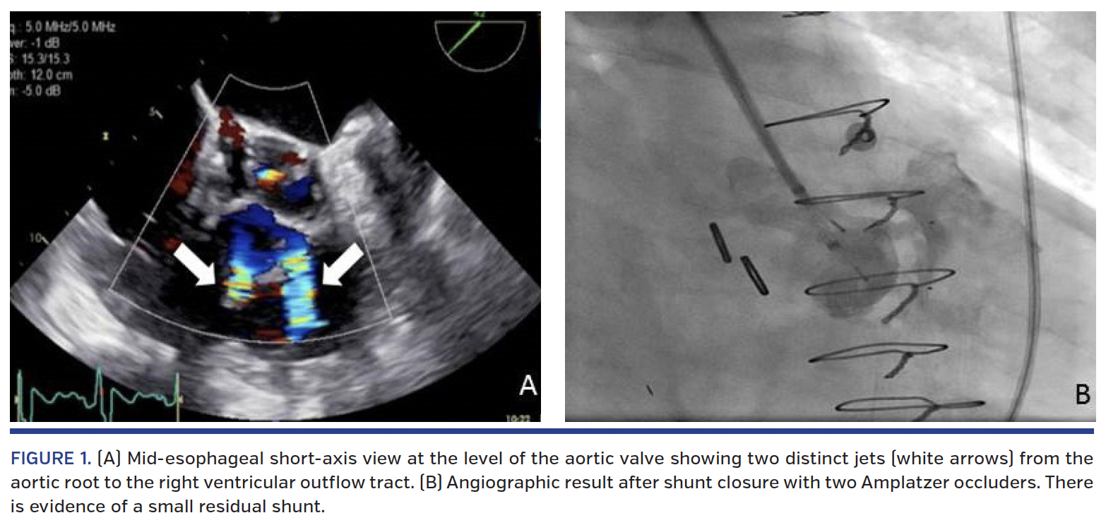
Aorto-right ventricular shunts have been described after surgical or transcatheter aortic valve replacement and as a complication of aortic valve endocarditis or trauma.1,2 In the treatment of intracardiac shunts, duct occluders have emerged as an alternative to surgery, and in many cases, as the preferred treatment option.3 The present case illustrates the successful transcatheter closure of an unusual double aortic root-to-RVOT fistula utilizing two Amplatzer duct occluders II.
View Videos 1-5 Here
References
1. Leu HB, Chang HH, Wu MH, Chen YH. Four-year follow-up of acquired aorto-right ventricular fistula after transcatheter aortic valve implantation. Eur Heart J. 2016;37:2679.
2. Gunarathne A, Hunt J, Gershlick A. Aorto-right atrial and right ventricular fistulae: a very rare complication of native bicuspid aortic valve endocarditis. Heart. 2013;99:1708.
3. Pineda AM, Mihos CG, Singla S, et al. Percutaneous closure of intracardiac defects in adults: state of the art. J Invasive Cardiol. 2015;27:561-572.
From 1The Columbia University Division of Cardiology at the Mount Sinai Heart Institute, Miami Beach, Florida; and 2the Cardiology Department, Nicklaus Children’s Hospital, Miami, Florida.
Disclosure: The authors have completed and returned the ICMJE Form for Disclosure of Potential Conflicts of Interest. The authors report no conflicts of interest regarding the content herein.
Manuscript accepted April 7, 2017.
Address for correspondence: Orlando Santana, MD, Director, Echocardiography Laboratory, Columbia University Division of Cardiology, Mount Sinai Heart Institute, 4300 Alton Road, Miami Beach, FL 33140. Email: osantana@msmc.com