Predictors of Recurrent Restenosis After Coronary Stenting: An Analysis of 197 Patients
April 2002
In recent years, coronary stent placement has become an established treatment for patients with coronary artery disease.1,2 However, in-stent restenosis has been reported to occur in 20–30% of patients, and the restenosis rate after treatment for in-stent restenosis is also high (> 30%), regardless of treatment modalities, including balloon angioplasty,3–5 rotational atherectomy,6,7 and repeat stenting. The effectiveness of stent therapy can be questioned, particularly in patients with recurrent restenosis after stent implantation. If the factors for recurrent restenosis can be predicted after stent implantation, other more effective strategies for treatment may be considered. In this study, we examined the incidence of recurrent restenosis in patients after Palmaz-Schatz (P-S) stent implantation to identify the factors affecting recurrent restenosis after stenting.
MATERIALS AND METHODS
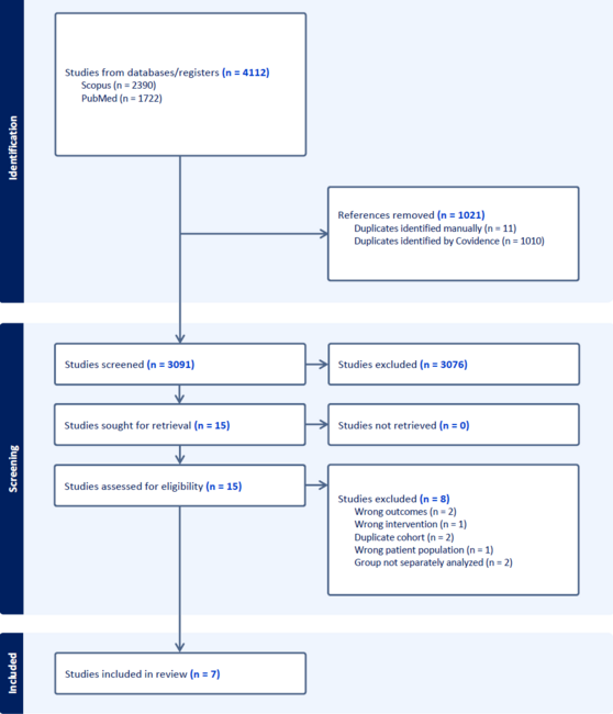
Study group. From January 1997 to December 1997, a total of 197 consecutive patients underwent successful coronary stent implantation using one P-S stent at Komatsushima Red Cross Hospital. The stent implantation technique has been described previously.1 Before stent implantation, 81 mg of aspirin, calcium blockers, and depending on the lesion morphology, nitrate or beta blockers were administered. All patients received 10,000 units of heparin intravenously prior to PTCA. After stent implantation, aspirin (162 mg) and ticlopidine (200 mg) were administered for at least 1 month. Coronary stents were implanted under fluoroscopic guidance. The balloon size and inflation pressure were at the operator’s discretion. Adequacy of the final result was based solely on the angiographic assessment. At the time of stenting, patients were asked to return for a 6-month follow-up angiogram, regardless of the presence or absence of symptoms. Coronary angiography was performed earlier if clinically indicated. Angiographic follow-up was performed at a mean of 5.8 ± 1.6 months after coronary stenting in 170 patients (86%). Restenosis was defined as a diameter stenosis >= 50% at the time of follow-up angiography. When in-stent restenosis was confirmed, repeat balloon angioplasty was performed. Three months after repeat balloon angioplasty, coronary angiography was again performed. If a second restenosis was confirmed, balloon angioplasty was repeated and then assessed by angiography 3 months later. After coronary stent implantation, 100 patients (Group A: 64 ± 8 years; M:F = 82:18) never restenosed, in Group B 49 patients (64 ± 9 years; M:F = 41:8) restenosed once, and 21 patients in Group C (65 ± 10 years; M:F = 11:10) restenosed more than twice. The 3 groups were analyzed.
Angiographic analysis. Quantitative analyses were performed independently by two experienced interventional cardiologists. Lesions were classified according to the American College of Cardiology/American Heart Association (ACC/AHA) classification system.8 Coronary calcification was defined by fluoroscopy. Angiographic coronary dissections after pre-dilation were identified according to the criteria of the National Heart, Lung, and Blood Institute Percutaneous Transluminal Coronary Angioplasty Registry9 and classified as either minor dissections (types A and B) or major dissections (types C–F). Patterns of in-stent restenosis were divided into focal and diffuse type according to angiography by Mehran et al.10 Before and after the intervention, as well as at follow-up angiography, efforts were made to control coronary vasomotor tone through intracoronary nitroglycerin administration. The analysis was performed using orthogonal end-diastolic cine frames demonstrating the stenosis in its most severe and nonforeshortened projections, using a computer-assisted, automated edge-detection algorithm (Cardio 500, Kontron Electronics, BILDANALYSE, München, Germany). The contrast-filled catheter was used as the calibration standard. Reference vessel size was determined from the post-procedural images.
Statistical analysis. Statistical analysis was performed with Statview version 5.0 (Abacus Concepts, Inc., Berkeley, California). All continuous variables are expressed as mean ± SD and categorical variables as percentages. Comparisons between groups were performed using the chi-square test to analyze differences in categorical variables, and the T test for continuous variables. A p value 20 mm), multiple vessel total occlusions, collateral circulation, and ostial and bifurcation lesions. Procedural factors include a post-procedure residual stenosis > 30%.13–18 At our institution, there were 21 patients of recurrent restenosis after coronary stenting between January through December of 1997. This study was undertaken to predict the factors that contribute to recurrent restenosis after coronary stenting. Our study demonstrates that there are several characteristics associated with recurrent restenosis after stent implantation: 1) female gender, 2) final diameter stenosis, and 3) diameter stenosis after predilatation.
Elezi et al.19 analyzed the clinical and angiographic outcome of diabetic patients following successful coronary stent placement, and compared these results with those achieved after stenting in nondiabetic patients. They found that diabetes is an independent risk factor for a poorer angiographic and clinical outcome. Furthermore, coronary stent placement did not appear to eliminate the excessive cardiovascular risk in diabetic patients after coronary intervention. Lau et al.20 also reported that there is a high incidence of in-stent restenosis in diabetic patients, particularly in small vessels. Diabetes is thought to be an important factor promoting neointimal proliferation. Excessive neointimal reaction in the setting of diabetes probably results from complex hormonal and biochemical alterations.21 These might cause accelerated smooth muscle cell proliferation after coronary stenting.22–24 But diabetes mellitus has not been a recurrent in-stent restenosis predictor in this study. This might be due to comparatively large reference diameter of the coronary artery assessed.
In this study, the diameter stenosis after predilation and after stenting was greater and the incidence of diffuse in-stent restenosis was higher after stenting in recurrent restenosis patients. Mehran et al.10 reported that diffuse type of ISR represents a spectrum of increasing severity (exaggerated neointimal hyperplasia), and the very high rate of subsequent revascularization after interventional therapy with currently available treatment modalities in patients with diffuse type ISR. Therefore, it is suggested that the quality of pre- and post-dilatation is related to the recurrence of restenosis after stenting. Serruys et al.25 reported that factors predicting in-stent restenosis include percent diameter stenosis after stent implantation and vessel diameter using intravascular ultrasound. After all, if stents are implanted in lesions without proper pre-dilation, the stent struts against the vessel wall become the incomplete apposition and diffuse type of in-stent restenosis is likely to occur. This may be a relevant point considering the current interest in direct stenting.26,27 Direct stenting is the method of stenting without balloon predilatation. It is reported that the in-hospital and long-term outcomes in patients undergoing a coronary intervention are equivalent when comparing stenting without balloon predilatation with balloon angioplasty followed by stenting.27 However, this study is the results of direct stenting for selected patients and all lesions are not suitable for direct stenting. Therefore, to avoid the recurrent in-stent restenosis, IVUS guidance before direct stenting is necessary to concern whether the target lesion could be dilatated properly with balloon or not.
Extensive data show that brachytherapy reduces recurrent in-stent restenosis.28–30 The report was the Coronary Radiation to Inhibit Intimal Proliferation Post Stenting (SCRIPS) Trial, in which most of the lesions were in-stent restenosis lesions.28 The WRIST Trial specifically addressed treatment of in-stent restenosis.29 Of note, more than half of the lesions had a reduction in intimal hyperplasia between radiation and follow-up. Therefore, brachytherapy should be considered for in-stent restenotic patients with recurrent predictors.
Study limitations. This was a retrospective analysis of the clinical and angiographic data derived from the group of consecutively patients after stenting. There is a tremendous limitation deriving from the small number of patients with recurrent restenosis analyzed in this study. The stents used are Palmaz-Schatz which are no longer widely used, so the results of this study are not necessary suitable for new generative stents. Diameter stenosis after predilatation might be a rather imprecise predictor because no specific predilatation protocol was implemented in this study. Coronary calcification is not estimated precisely on account of the fact that calcification is difficult to quantify angiographically. No other interventional techniques for in-stent restenosis, such as cutting balloon and rotablator, were used in this study. Thus, our results should be restricted to angiography-guided stenting and balloon angioplasty for in-stent restenosis. Whether these tools can lower the probability of recurrent restenosis after stenting remains unknown. However, our findings should be considered preliminary and require further verification from large, prospective trials.
Conclusion
This comparative study clearly demonstrates that there are several characteristics that predict recurrent in-stent restenosis. These include 1) female gender, 2) final diameter stenosis, and diameter stenosis after predilatation.
1. Serruys PW, de Jaegere P, Kiemeneij F, et al. A comparison of balloon-expandable-stent implantation with balloon angioplasty in patients with coronary artery disease: Benestent Study Group. N Engl J Med 1994;331:489–495.
2. Fischman DL, Leon MB, Baims DS, et al. A randomized comparison of coronary-stent placement and balloon angioplasty in the treatment of coronary artery disease. N Engl J Med 1994;331:496–501.
3. Eltchaninoff H, Konig R, Tron C, et al. Balloon angioplasty for the treatment of coronary in-stent restenosis: immediate results and 6-months angiographic recurrent restenosis rate. J Am Coll Cardiol 1997;30:186–192.
4. Reimers B, Moussa I, Akiyama T, et al. Long-term clinical follow-up after successful repeat percutaneous intervention for stent restenosis. J Am Coll Cardiol 1997;30:186–192.
5. Bauters C, Banos JL, Van Belle E, et al. Six-month angiographic outcome after successful repeat percutaneous intervention for in-stent restenosis. Circulation 1998;97:318–321.
6. Lee SG, Lee CW, Cheong SS, et al. Immediate and long-term outcomes of rotational atherectomy versus balloon angioplasty alone for treatment of diffuse stent restenosis. Am J Cardiol 1988;82:140–143.
7. Dauerman HL, Baims DS, Cutlip DE, et al. Mechanical debulking versus balloon angioplasty for the treatment of diffuse in-stent restenosis. Am J Cardiol 1998;82:277–284.
8. Ryan TJ, Faxon DP, Gunnar RM, et al. Guidelines for percutaneous transluminal coronary angioplasty: A report of the American College of Cardiology/American Heart Association Task Force on assessment of Diagnostic and Therapeutic Cardiovascular Procedures. Circulation 1988;78:486–502.
9. Huber MS, Mooney JD, Madison J, Mooney MR. Use of morphologic classification to predict clinical outcome after dissection from coronary angioplasty. Am J Cardiol 1991;68:467–471.
10. Mehran R, Dangas G, Abizaid AS, et al. Angiographic patterns of in-stent restenosis: Classification and implications for long-term outcome. Circulation 1999;100:1872–1878.
11. Schatz RA, Baim DS, Leon M, et al. Clinical experience with the Palmaz-Schatz coronary stent: Initial results of a multicenter study. Circulation 1991;83:148–161.
12. Desmarais RL, Sarembock IJ, Ayers CR, et al. Elevated serum lipoprotein(a) is a risk factor for clinical recurrence after coronary balloon angioplasty. Circulation 1995;91:1403–1409.
13. Buters C, Fadden EP, Lablanche JM, et al. Restenosis rate after multiple percutaneous transluminal coronary angioplasty procedure at the same site: A quantitative angiographic study in consecutive patients undergoing a third angioplasty procedure for a second restenosis. Circulation 1993;88:969–974.
14. Teirstein PS, Hoover CA, Ligon RW, et al. Repeat coronary angioplasty: efficacy of a third angioplasty for a second restenosis. J Am Coll Cardiol 1989;13:291–296.
15. Dangas G, Fuster V. Management of restenosis after coronary intervention. Am Heart J 1996;132:428–436.
16. Dimas AP, Grigera F, Arora RR, et al. Repeat coronary angioplasty as treatment for restenosis. J Am Coll Cardiol 1992;19:1310–1314.
17. Hirshfeld JW, Schwartz JS, Jugo R, et al. Restenosis after coronary angioplasty: a multivariate statistical model to relate lesion and procedure variables to restenosis. J Am Coll Cardiol 1991;18:647–656.
18. Foley DP, Melkert R, Serruys PW, et al. Influence of coronary vessel size on renarrowing process and late angiographic outcome after successful balloon angioplasty. Circulation 1994;90:1239–1251.
19. Elezi S, Kastrati A, Pache J, et al. Diabetes mellitus and the clinical and angiographic outcome after coronary stent placement. J Am Coll Cardiol 1998;32:1866–1873.
20. Lau KW, Ding ZP, Johan A, Lim YL. Midterm angiographic outcome of single-vessel intracoronary stent placement in diabetic versus nondiabetic patients: A matched comparative study. Am Heart J 1998;136:150–155.
21. Aronson D, Bloomgarden Z, Rayfield EJ. Potential mechanisms promoting restenosis in diabetic patients. J Am Coll Cardiol 1996;27:528–535.
22. Stout RW, Bierman EL, Ross R. Effect of insulin on the proliferation of cultured primate arterial smooth muscle cell. Circ Res 1975;36:319–327.
23. Pfefile B, Ditschuneit H. Effect of insulin on growth of cultured human arterial smooth muscle cells. Diabetologica 1981;20:155–158.
24. Bornfeldt KE, Raines EW, Nakano T, et al. Insulin like growth factor-1 and platelet derived growth factor-BB induce direct migration of human smooth muscle cells via signaling pathways that are distinct from those of proliferation. J Clin Invest 1994;93:1266–1274.
25. Serruys PW, Kay P, Disco C, et al. Periprocedural quantitative coronary angiography after Palmaz-Schatz implantation predicts the restenosis rate at six months: Results of a meta-analysis of the Belgan Netherlands Stent Study (BENESTENT) I, BENESTENT II pilot, BENESTENT II and MUSIC trials. J Am Coll Cardiol 1999;34:1067–1074.
26. Briguori C, Sheiban I, De Gregorio J, et al. Direct coronary stenting without predilation. J Am Coll Cardiol 1999;34:1910–1915.
27. Wilson SH, Berger PB, Mathew V, et al. Immediate and late outcomes after direct stent implantation without balloon predilation. J Am Coll Cardiol 2000;35:937–943.
28. Teirstein PS, Massullo V, Jani S, et al. Catheter-based radiotherapy to inhibit restenosis after coronary stenting. N Engl J Med 1997;336:1697–1703.
29. Waksman R, White RL, Chan RC, et al. Intracoronary gamma radiation therapy after angioplasty inhibits recurrence in patients with in-stent restenosis. Circulation 2000;101:2165–2171.
30. Ahmed JM, Mintz GS, Waksman R, et al. Serial intravascular ultrasound assessment of the efficacy of intracoronary g-radiation therapy for preventing recurrence in very long, diffuse, in-stent restenosis lesions. Circulation 2001;104:856–859.