Key features of the surgery center include two new cardiac catheterization/ electrophysiology labs, dedicated pre‑operative and post‑anesthesia care space and on‑site sterile processing and administrative areas.
Key features of the surgery center include two new cardiac catheterization/ electrophysiology labs, dedicated pre‑operative and post‑anesthesia care space and on‑site sterile processing and administrative areas.
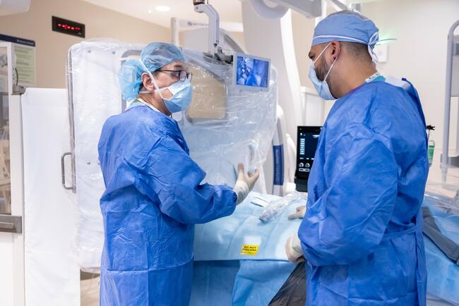
On June 1, Dr. Rizik and Dr. Paul Sorajja, director of interventional cardiology and structural heart at Banner, performed the first lead-free cardiac catheterization procedure in Arizona, a historic milestone that marks the beginning of a...
On June 1, Dr. Rizik and Dr. Paul Sorajja, director of interventional cardiology and structural heart at Banner, performed the first lead-free cardiac catheterization procedure in Arizona, a historic milestone that marks the beginning of a...
Published in JCCT and endorsed by the ACC, “Fractional flow reserve in coronary computed tomography angiography” provides evidence-based guidelines for the acquisition, interpretation and reporting of FFR-CT.
Published in JCCT and endorsed by the ACC, “Fractional flow reserve in coronary computed tomography angiography” provides evidence-based guidelines for the acquisition, interpretation and reporting of FFR-CT.
Advanced NanoTherapies (ANT)'s SirPlux Duo, a dual-drug device with sirolimus and paclitaxel that uses a proprietary nanoparticle delivery system to drive active cellular uptake within the vessel wall.
Advanced NanoTherapies (ANT)'s SirPlux Duo, a dual-drug device with sirolimus and paclitaxel that uses a proprietary nanoparticle delivery system to drive active cellular uptake within the vessel wall.
The award, named after Andreas Grüntzig, the pioneering German cardiologist who developed the first balloon angioplasty, is the highest honor presented by the Percutaneous Cardiovascular Registry (PCR).
The award, named after Andreas Grüntzig, the pioneering German cardiologist who developed the first balloon angioplasty, is the highest honor presented by the Percutaneous Cardiovascular Registry (PCR).
Key takeaways:
Project combines a wide range of data from more than 2,200 breast cancer patients to help the development of personalised AI-driven tools.
Researchers have developed two dedicated applications to monitor both physical...
Key takeaways:
Project combines a wide range of data from more than 2,200 breast cancer patients to help the development of personalised AI-driven tools.
Researchers have developed two dedicated applications to monitor both physical...
Your heart already has its own built-in bypass system that predicts whether the heart muscle is alive or dead, according to new research from the University of East Anglia and Norfolk and Norwich University Hospital.
A new study reveals...
Your heart already has its own built-in bypass system that predicts whether the heart muscle is alive or dead, according to new research from the University of East Anglia and Norfolk and Norwich University Hospital.
A new study reveals...
The Vertex system leverages the company’s patented Transforming Fixation (TFX) platform technology in a novel catheter system designed to navigate flexibly through the right heart and then stabilize on-demand in the pulmonary arteries for...
The Vertex system leverages the company’s patented Transforming Fixation (TFX) platform technology in a novel catheter system designed to navigate flexibly through the right heart and then stabilize on-demand in the pulmonary arteries for...
A 54-year-old man with coronary artery disease with multiple prior percutaneous coronary interventions, hypertension, paroxysmal atrial fibrillation on apixaban, hyperlipidemia, prior cerebrovascular accident with residual right-sided...
A 54-year-old man with coronary artery disease with multiple prior percutaneous coronary interventions, hypertension, paroxysmal atrial fibrillation on apixaban, hyperlipidemia, prior cerebrovascular accident with residual right-sided...
A 54-year-old man with a history of alcohol use, smoking, and hypertension presented with unstable angina. Baseline electrocardiogram and transthoracic echocardiography were unremarkable.
A 54-year-old man with a history of alcohol use, smoking, and hypertension presented with unstable angina. Baseline electrocardiogram and transthoracic echocardiography were unremarkable.
A 30-year-old man presented with acute onset chest pain followed by an episode of syncope. He had no prior history of hypertension, diabetes, or any other known cardiac disease.
A 30-year-old man presented with acute onset chest pain followed by an episode of syncope. He had no prior history of hypertension, diabetes, or any other known cardiac disease.
Miguel Ángel Montilla-Garrido, MD; Santiago Jesús Camacho-Freire, MD, PhD, Prof; Elena Izaga-Torralba, MD, PhD; Carmen Lluch-Requerey, MD; Jessica Roa-Garrido, MD; Oscar Lagos-De Grande, MD; Antonio Enrique Gómez-Menchero, MD
The study evaluated the indication for the use, safety, and efficacy of the OPN NC balloon (SIS Medical AG) in real-world settings.
The study evaluated the indication for the use, safety, and efficacy of the OPN NC balloon (SIS Medical AG) in real-world settings.
Pradyumna Agasthi, MD; Kaitlyn Krebushevski, MD; Allison K. Cabalka, MD; Jason H. Anderson, MD
The authors report use of a thoracic branch endoprostheses to treat recurrent coarctation of the aorta in a patient presenting with an aortic pseudoaneurysm and a peak-to-peak gradient of 29 mm Hg between the ascending and descending aorta.
The authors report use of a thoracic branch endoprostheses to treat recurrent coarctation of the aorta in a patient presenting with an aortic pseudoaneurysm and a peak-to-peak gradient of 29 mm Hg between the ascending and descending aorta.
A 59-year-old woman with end-stage renal disease undergoing maintenance hemodialysis through sequential right and left internal jugular venous catheters presented with progressive facial and neck swelling, dyspnea, and a non-functioning,...
A 59-year-old woman with end-stage renal disease undergoing maintenance hemodialysis through sequential right and left internal jugular venous catheters presented with progressive facial and neck swelling, dyspnea, and a non-functioning,...
Eric J. Kim, MD, MBE; Yash Prakash, MD; Hassan Beesley, MD; Grigory Manyak, MD; Mihir Prakash; Lakshay Chopra, MD; Akarsh Sharma, MD; Carlo Mannina, MD; George D. Dangas, MD, PhD; Lucy M. Safi, DO; Sahil Khera, MD, MPH; Samin K. Sharma, MD; Annapoorna S. Kini, MD; Gilbert H.L. Tang, MD, MSc, MBA; Stamatios Lerakis, MD, PhD
The study examined the association between chronic kidney disease (CKD) and right-sided extravalvular damage within low-flow, low-gradient aortic stenosis and across each stage of CKD.
The study examined the association between chronic kidney disease (CKD) and right-sided extravalvular damage within low-flow, low-gradient aortic stenosis and across each stage of CKD.
In this 2026 SCAI session, Dr Saibal Kar explored how LAAO is moving beyond its traditional use in patients with nonvalvular AFib who are ineligible for anticoagulation.
In this 2026 SCAI session, Dr Saibal Kar explored how LAAO is moving beyond its traditional use in patients with nonvalvular AFib who are ineligible for anticoagulation.
The randomized EQUAL trial showed that smartwatch-based atrial fibrillation (AF) screening increased detection rates more than 4-fold vs usual care in older adults with elevated stroke risk.
The randomized EQUAL trial showed that smartwatch-based atrial fibrillation (AF) screening increased detection rates more than 4-fold vs usual care in older adults with elevated stroke risk.