Vascular Disease
Treatment of Iatrogenic Renal Artery Perforation with a Covered Stent and Subsequent Rheolytic Thrombectomy
July 2005
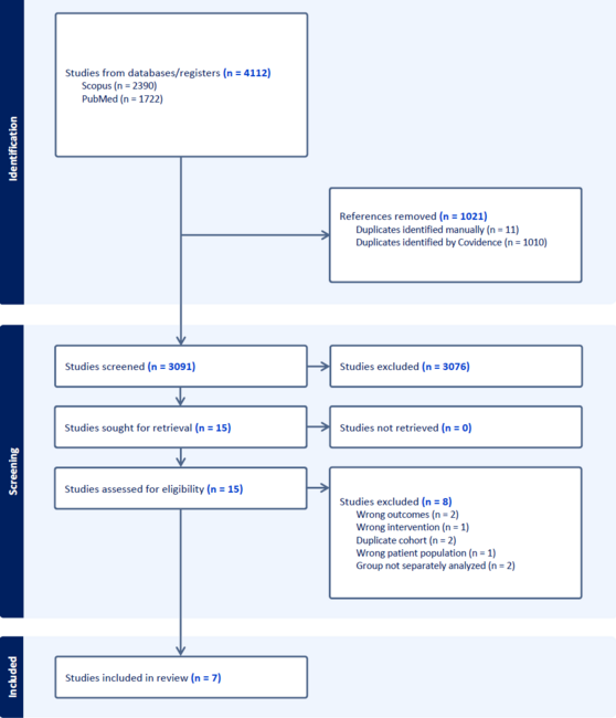
Iatrogenic renal artery perforation is a rare, but well-described complication of percutaneous renal artery intervention.1–3 Treatment has traditionally involved surgical intervention including renal artery reconstruction, bypass grafting, and even nephrectomy.4,5 More recently, several non-surgical techniques have been described, including balloon tamponade, custom-made stent graft deployment, and use of an autologous vein-covered stent.6,7 The JoStent® (JoMed, Rancho Cordova, California) has been approved by the FDA under humanitarian device exemption for the treatment of coronary artery perforations. Although experience in a variety of clinical settings has been described,8 use of the Jostent for renal artery perforations has not been reported. We describe a case in which we successfully treated a renal artery perforation with balloon tamponade and deployment of the Jostent, with subsequent rheolytic thrombectomy using the AngioJet thrombectomy catheter (Possis Medical, Minneapolis, Minnesota).
Case Report. The patient is a 73-year-old female with a past medical history significant for coronary artery disease, hypertension, diabetes mellitus type II, chronic renal insufficiency, and morbid obesity. In addition, she had known right renal artery stenosis (documented by angiography) that was managed medically. The left renal artery was very small and supplied an atrophic left kidney. Over the past year, her hypertension became increasingly resilient to medications and the decision was made to perform percutaneous right renal artery intervention.
An 8 French (Fr) sheath was inserted in the right femoral artery and and the right renal artery was engaged with an 8 Fr RDC catheter (Cordis Corporation, Miami, Florida). Selective angiography revealed an 80% lesion involving the ostium of the right renal artery (Figure 1). The patient received 5,000 units of heparin, and a 190 cm .014 inch S’port™ wire (Guidant Corporation, Indianapolis, Indianapolis) was advanced across the lesion into the distal renal artery. The lesion was directly stented with a 6.0 mm x 24 mm Genesis™ stent (Cordis Corp.) to 12 atmospheres, and subsequently post-dilated with a 7.0 mm x 15 mm Viatrac® balloon (Guidant Corp.) to 8 atmospheres. Final angiography revealed a 0% residual lesion (Figure 2). The patient’s activated clotting time at the end of the procedure was 285 seconds.
Approximately 2 hours after the intervention, the patient developed severe right flank and back pain. She was emergently returned to the angiography suite. The right renal artery was again engaged with an 8 Fr RDC catheter. The artery was widely patent with normal flow but there was an area of dye extravasation distal to the previously placed stent (Figure 3). Protamine was not administered given the concern of anaphylaxis in this patient who previously received protamine-containing insulin.
Consideration was given to placing a large (> 5 mm) perfusion balloon or covered stent; however, attempts at engaging the right renal artery with multiple larger guides were unsuccessful. We re-engaged the artery with the 8 Fr RDC catheter. A 190 cm .014 inch Marker wire (Cordis Corp.) was advanced into the distal renal artery. A 4.5 mm x 19 mm Jostent (the largest such covered stent in our supply) was advanced across the area of extravasation and inflated to 16 atmospheres. The Jostent was post-dilated with a 6.0 mm x 15 mm Viatrac balloon to 12 atmospheres. The post-deployment angiogram showed significant improvement in the perforation but a small leak persisted. A 4.0 mm x 20 mm ACS Esprit™ perfusion balloon (Guidant Corp.; the largest in our supply) was then advanced across the stented area and inflated to 8 atmospheres. Given that the right kidney was most likey the sole functioning kidney, the patient was sent for a CT scan of the abdomen and pelvis to rule out any significant organ compression that may require surgical intervention. The scan revealed the presence of a moderate-sized retroperitoneal bleed which did not compress the renal artery or kidney.
The patient was returned to the angiography suite, and the perfusion balloon was deflated. Repeat angiography confirmed that the perforation had been sealed. Unfortunately, there was now a large, mobile filling defect within the artery consistent with thrombus (Figure 4). A 300 cm 0.014 inch S’port wire was advanced into the distal artery. A 4 Fr AngioJet catheter was passed through the area of thrombus and there was complete resolution of the filling defect with excellent antegrade flow in the renal artery (Figure 5). A total of 325 ml of contrast was utilized, and over the next few days, her creatinine increased from a baseline of 1.9 to 3.2. This subsequently improved to 2.1 prior to discharge. Her hypertension was well-controlled with beta blockers, hydralazine, and clonidine. The patient was discharged home in good condition 6 days after the initial procedure.
Discussion. Percutaneous intervention has now become a common practice in the treatment of renal artery stenosis, with efficacy and safety similar to or better than surgery.9 Advances in both interventional equipment and operator technique have lowered complication rates over the past decade.3 Despite these advances, the risk of renal artery perforation still exists. Perforation can occur from the guidewire (especially hydrophilic wires), angioplasty balloon, stent, and even from the guiding catheter itself. In our case, the perforation was most likely secondary to direct placement of the stent, as this was the site of dye extravasation and there was minimal difficulty with initial wiring of the vessel. Quantitative measurements of the vessel, or pre-dilatation prior to stent placement, may have helped to avoid this complication.
Potential therapies for an iatrogenic renal artery perforation include deployment of a covered stent and inflation of a perfusion balloon, both of which were utilized in this case. Covered stents have been most widely studied in saphenous vein grafts, albeit with some lingering issues regarding potential late thrombosis.10 These stents, however, have proven reliable in treating perforations of both the iliac11 and coronary arteries.12,13 In addition, they have shown to be effective in treating a variety of other peripheral vascular injuries.14 Finally, the ability to rapidly deploy the covered stent coupled with the relative ease of its use make it an attractive option when compared to surgical intervention.
In this case, the amount of dye extravasation after coverd stent deployment was markedly reduced; however, sealing of the renal artery remained incomplete. This was most likely secondary to the size mismatch between the available covered stent and the larger renal artery, despite having post-dilated the covered stent with a 6 mm balloon. Prolonged inflation of a perfusion balloon was performed to fully seal the artery, and this technique has been described in both the treatment of coronary15 and renal6 perforations. The availability of a larger covered stent may have obviated the need for the prolonged balloon inflation.
The extended balloon inflation in our patient likely facilitated formation of thrombus distal to the stent.This rapidly resolved after treatment with the Angiojet catheter. Rheolytic thrombectomy with the Angiojet is now well-established in saphenous vein grafts,16 and there is an increasing amount of evidence for its utility in native coronary and peripheral thrombosis.17–19 Use of this device in renal artery thrombosis has also been reported.20 Another theoretical option is thrombolytic infusion, but this was not considered here given the recent perforation.
Even with newer equipment and techniques, the risk of arterial perforation still exists when performing renal artery intervention. This case demonstrates the possible role of a covered stent in the management of this potentially catastrophic complication.
friedelmd@yahoo.com
1. Henry M, Amor M, Henry I, et al. Stents in the treatment of renal artery stenosis: Long-term follow-up. J Endovasc Surg 1999;6:42–51.
2. Mahler F, Triller J, Weidmann P, Nachbur B. Complications in percutaneous transluminal dilatation of renal arteries. Nephron 1986;44(Suppl 1):60–63.
3. Zeller T, Frank U, Muller C, et al. Technological advances in the design of catheters and devices used in renal artery interventions: Impact on complications. J Endovasc Ther 2003;10:1006–1014.
4. Dean RH, Callis JT, Smith BM, Meacham PW. Failed percutaneous transluminal renal angioplasty: Experience with lesions requiring operative intervention. J Vasc Surg 1987;6:301–307.
5. McCann RL, Bollinger RR, Newman GE. Surgical renal artery reconstruction after percutaneous transluminal angioplasty. J Vasc Surg 1988;8:389–394.
6. Morris CS, Bonnevie GJ, Najarian KE. Nonsurgical treatment of acute iatrogenic renal artery injuries occurring after renal artery angioplasty and stenting. Am J Roentgenol 2001;177:1353–1357.
7. Bates MC, Shamsham FM, Faulknier B, Crotty B. Successful treatment of iatrogenic renal artery perforation with an autologous vein-covered stent. Catheter Cardiovasc Interv 2002;57:39–43.
8. Gercken U, Lansky AJ, Buellesfeld L, et al. Results of the Jostent coronary stent graft implantation in various clinical settings: Procedural and follow-up results. Catheter Cardiovasc Interv 2002;56:353–360.
9. Geroulakos G, Missouris C, Mitchell A, Greenhalgh RM. Endovascular treatment of renal artery stenosis. J Endovasc Ther 2001;8:177–185.
10. Schachinger V, Hamm CW, Munzel T, et al. A randomized trial of polytetrafluoroethylene membrane-covered stents compared with conventional stents in aortocoronary saphenous vein grafts. J Am Coll Cardiol 2003;42:1360–1369.
11. Allaire E, Melliere D, Poussier B, et al. Iliac artery rupture during balloon dilatation: What treatment? Ann Vasc Surg 2003;17:306–314. Epub 2003 Apr 28.
12. Gunning MG, Williams IL, Jewitt DE, et al. Coronary artery perforation during percutaneous intervention: Incidence and outcome. Heart 2002;88:495–498.
13. Bates MC, Shamsham FM, Faulknier B, Crotty B. Artery perforation during catheterization: Fighting with a catastrophe. Catheter Cardiovasc Interv 2002;57:44–46.
14. Baltacioglu F, Cimsit NC, Cil B, et al. Endovascular stent-graft applications in latrogenic vascular injuries. Cardiovasc Intervent Radiol 2003;26:434–439.
15. Fukutomi T, Suzuki T, Popma JJ, et al. Early and late clinical outcomes following coronary perforation in patients undergoing percutaneous coronary intervention. Circulation 2002;66:349–356.
16. Kuntz RE, Baim DS, Cohen DJ, et al. A trial comparing rheolytic thrombectomy with intracoronary urokinase for coronary and vein graft thrombus (the Vein Graft AngioJet Study [VeGAS 2]). Am J Cardiol 2002;89:326–330.
17. Rinfret S, Katsiyiannis PT, Ho KK, et al. Effectiveness of rheolytic coronary thrombectomy with the AngioJet catheter. Am J Cardiol 2002;90:470–476.
18. Singh M, Tiede DJ, Mathew V, et al. Rheolytic thrombectomy with Angiojet in thrombus-containing lesions. Catheter Cardiovasc Interv 2002;56:1–7.
19. Ansel GM, George BS, Botti CF, et al. Rheolytic thrombectomy in the management of limb ischemia: 30-day results from a multicenter registry. J Endovasc Ther 2002;9:395–402.
20. Greenberg JM, Steiner MA, Marshall JJ. Acute renal artery thrombosis treated by percutaneous rheolytic thrombectomy. Catheter Cardiovasc Interv 2002;56:66–68.