


Early and Mid-Term Outcomes of Femoro-Ilio-Caval Vein Stent Implantation
Abstract
Objective. We sought to investigate mid-term clinical outcomes and identify risk factors in one of the largest comprehensive series reported of femoro-ilio-caval (FIC) vein stent placement. Background. Endovascular intervention with balloon angioplasty and stenting of the iliac and common femoral veins has become first-line treatment for symptomatic deep venous outflow obstruction. Methods. We conducted a single-center, retrospective analysis of 180 patients who underwent FIC stent implantation between May 2017 and May 2019; 327 procedures were performed. Our primary objective was to evaluate a composite of stent thrombosis and stent restenosis. Secondary outcomes included individual predictors of in-stent restenosis (ISR) and in-stent thrombosis (IST), primary and secondary patency, access-site complications, major bleeding, pulmonary embolism, cardiovascular death, any death, intracranial bleeding, all-cause mortality, and components of major adverse cardiac and cerebrovascular events (MACCE) in a 24-month period. Results. A total of 327 procedures were performed for 180 patients. At 2-year follow up, 78.3% of cases remained free of any complication. Primary outcome occurred in 53 procedures (16.2%) and was highest at early (<30 days) follow-up. Primary patency at 2-year follow-up was 78.43%. There were no deaths, 1 patient (0.3%) had a subdural hematoma, and 3 patients (0.9%) had MACCE. Age and post-thrombotic syndrome (PTS) were significant predictors of primary outcome. PTS and Venous Clinical Severity score (VCSS) ≥10 were found to have higher rates of thrombosis. Active smokers, the elderly, history of deep vein thrombosis (DVT), and VCSS ≥10 had a statistically significant elevated risk of ISR. Conclusion. Endovascular treatment with stent implantation for non-thrombotic iliac vein lesion and PTS is safe, with low morbidity, zero mortality, low complications, and persistent improvement of symptoms. Age and PTS were significant predictors of primary outcome.
J INVASIVE CARDIOL 2021;33(7):E497-E505. doi:10.25270/jic/20.00507
Key words: deep venous outflow obstruction, iliac, in-stent restenosis, in-stent thrombosis, non-thrombotic iliac vein lesion, post-thrombotic syndrome, pulmonary embolism
Introduction
Femoro-ilio-caval (FIC) occlusive or stenotic lesions may arise from two separate entities, either post-thrombotic syndrome (PTS) or non-thrombotic iliac vein lesion (NIVL).1 PTS commonly develops in up to 50% of patients experiencing deep venous thrombosis (DVT).2 NIVL includes compression such as May-Thurner syndrome,3 iliac vein compression, and Cockett4 syndrome. Less common causes of FIC obstruction include congenital venous anomalies, iatrogenic, fibrosis, and tumors.5 These conditions lead to chronic venous disease (CVD) with debilitating lower-extremity symptoms such as venous claudication, venous ulcers, hyperpigmentation, varicosities, chronic pelvic pain,6-8 and swelling that ultimately progress to reduction in quality of life (QOL).1,9
Over the last 2 decades, FIC venous stent implantation has become the preferred treatment option for deep venous obstruction and replaced surgery as standard of care.10,11 Currently, there are several stent platforms available with favorable engineering for the venous system or specifically designed for deep vein stenting and treatment of FIC obstruction: Wallstent (off-label use; Boston Scientific), Venovo (Bard),12 sinus-Venous (OptiMed),13,14 sinus-Obliquus (OptiMed),15,16 Zilver Vena (Cook Medical),17 and Veniti Vici (Boston Scientific).9,18,19 Prior systematic reviews have shown that stenting of FIC veins is associated with low complication rates, high technical success rates, and improvement in QOL.20-22 Despite the lack of randomization and long-term data, the American Heart Association (AHA) recommends treatment for PTS with endovascular percutaneous recanalization for symptomatic patients with post-thrombotic occlusion of the inferior vena cava (IVC) or iliofemoral veins.23 In the current study, we report procedural, short-term, and mid-term clinical outcomes in one of the largest series of FIC venous stenting.
Methods
This was a single-center, retrospective analysis of 180 consecutive patients with a total number of 327 procedures performed from May 2017 to May 2019. Duplex ultrasound (DU) of bilateral FIC was available for all patients regardless of bilateral or unilateral symptoms of venous outflow obstruction due to NIVL or PTS. Patients who met clinical and DU criteria for FIC compression underwent further invasive diagnostic study and stenting.
Clinical inclusion criteria consisted of lower-extremity edema, pain, heaviness, cramps, or venous claudication (worsening of pain with ambulation that alleviated with rest) affecting QOL and daily activity, and physical exam findings of clinical, etiology, anatomy, and pathophysiology (CEAP) classification score ≥3. Ultrasound techniques have been previously published and thoroughly described.24 Briefly, a narrowing of the vein diameter, a mosaic color pattern that indicates a post-stenosis turbulence, slow flow, abnormal Doppler waveform, and dilation or dampened flow proximal or distal to the area of stenosis were considered findings suggestive of significant stenosis. A reduction in vein caliber of 50% measured with calipers, a diameter of <8 mm for common iliac vein (CIV), <7 mm for external iliac vein (EIV), and <6 mm for common femoral vein (CFV) were considered to be compressed with or without abnormalities in color flow or waveforms. In addition, a velocity ratio >2.5 was suggestive of significant compression.25
Invasive diagnosis to evaluate FIC vein compression included a gold-standard intravascular ultrasound (IVUS) and venography. A 50% reduction of the FIC vein in cross-sectional area or diameter compared with a healthy area within the same segment on IVUS was used to confirm the diagnosis. The stenotic lesion was treated with an appropriately sized stent. Intravenous hydration was given with leg raise to ensure adequate hydration in most patients. Valsalva maneuver was performed if the operator thought the pelvic veins were small in caliber, if there was a discrepancy between the venogram and IVUS, or if the IVC looked collapsed despite hydration.26 Primary stenting was most commonly performed with ≥1 Wallstents. Stents were oversized by 15%-20% to allow for proper apposition. Normal caliber area of vessels on IVUS were to be 200, 150, and 125 mm2 for CIV, EIV and CFV, respectively.27 We defined technical success as antegrade flow and residual luminal stenosis of <30% at the end of the procedure by venogram and IVUS along with successful recanalization and stent placement without major periprocedural complication. We did not require performance of any hybrid cases. Post procedure, all patients were recommended to wear elastic stockings for more than 1 year.
To evaluate patency of the treated venous segment post intervention, routine surveillance follow-up DU was recommended at 1 and 4 weeks, followed by 3, 6, 9-15, and 18-24 months. Primary patency was defined as percentage of patients after successful initial treatment with stent placement not having compromised flow in the stent lumen requiring reintervention. Secondary patency was defined as flow in the stent lumen after repeat intervention of the target lesion. Follow-up DU patency was assessed as mild, moderate, severe in-stent restenosis (ISR), or in-stent thrombosis (IST). We categorized ISR of the treated venous segment as mild (luminal obstruction <50%), moderate (50%-70%), or severe (>70%) on venous cross-sectional area (CSA) on DU. In the treated segment, a luminal venous obstruction >50% was defined as ISR. We defined early ISR or IST as restenosis or thrombotic occlusion within 30 days of procedure and late ISR or IST as ≥30 days. Venography and IVUS were performed in cases of recurrent stenosis based on the ultrasound assessment and worsening symptoms. Reintervention was performed in those patients with confirmed diagnosis by IVUS. Symptom improvement was determined by pain and edema at follow-up. Patients with mild to moderate ISR were treated conservatively and had surveillance DU.
Baseline patient characteristics are shown in Table 1. Primary outcome was defined as moderate and severe ISR and/or IST. Secondary outcomes included individual predictors of ISR and IST, primary and secondary patency, access-site complication (hematoma or arteriovenous fistula [AVF] formation, major bleeding, pulmonary embolism [PE], cardiovascular death, intracranial bleeding, acute kidney injury [AKI], all-cause mortality and components of major adverse cardiac and cerebrovascular events [MACCE], defined as composite of death, myocardial infarction [MI], cerebrovascular accident [CVA], or stroke). We defined major bleeding according to the Bleeding Academic Research Consortium (BARC) classification as any bleeding ≥2. We defined AKI as rise in serum creatinine by ≥1.5 times (≥50% above baseline) post procedure. For patients with NIVL, post-procedure dual-antiplatelet therapy (DAPT) was prescribed for 3-6 months followed by lifelong single-antiplatelet therapy (SAPT). After initial loading dose of 600 mg clopidogrel, we prescribed a maintenance dose of 75 mg for 3-6 months along with 81 mg aspirin daily. For PTS, we prescribed SAPT in conjunction with an anticoagulant, either a vitamin K antagonist (VKA) or a novel oral anticoagulant (NOAC) for at least 3 months.28-30 If evidence of ISR or IST was found, we added an anticoagulant to SAPT at the time of diagnosis by DU. Written informed consent was obtained from each patient after detailed explanation of risks and benefits of the procedure. An institutional review board permission for retrospective analysis and publication of this study was approved.
Procedural technique. Ultrasound-guided access of the mid-thigh femoral vein (FV) was obtained with an 8 Fr Pinnacle Precision kit (Terumo) with a target access in the distal aspect of the proximal FV. After wire confirmation with ultrasound and fluoroscopy, the sheath was inserted. Once adequate access was confirmed, intravenous heparin with an initial bolus of 4000-5000 IU was administered to achieve a target activated clotting time (ACT) of 250-300 seconds. If bilateral access was not desired, contralateral venogram and IVUS were performed using a 5 Fr Contralateral catheter (Boston Scientific) that was advanced to the IVC over a 0.035˝ wire and used to cross over. It was then positioned in the contralateral FV. With the catheter in the contralateral side, we performed bilateral venogram with simultaneous injection from the sheath and the catheter. If needed, an IVUS of the contralateral side could be performed by exchanging the catheter over a 0.035˝ wire for the IVUS catheter (Vision PV 0.035 digital IVUS catheter; Philips).
Planimetric evaluation of the luminal areas of the CFV, EIV, CIV, and IVC were made. If an intervention was needed, the 8 Fr sheath was exchanged for a 10 Fr sheath and a second IVUS measurement was made. At this point, keeping the table in position and using an erasable marker, we marked the beginning and end of the target lesion. Depending upon the degree of stenosis and fibrosis, we proceeded to an initial balloon dilation followed by stenting. We performed predilation in all thrombotic, Rokitansky stenosis,31 and severely compressed NIVLs. We recommend postdilation balloon32 and final IVUS to establish adequate stent expansion, to minimize residual lesions, and to ensure adequate transition from the stented segment to the native vein with a goal of <30% difference in area between the stented segment and the native vein. To avoid potential early ISR, we extended stents 3-5 mm into the IVC if iliocaval confluence was obstructive. Common stent sizes used were 18 x 90 mm and 16 x 90 mm, with at least 5 mm overlap to ensure adequate coverage of the target lesion.
Statistical analysis. Baseline characteristics were analyzed and summarized using descriptive statistics, mean ± standard deviation for continuous parametric variables, median and interquartile range (IQR) for non-parametric data, and frequency (percentage) for categorical or nominal variables. Baseline continuous variables were compared between the 2 groups using t-test or non-parametric equivalent and Chi-square test (exact test in the case of sparse data) was employed for comparison of categorical and nominal variables. Outcomes were assessed with multiple regression methods (linear for continuous outcomes, logistic for categorical outcomes, and Cox regression for time to event analyses), adjusting for risk factors as described below. Statistical significance was defined at a computed P-value ≤.05. Kaplan-Meier survival analysis was used to estimate patency rates and compare PTS with NIVL. Data were analyzed using a dedicated statistical analysis software (STATA, version 13.0; Statacorp).
Results
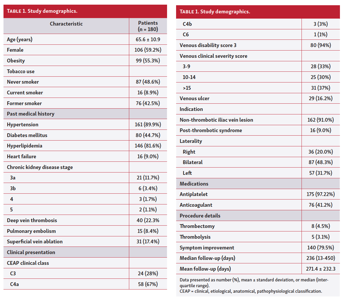
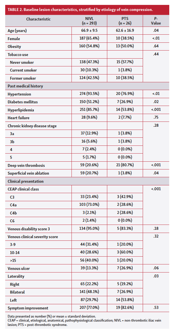
Baseline demographic and anatomic characteristics for 180 patients are presented in Table 1. Among 180 patients, 106 (59.2%) were female, mean age was 65.6 years, and 55.3% were obese (defined as body mass index [BMI] >30.0 kg/m2). History of DVT was present in 22.3% of the study population. CEAP classification >C3 was present in 71%, VDS score of 3 was present in 94%, and a VCSS of ≥10 was present in 67% of patients. Nine percent of patients presented with PTS, while 91% presented with NIVL. Lower-extremity venous ulcers were recorded in 16.2% of patients, while bilateral lower-extremity symptoms were found in 48.3%. A comparison between NIVL and PTS patients was performed. In general, PTS patients were younger, more likely to be male, had higher rate of prior DVT, and had unilateral symptoms. NIVL patients were more likely to have hypertension, diabetes mellitus, hyperlipidemia, and CEAP ≥4 (Table 2). Immediate technical success was achieved in all limbs; all patients had successful recanalization and angioplasty. Mean and median follow-up periods were 271 ± 232.3 days and 236 days (IQR, 13-450 days), respectively. Symptom improvement was reported in 140 patients (79.5%) at follow-up.
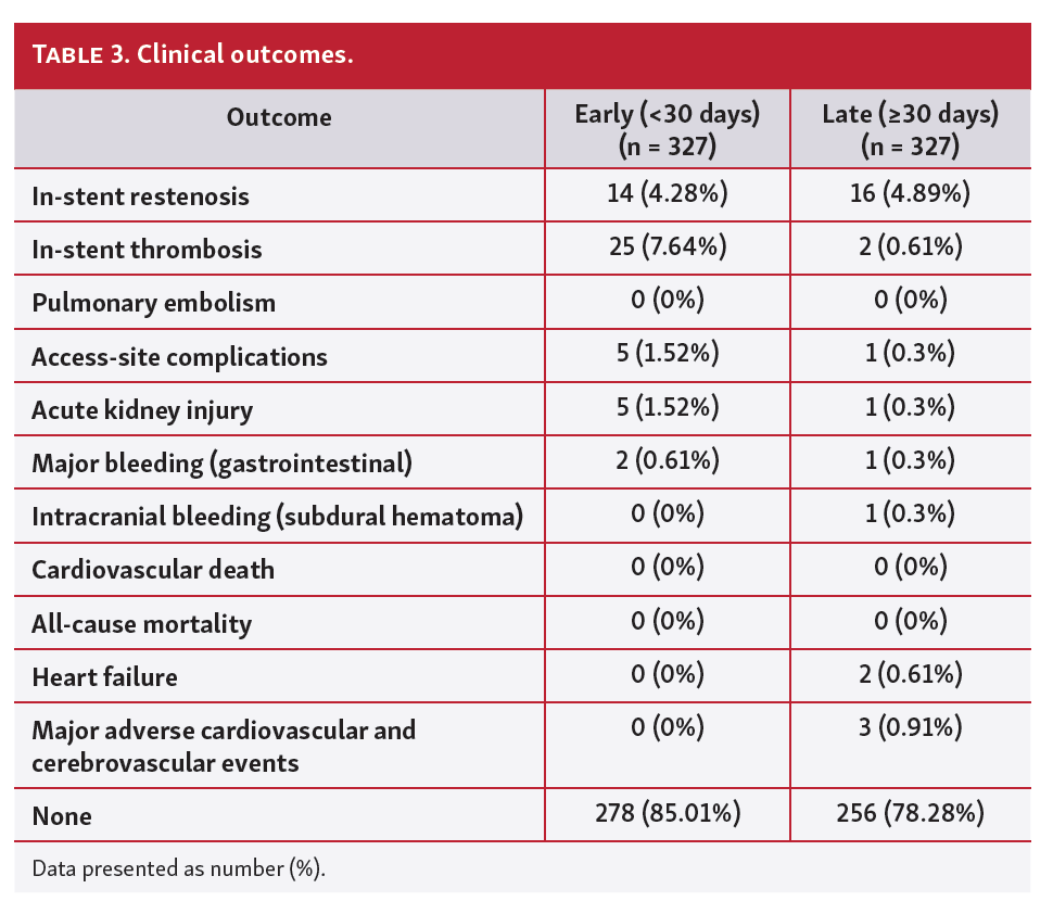
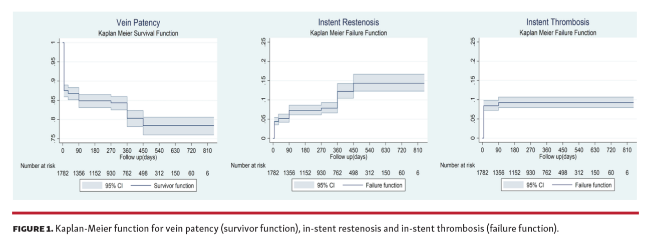
Among 327 procedures, primary outcome occurred in 53 procedures (16.2%), with 30 ISRs, 27 ISTs, and 4 procedures ultimately resulting in both ISR and IST. VCSS of ≥10 and symptom improvement (80.6% vs 64%, respectively; P=.01) were statistically different between patients who developed primary outcome and those who didn’t. Secondary outcomes are described as early and late events (Table 3). At 1 month, 85.01% were free of any complications and 78.28% had an uneventful 2-year follow-up. There was a total of 30 ISRs (14 occurred early, and 16 late) and 27 ISTs (25 early and 2 late). Among early ISR, 12 occurred within 1 week and all early ISTs occurred within 1 week post procedure. Very late (>1 year) ISR was seen in 2.43% of patients and there was no very late IST (Figure 1). Primary patency at 2-year follow-up was 78.43%. Secondary patency was 79% (secondary ISR and IST were 17% and 4%, respectively). Among the 27 IST patients, 23 (85.2%) had NIVL and 4 (14.8%) had PTS. The PTS patients were less likely to be on antiplatelet medications than NIVL patients (0/4 [0%] vs 23/23 [100%]; P<.05]; however, they were more likely to be on anticoagulation than NIVL patients in those who developed IST (4/4 [100%] vs 8/23 [34.8%]; P<.05]. There were 6 access-site complications (5 early and 1 late), 6 AKIs (5 early and 1 late), and 3 major bleeds (2 early and 1 late). There were no deaths, 1 (0.3%) patient had a subdural hematoma (while being treated with a NOAC), 2 patients had AVF formation, and 3 patients (0.9%) had MACCE. There was 1 case of balloon fracture and dislodgment requiring balloon retrieval using a snare. Similar to prior reports,33-40 we had 1 case of stent migration into the right ventricle requiring successful surgical removal.
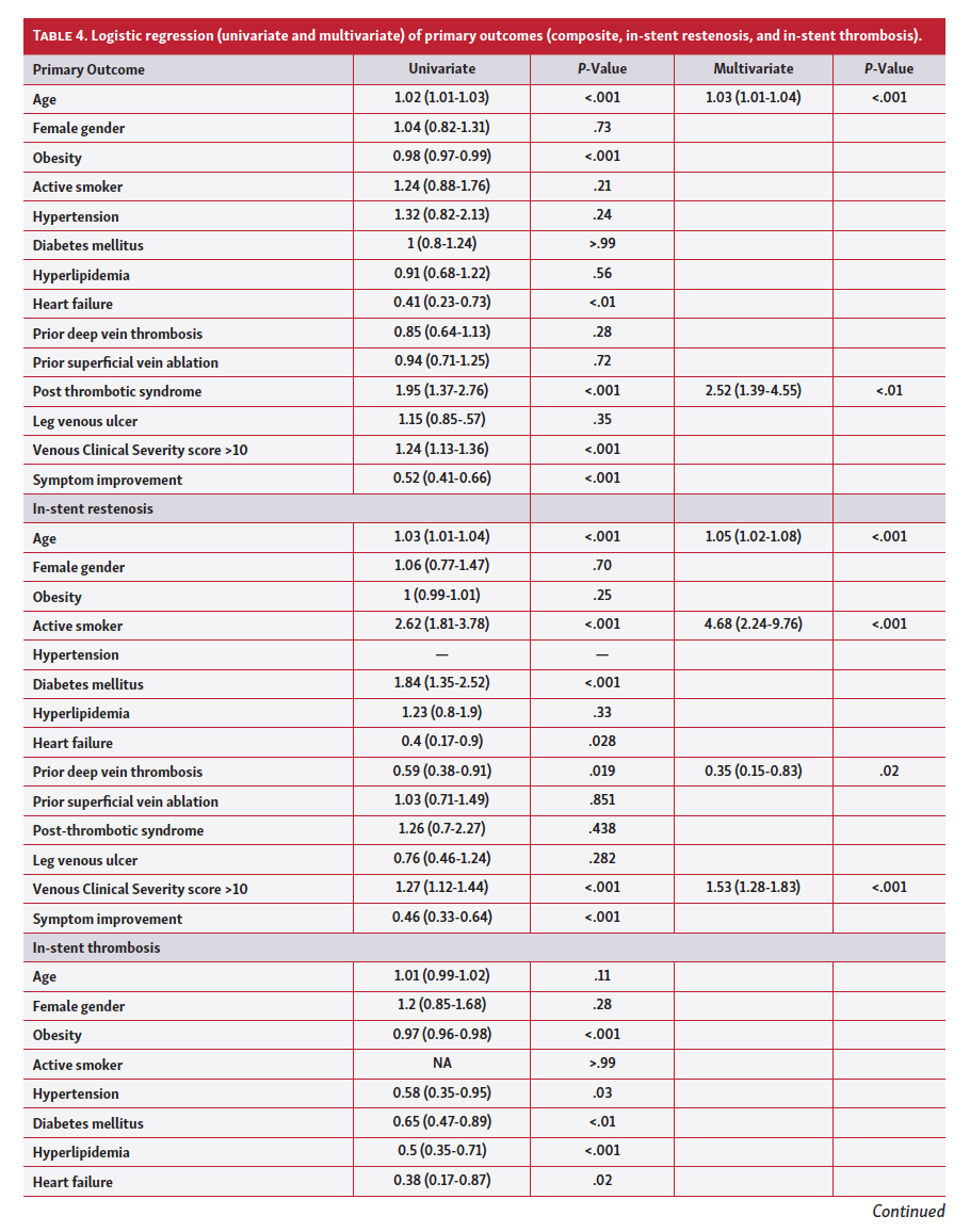
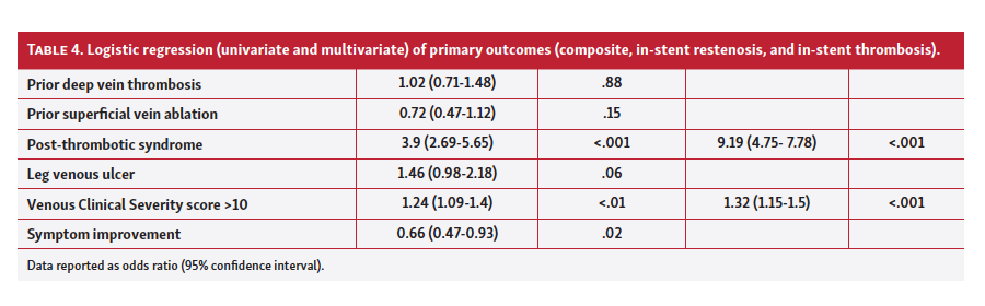
To identify factors associated with individual components for primary outcome, logistic regression was performed (Table 4A and 4B). In a multivariate analysis, significant predictors associated with development of primary outcome were age (hazard ratio [HR] 1.03; 95% confidence interval [CI], 1.01-1.04) and PTS (HR, 2.52; 95% CI, 1.39-4.55). Predictors significant for development of ISR were age (HR 1.05; 95% CI, 1.02-1.08), active smoking (HR, 4.68; 95% CI, 2.24-9.76), prior DVT (HR, 0.35; 95% CI, 0.15-0.83; P=.02) and VCSS ≥10 (HR, 1.53; 1.28 - 1.83)]. PTS (HR, 9.19; 95% CI, 4.75-17.78) and VCSS ≥10 (HR, 1.32; 95% CI, 1.15-1.5) were associated with IST. We also performed an exploratory subgroup analysis to compare components of primary outcome and vein patency between patients with NIVL and PTS. Vein patency was statistically different (log rank P<.001) due to difference in IST (log rank P<.001) and ISR was not different (log rank P=.43).
Discussion
We describe results of 180 patients treated for FIC venous obstruction either for PTS or NIVL with a 2-year follow-up. Stent implantation was performed for highly symptomatic patients who had FIC obstruction on DU and failed medical therapy. Our overall primary and secondary patency rates were 78.43% and 79%, respectively. Primary outcome, defined as composite of ISR and IST, occurred in 16% at a 24-month follow-up. Overall complications were low, with the most common being ISR. Stent thrombosis primarily occurred early and was highest within 1 week of stent implantation, and subsequently stabilized over time. We hypothesize that this may have been due to initial medication non-compliance and non-adherence. Beyond 1 year, there was freedom from very late stent thrombosis and ISR was found in 2.43%. At 1 month, we found that 85.01% were free of any complication. At the completion of mid-term follow-up of 24 months, 78.28% were free of any complication.
Similar to our findings, 2 large observational studies reported FIC stenting as a safe and highly effective treatment for symptomatic patients. Jayaraj et al demonstrated that stent occlusion is a rare occurrence, with only a 3% stent occlusion rate and no significant adverse event or mortality after an 18-year follow-up.41 In a study with 8-year follow-up of 982 lesions, Neglen et al concluded a beneficial clinical outcome with venous stenting. Authors demonstrated low morbidity and mortality, high patency rates, low ISR, and major symptom relief.42 Moreover, a meta-analysis of 2869 patients demonstrated that stent placement for FIC venous system was safe, and there were high technical success rates. At 1 year, for patients with NIVL, primary and secondary patency rates were 96% and 99%, respectively. Lower primary and secondary rates were found in thrombotic patients.21
Dedicated venous stents have been studied, although these findings were in small cohorts; large prospective trials will determine whether the differences in stent types will have a clinical impact. Based on these studies, over the last few years, dedicated venous stents were introduced to our clinical practice. In a systemic review, dedicated venous stents (740 patients) and standard venous stents (3072) were evaluated. With the use of standard stents (off-label), median primary and secondary patency rates were 71% and 91%, respectively. At 12 months, overall primary patency for dedicated venous stents was 78.8% with lower patency rates in PTS (73%).43 With the use of standard stents, we achieved an overall primary and secondary patency of 78.43% and 79%, respectively. Raju et al described findings of 1500 patients with FIC obstruction who underwent standard venous stent (predominantly Wallstent) placement. They reported <1% morbidity with no deaths or PEs, and patency rates ranging from 90%-100% for NIVL and 74%-89% for PTS at 3-5 years. IST was discovered to be extremely rare in patients with NIVL disease, coinciding with our multivariate analysis of patients with PTS having an elevated risk of IST. The authors concluded that iliac vein stenting is a safe and effective treatment option for FIC obstruction.44
Currently, there is little information in the guidelines for techniques related to the performance of iliac vein stenting. Raju and Neglen et al recommended extending stents into the IVC when treating focal obstruction adjacent to the confluence of the CIV to avoid possible stent migration and potential complication with ISR or thrombosis with missed proximal lesions.45,46 We followed their technique of stent extension into the IVC if the iliocaval confluence was obstructive and ensured there was at least 5 mm of overlap when placing 2 or more stents.5 Alsheekh et al demonstrated no significant difference in stent thrombosis when covering only the lesion and not stenting into the IVC; hence, putting into question the need for routine stent extension into the IVC as it increases time, cost, and radiation exposure.47 Complications of stent thrombosis and ISR have been investigated in prior literature to evaluate whether the stenting technique contributes to outcome. However, the precise factor in technique that may contribute to etiology of these complications remains unknown.
Newer and revolutionary dedicated venous stent differences, techniques, and patient confounders need further investigation as possible risk factors for ISR and IST. Access site of either the CFV or the FV had a significant difference in IST with either approach.48 Jayaraj et al demonstrated that a potential cause of stent occlusion after stent placement is possibly due to missed lesions or intrinsically poor inflow. Additionally, degree of stenosis on IVUS for PTS patients did not affect stent patency rates.41 Furthermore, patency rates for stenting across the inguinal ligament does not affect long-term patency, and there was no risk in stent fracture or narrowing due to external compression.49 The mechanism of FIC in obese patients is similar to the mechanism in non-obese patients.50 We also did not observe statistical significance for obesity as a possible mechanism attributing to ISR or IST. In a prior study, risk factors for ISR were thrombophilia and presence of thrombotic disease. However, limbs that eventually occluded had similar risk factors, and a conclusion was not reached regarding causation.51 To the best of our knowledge, we report the first-ever clinical predictors of IST and ISR. We found age and PTS to be significant predictors for development of primary outcome. Additionally, higher rates of IST were reported in patients with PTS. Independent predictors associated with ISR were active smoking, history of DVT, age, and VCSS ≥10. We suggest considering risk factors for ISR in patients with a history of DVT, active smokers, VCSS ≥10, and the elderly.
Improvement in symptoms after intervention has been in question and is a topic often criticized by clinicians. Approximately 80% of our patients demonstrated symptom improvement post intervention. Compared with NIVL, the PTS group reported more symptom relief post intervention despite NIVL patients being more symptomatic at baseline. Raju et al found post intervention that patients with NIVL achieved complete pain relief, overall complete clinical relief, ulcer healing, and relief in swelling in 82%, 75%, 67%, and 47%, respectively.1 In a randomized, controlled trial, there was improvement in VAS, VCSS, and QOL in limbs treated with stents compared with medical therapy alone.52 In an 11-year follow-up of 528 limbs with stent implantation for FIC obstruction, there was improvement in pain, swelling, and healed ulcers (78%, 55% and 54%, respectively).53 Moreover, Razavi et al9 evaluated results of the dedicated venous Veniti Vici stent prospectively with a 12-month improvement in QOL, VCSS, and visual analogue scale (VAS) score. However, a meta-analysis found that most studies were not able to report symptom relief. There was significant heterogeneity in studies for relief of pain and edema. Among the studies with NIVL, there was complete symptom relief in 69%-82% for pain and 64%-68% for edema. Although symptom relief data were reported inconsistently, the majority of patients experienced complete resolution of symptoms post procedure.21
Study limitations. Although there is considerable evidence of observational studies, well-designed, large, randomized, controlled trials are imperative to further evaluate our findings. Even though favorable results were shown, some limitations are important to emphasize. Most notably, recall bias is possible given the retrospective nature of the investigation. In addition, the study lacks a full assessment of QOL, CEAP, VCSS, and VAS scores, and ulcer resolution on follow-up. We did not study stent type as a variable in this model to predict the primary outcome as >95% of the stents used were Wallstents. The lesions in the current study were predominantly NIVL (91%); therefore, the results may not be applicable to patients with PTS. During the last year of follow-up, the number of procedures and patients increased, hence decreasing numbers on longer follow-up.
Conclusion
Endovascular treatment with stent implantation for NIVL and PTS is safe, with low morbidity, zero mortality, low complications, and improvement of symptoms. Age and PTS were significant predictors of primary outcome. Patients with PTS and VCSS ≥10 were found to have higher rates of thrombosis. Lastly, active smokers, the elderly, those with history of DVT, and patients with VCSS ≥10 had a statistically significant elevated risk of ISR. Further randomized, controlled trials are needed to validate our findings.
Affiliations and Disclosures
From the 1Mount Sinai Heart at Mount Sinai Beth Israel, Department of Cardiovascular Diseases, New York, New York; 2Cleveland Clinic Indian River Hospital, Department of Cardiovascular Diseases, Vero Beach, Florida; and3 Icahn School of Medicine at Mount Sinai (Beth Israel), Department of Internal Medicine, New York, New York.
Disclosure: The authors have completed and returned the ICMJE Form for Disclosure of Potential Conflicts of Interest. The authors report no conflicts of interest regarding the content herein. Manuscript accepted December 1, 2020.
Address for correspondence: Robert Sogomonian, MD, Icahn School of Medicine at Mount Sinai (Beth Israel/West), 281 1st Avenue, New York, NY 10003. Email: Robert.sogomonian@ mountsinai.org
References
1. Raju S, Neglen P. High prevalence of nonthrombotic iliac vein lesions in chronic venous disease: a permissive role in pathogenicity. J Vasc Surg. 2006;44:136-143.
2. Prandoni P, Kahn SR. Post-thrombotic syndrome: prevalence, prognostication and need for progress. Br J Haematol. 2009;145:286-295.
3. May R, Thurner J. The cause of the predominantly sinistral occurrence of thrombosis of the pelvic veins. Angiology. 1957;8:419-427.
4. Cockett FB, Thomas ML. The iliac compression syndrome. Br J Surg. 1965;52:816-821.
5. Jayaraj A, Crim W, Knight A, Raju S. Characteristics and outcomes of stent occlusion after iliocaval stenting. J Vasc Surg Venous Lymphat Disord. 2019;7:56-64.
6. Santoshi RKN, Lakhanpal S, Satwah V, Lakhanpal G, Malone M, Pappas PJ. Iliac vein stenosis is an underdiagnosed cause of pelvic venous insufficiency. J Vasc Surg Venous Lymphat Disord. 2018;6:202-211.
7. Blattler W, Blattler IK. Relief of obstructive pelvic venous symptoms with endoluminal stenting. J Vasc Surg. 1999;29:484-488.
8. Daugherty SF, Gillespie DL. Venous angioplasty and stenting improve pelvic congestion syndrome caused by venous outflow obstruction. J Vasc Surg Venous Lymphat Disord. 2015;3:283-289.
9. Razavi M, Marston W, Black S, Bentley D, Neglen P. The initial report on 1-year outcomes of the feasibility study of the Veniti Vici venous stent in symptomatic iliofemoral venous obstruction. J Vasc Surg Venous Lymphat Disord. 2018;6:192-200.
10. Bruhlmann W. The treatment of stenosis of central veins using percutaneous implantation of self-expanding metal stents. Praxis (Bern 1994). 1994;83:1151-1156.
11. Nazarian GK, Austin WR, Wegryn SA, et al. Venous recanalization by metallic stents after failure of balloon angioplasty or surgery: four-year experience. Cardiovasc Intervent Radiol. 1996;19:227-233.
12. Lichtenberg MKW, de Graaf R, Stahlhoff WF, Ozkapi A, Rassaf T, Breuckmann F. Venovo venous stent in the treatment of non-thrombotic or post-thrombotic iliac vein lesions - short-term results from the Arnsberg venous registry. Vasa. 2019;48:175-180.
13. de Wolf MA, de Graaf R, Kurstjens RL, Penninx S, Jalaie H, Wittens CH. Short-term clinical experience with a dedicated venous nitinol stent: initial results with the sinus-venous stent. Eur J Vasc Endovasc Surg. 2015;50:518-526.
14. van Vuuren T, Wittens C, de Graaf R. Stent extension below the common femoral vein in extensive chronic iliofemoral venous obstructions. J Vasc Interv Radiol. 2018;29:1142-1147.
15. Lichtenberg M, de Graaf R, Stahlhoff WF, Ozkapi A, Simon M, Breuckmann F. Patency rates, safety and clinical results of the sinus-Obliquus venous stent in the treatment of chronic ilio-femoral venous outflow obstruction - data from the Arnsberg venous registry. Vasa. 2019;48:270-275.
16. Stuck AK, Kunz S, Baumgartner I, Kucher N. Patency and clinical outcomes of a dedicated, self-expanding, hybrid oblique stent used in the treatment of common iliac vein compression. J Endovasc Ther. 2017;24:159-166.
17. O'Sullivan GJ, Sheehan J, Lohan D, McCann-Brown JA. Iliofemoral venous stenting extending into the femoral region: initial clinical experience with the purpose-designed Zilver Vena stent. J Cardiovasc Surg (Torino). 2013;54:255-261.
18. Black S, Gwozdz A, Karunanithy N, et al. Two year outcome after chronic iliac vein occlusion recanalisation using the Vici Venous Stent(R). Eur J Vasc Endovasc Surg. 2018;56:710-718.
19. Shamimi-Noori SM, Clark TWI. Venous stents: Current status and future directions. Tech Vasc Interv Radiol. 2018;21:113-116.
20. Seager MJ, Busuttil A, Dharmarajah B, Davies AH. Editor's Choice – A systematic review of endovenous stenting in chronic venous disease secondary to iliac vein obstruction. Eur J Vasc Endovasc Surg. 2016;51:100-120.
21. Razavi MK, Jaff MR, Miller LE. Safety and effectiveness of stent placement for iliofemoral venous outflow obstruction: systematic review and meta-analysis. Circ Cardiovasc Interv. 2015;8:e002772.
22. Qiu P, Zha B, Xu A, et al. Systematic review and meta-analysis of iliofemoral stenting for post-thrombotic syndrome. Eur J Vasc Endovasc Surg. 2019;57:407-416.
23. Kahn SR, Comerota AJ, Cushman M, et al. The postthrombotic syndrome: evidence-based prevention, diagnosis, and treatment strategies: a scientific statement from the American Heart Association. Circulation. 2014;130:1636-1661.
24. Sloves J, Almeida JI. Venous duplex ultrasound protocol for iliocaval disease. J Vasc Surg Venous Lymphat Disord. 2018;6:748-757.
25. Metzger PB, Rossi FH, Kambara AM, et al. Criteria for detecting significant chronic iliac venous obstructions with duplex ultrasound. J Vasc Surg Venous Lymphat Disord. 2016;4:18-27.
26. Brinegar KN, Sheth RA, Khademhosseini A, Bautista J, Oklu R. Iliac vein compression syndrome: Clinical, imaging and pathologic findings. World J Radiol. 2015;7:375-381.
27. Raju S, Buck WJ, Crim W, Jayaraj A. Optimal sizing of iliac vein stents. Phlebology. 2018;33:451-457.
28. Endo M, Jahangiri Y, Horikawa M, et al. Antiplatelet therapy is associated with stent patency after iliocaval venous stenting. Cardiovasc Intervent Radiol. 2018;41:1691-1698.
29. Attaran RR, Ozdemir D, Lin IH, Mena-Hurtado C, Lansky A. Evaluation of anticoagulant and antiplatelet therapy after iliocaval stenting: factors associated with stent occlusion. J Vasc Surg Venous Lymphat Disord. 2019;7:527-534.
30. Padmos LJ, Garcia D. May-Thurner syndrome and thrombosis: A systematic review of antithrombotic use after endovascular stent placement. Res Pract Thromb Haemost. 2018;3:70-78. Epub Jan 2019.
31. Raju S, Davis M. Anomalous features of iliac vein stenosis that affect diagnosis and treatment. J Vasc Surg Venous Lymphat Disord. 2014;2:260-267.
32. Shammas NW, Shammas GA, Jones-Miller S, Radaideh Q. Safety of the Atlas Gold Balloon in treating iliofemoral veins: experience from a single center. J Invasive Cardiol. 2018;30:401-405.
33. Ashar RM, Huettl EA, Halligan R. Percutaneous retrieval of a Wallstent from the pulmonary artery following stent migration from the iliac vein. J Interv Cardiol. 2002;15:101-106.
34. Caton MT Jr, Brown JM, Steigner ML. Iliac stent migration to the right ventricular outflow tract. Circ Cardiovasc Imaging. 2018;11:e008520.
35. Ogami T, Zimmermann E, Zhu R, Worku B, Avgerinos DV. Embolization of an iliac vein stent to the right atrium. J Card Surg. 2018;33:855-856.
36. Holst T, Grosswendt T, Laham MM, Roosta-Azad M, Zandi A, Kamler M. Acute stent migration into the right ventricle in a patient with iliac vein stenting. Thorac Cardiovasc Surg Rep. 2018;7:e7-e8.
37. El Feghaly M, Soula P, Rousseau H, et al. Endovascular retrieval of two migrated venous stents by means of balloon catheters. J Vasc Surg. 1998;28:541-546.
38. Mullens W, De Keyser J, Van Dorpe A, et al. Migration of two venous stents into the right ventricle in a patient with May-Thurner syndrome. Int J Cardiol. 2006;110:114-115.
39. Slonim SM, Dake MD, Razavi MK, et al. Management of misplaced or migrated endovascular stents. J Vasc Interv Radiol. 1999;10:851-859.
40. Gabelmann A, Kramer SC, Tomczak R, Gorich J. Percutaneous techniques for managing maldeployed or migrated stents. J Endovasc Ther. 2001;8:291-302.
41. Jayaraj A, Buck W, Knight A, Johns B, Raju S. Impact of degree of stenosis in May-Thurner syndrome on iliac vein stenting. J Vasc Surg Venous Lymphat Disord. 2019;7:195-202.
42. Neglen P, Hollis KC, Olivier J, Raju S. Stenting of the venous outflow in chronic venous disease: long-term stent-related outcome, clinical, and hemodynamic result. J Vasc Surg. 2007;46:979-990.
43. Williams ZF, Dillavou ED. A systematic review of venous stents for iliac and venacaval occlusive disease. J Vasc Surg Venous Lymphat Disord. 2019;8:145-153. Epub 2019 Nov 5.
44. Raju S. Best management options for chronic iliac vein stenosis and occlusion. J Vasc Surg. 2013;57:1163-1169.
45. Neglen P, Raju S. Balloon dilation and stenting of chronic iliac vein obstruction: technical aspects and early clinical outcome. J Endovasc Ther. 2000;7:79-91.
46. Murphy EH, Johns B, Varney E, Buck W, Jayaraj A, Raju S. Deep venous thrombosis associated with caval extension of iliac stents. J Vasc Surg Venous Lymphat Disord. 2017;5:8-17.
47. Alsheekh A, Hingorani A, Aurshina A, Kibrik P, Chait J, Ascher E. Iliac vein stent placement and the iliocaval confluence. Ann Vasc Surg. 2020;63:307-310. Epub 2019 Oct 21.
48. Chait J, Alsheekh A, Aurshina A, et al. Effect of venous access site on postintervention stent thrombosis for nonthrombotic iliac vein stenting. J Vasc Surg Venous Lymphat Disord. 2020;8:84-88. Epub 2019 Jun 21.
49. Neglen P, Tackett TP Jr, Raju S. Venous stenting across the inguinal ligament. J Vasc Surg. 2008;48:1255-1261.
50. Raju S, Darcey R, Neglen P. Iliac-caval stenting in the obese. J Vasc Surg. 2009;50:1114-1120.
51. Neglen P, Raju S. In-stent recurrent stenosis in stents placed in the lower extremity venous outflow tract. J Vasc Surg. 2004;39:181-187.
52. Rossi FH, Kambara AM, Izukawa NM, et al. Randomized double-blinded study comparing medical treatment versus iliac vein stenting in chronic venous disease. J Vasc Surg Venous Lymphat Disord. 2018;6:183-191.
53. Raju S, Darcey R, Neglen P. Unexpected major role for venous stenting in deep reflux disease. J Vasc Surg. 2010;51:401-408.


