Addressing Talar Avascular Necrosis With A Total Talar Implant
In an intriguing case study involving a young patient with a severely comminuted talar fracture, these authors discuss employing a total talar implant and potential benefits including limb length preservation, early weightbearing and significant pain relief.
Avascular necrosis of the talus is a devastating complication, which usually results from talar fractures with most cases being traumatically-induced.1 The unique arterial blood supply to the talus predisposes it to avascular necrosis. This can then lead to joint depression, fragmentation and intra-articular collapse, causing pain and instability of the ankle and subtalar joints. Surgical treatment options include talectomy, tibiocalcaneal arthrodesis or pantalar arthrodesis.2 Currently, there is limited research involving the use of a total talar implant for treatment of this condition.
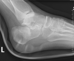
In a recent case, a 19-year-old African-American female presented to the emergency room with a severely comminuted left talar fracture after a pedestrian-motor vehicle accident approximately five hours earlier. The patient had severe pain to the left ankle. She related no significant past medical history and reported smoking about half of a pack of cigarettes per day for a few years.
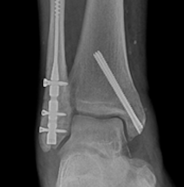
On physical exam, we noted skin tenting to the lateral ankle as well as a varus deformity of the foot. Examination also revealed intact neurovascular status and diffuse tenderness to the ankle. Radiographs and computed tomography (CT) showed a severely comminuted fracture of the talus (see first three images above). An attempt at closed reduction in the emergency room was unsuccessful. The patient subsequently underwent closed reduction in the operating room with application of an external fixator and a definitive plan for pantalar fusion depending on the viability of the talus in the near future (see fourth image above).
About four months after the initial procedure, the patient developed an infection of the proximal tibia pin site, resulting in surgical debridement with adjustment of the external fixator and insertion of antibiotic beads. Intraoperative bone biopsies did not reveal osteomyelitis but tissue cultures showed bacterial growth. The patient received intravenous antibiotics for four weeks.
Approximately seven months after the initial injury, the patient developed avascular necrosis of the talus (see fifth image above). The talus was completely necrosed and the patient declined fusion as she is young and desired to still have motion in the ankle. We also discussed an alternative of pantalar arthrodesis, which she also refused. The patient then agreed to a total talectomy with insertion of a cobalt talus implant (see sixth image above). If the implant should fail, we informed the patient that she may require a talocalcaneal arthrodesis.
Key Points In The Surgical Approach With A Total Talar Implant
For the procedure, the operative team ensured supine positioning of the patient and inflation of a proximal thigh tourniquet. After a six cm anterior ankle incision just medial to the tibialis anterior tendon, we performed blunt dissection down to the level of the capsule, taking care to retract the anterior neurovascular structures. Once we retracted the tibialis anterior tendon, extensor tendon and the neurovascular structures, we performed a full-thickness capsulotomy starting from the distal tibia down to the level of the talus. Next, in order to expose the talus, we elevated full-thickness periosteal flaps medially and laterally.
The talus appeared brown, unhealthy, severely fragmented and not anatomically stable. We then removed the talus. Prying it out with a Cobb elevator in pieces, we started with the talar head and neck, and then removed the body, which was already fractured from the initial injury. After multiple flushes, we excised any remaining fragments.
After complete removal of the talus, we examined the distal tibial plafond, the posterior and anterior facets of the calcaneus, the medial malleolus, fibula and articular surfaces and determined these structures were still viable. Using a bone rongeur, we removed any retained soft tissue attachments from the tibial plafond and calcaneus.
Utilizing the engineered 4WEB® XL talus implant, we placed trial implants where the talus previously resided. We chose the 33.5 mm implant from the 4Web XL options. After removing the final implant from the sterile package, we placed it in a bacitracin-diluted sterile saline solution. We then irrigated the open surgical site copiously with normal sterile saline solution and subsequent use of bacitracin-diluted sterile saline solution.
An engineer created this implant with a 3-D reconstruction based on a CT of the contralateral talus. They created a computerized 3-D mirror image of the right talus and the attending surgeon participated in a live online interactive meeting with the engineer. This allowed the surgeon to see how the 3-D image fit in the ankle. The engineers produced two size adjustments; small and large. The larger implant fit the best intraoperatively.
We augmented the lateral and medial ligaments of the ankle joint with FiberTape® (Arthrex) to stabilize the talus implant. After press fitting the final talus implant into the ankle joint, we confirmed sound anatomical alignment with intraoperative fluoroscopy. At this time, evaluation revealed anatomical range of motion.
The patient remained non-weightbearing in a posterior splint for four weeks. She transitioned to partial weightbearing with a cast at one month postoperatively and transitioned to a fracture boot four weeks later. At the three-month postoperative visit, the patient had mild pain with palpation and stiffness with range of motion of the ankle. She then transitioned to an ankle foot orthosis (AFO) at five months postoperatively and started physical therapy. She is now back to work with her AFO and currently complains of symptoms of Achilles tendonitis.
Concluding Thoughts
Historically, the Blair arthrodesis has been the preferred surgical treatment option for avascular necrosis of the talus.3 The main disadvantage to this arthrodesis is limb length loss. These procedures have a long rehabilitation time with a high rate of complications, including non-union or pseudarthrosis. On the other hand, total talus implantation allows for limb length preservation, early weightbearing, range of motion and pain relief.3
We sought total talus implantation as the most suitable treatment choice for this patient due to the preservation of surrounding bones and articular surfaces. This case study discusses a viable alternative surgical option for the treatment of avascular necrosis. Traumatic talus injuries and avascular necrosis of the talus can be extremely challenging cases with high rates of complications. There are very few published cases presenting the results of a talar implant for avascular necrosis. Long-term outcomes are necessary to evaluate the longevity and success of these implants.
Dr. Sharpe is a third-year resident in the Podiatric Surgery Residency Program at Ascension Providence Hospital in Southfield, Mich.
Dr. Yoo is a third-year resident in the Podiatric Surgery Residency Program at Ascension Providence Hospital in Southfield, Mich.
Dr. Austin is a third-year resident in the Podiatric Surgery Residency Program at Ascension Providence Hospital in Southfield, Mich.
Dr. Khalil is a Diplomate of the American Board of Foot and Ankle Surgery in both foot and rearfoot/reconstructive surgery. He is an Attending Physician for the Podiatric Residency Program at Ascension Providence Hospital in Southfield Mich. and is in private practice in Wyandotte and Oak Park, Mich.
References
- Ando Y, Yasui T, Isawa K, Tanaka S, Tanaka Y, Takakura Y. Total talar replacement for idiopathic necrosis of the talus: a case report. J Foot Ankle Surg. 2016;55(6):1292-1296.
- Cohen M, Kazak M. Tibiocalcaneal arthrodesis with a porous Tantalum spacer and locked intramedullary nail for post-traumatic global avascular necrosis of the talus. J Foot Ankle Surg. 2015;54(6):1172-1177.
- Dennison MG, Pool RD, Simonis RB, Singh BS. Tibiocalcaneal fusion for avascular necrosis of the talus. J Bone Joint Surg Br. 2001;83(2):199-203.