Freiberg’s Osteochondrosis: A New Surgical Approach to a Relatively Uncommon Condition
In 1914, Freiberg described a condition that most commonly affected the second and third metatarsals, which became known as Freiberg’s osteochondrosis.1 In this condition, trauma and vascular insufficiency impact the metatarsal physis. Conditions such as diabetes mellitus, rheumatoid arthritis, and systemic lupus erythematosus may contribute to the vascular insufficiency. Hypercoagulability can also contribute to this condition. Freiberg’s osteochondrosis remains a relatively infrequent presentation to most surgeons’ practices. Bilateral involvement occurs less than 10% of the time, and females experience this condition more than males at a 5:1 ratio.2,3
Previously, established classification systems did not usefully guide surgical treatment of Freiberg’s osteochondrosis. Smillie introduced what became the mainstay classification system for this condition in 1957.4 This description, based on radiographic findings, served as the historic framework on which surgeons could base treatment. However, this posed challenges, since radiographs alone do not reveal the full extent of the pathology in these types of cases.
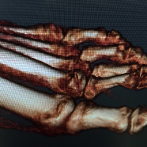
Hoggett and colleagues described a new classification for Freiberg’s disease in a 2022 publication.3 They based their classification on thin-sliced computed tomography (CT) studies to evaluate the “zone” of the defect. The authors conducted a review of 24 CT scans over a 10-year period. They identified a sagittal imaging slice that displayed the widest portion of the proximal articular margins of the proximal phalanx and divided it anatomically into 2 articular zones—dorsal (zone 1) and plantar (zone 2). Eighteen patients that they studied had disease confined to zone 1, making them candidates for osteotomy, and 6 had pathology in both zones, which the authors deemed a more appropriate scenario for debridement.
A recently published February 2024 study elaborates further on this zone-based paradigm, reviewing 80 cases with CT images.5 These authors propose a 5-segment classification system and found excellent inter- and intra-observer agreement between evaluators in their study. However, this article will focus on the system outlined by Hoggett and team, as the 5-segment system necessitates more study on clinical and surgical applications.3

Options to Manage Freiberg’s Osteochondrosis
Management options for patients can be divided into operative and nonoperative care. Nonoperative care may include the use of oral analgesics, nonsteroidal anti-inflammatories, episodic corticosteroids, activity modification, and utilization of rigid functional orthotic devices with metatarsal offloading padding. In consideration for surgical intervention, there are, in our experience, 3 sources of pain to assess that may suggest a surgical pathway is best:
- impingement;
- ischemic necrotic bone (as determined by magnetic resonance imaging); and
- osteoarthritic pain, loose cartilage flaps, osteophytes, and joint mice.
Regarding surgical management of Freiberg’s osteonecrosis, Alhadhoud and colleagues conducted a systematic review of the numerous available surgical interventions.6 This team concluded that more research is necessary in order to clearly validate procedural options and guide surgeons. However, in considering surgical treatment options, in our experience, it is imperative for the discussion to focus on zones of involvement as described by Hoggett and colleagues.3
Management of Zone 1 Pathology
Cheilectomy is one potential option for managing Freiberg’s osteochondrosis in zone 1. Surgeons will undertake several steps in performing this procedure. One should evaluate synovial proliferation, and debride and excise loose dorsal cartilage flaps. Debridement of loose ossicles, spurs, and osteophytes takes place, with care taken to preserve the hyaline articular cartilage peripherally.
Furthermore, cheilectomy is perhaps best reserved for patients whose symptoms primarily relate to pain from dorsal impingement of periarticular osteophytes. The literature supporting cheilectomy demonstrates consistently good outcomes; however, the majority of research that supports cheilectomy are of lower quality—noted to be either Level III or IV studies.7,8 However in these articles, overall success was greater than 90%. There is ease in performance of the procedure and it requires minimal operating room time and resources. Cheilectomy also has good surgeon reliability in performance of the procedure.
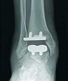
A dorsal closing wedge osteotomy for this Freiberg’s osteochondrosis entails a bisection of the metatarsal, which creates a 30-degree arc to rotate the plantar cartilage at zone 1.3,9 This maneuver rotates healthy articular cartilage into the functional arc of joint excursion by revising the metatarsal head’s sagittal plane anatomy.
Options for Zone 2 Conditions
For zone 2 Freiberg’s osteochondrosis, surgeons may consider arthroplasty as an option. In these cases, one would perform chondroplasty to debride loose osteochondral flaps and achieve stable cartilage margins.
Preoperative MRI images can help surgeons determine the extent of necrotic bone and intramedullary edema. In turn, the surgeon can then execute debridement to remove the edematous bone per the MRI.
Additionally, one may seek to improve neoangiogenesis with microfracture, opening the medullary canal and creating an enhanced vascular environment.9
Treatment as a nonunion may also be a choice, utilizing microfracture to open the medullary canal and canal pedaling residual cortical bone.
In taking this on, one should determine whether the cortex is contained or uncontained. A contained lesion is one that is surrounded by intact cartilage, while an uncontained lesion is one that extends beyond the cartilaginous margin.
For a contained cortical shell, one may fill the medullary canal with cortical-cancellous auto- or allograft.
For an uncontained cortical shell, conversion to a contained cortical shell can take place by debriding back to intact cortical bone. The surgeon can then backfill the remaining open medullary canal with impacted cortical-cancellous bone graft.
Once one achieves a contained bony lesion, one can utilize autologous matrix-induced chondrogenesis (AMIC) or apply demineralized bone matrix with a mixture of cancellous bone chips to the contained construct.10 Rajeev and colleagues conducted a retrospective analysis of small subset of individuals (only 10 patients) with Freiberg’s disease that underwent treatment with open debridement, microfracture, bone grafting, and application of AMIC.10
Zone 2 Options Based on Patient Age
Yoshimura and colleagues have a stepwise approach for staging nonoperative versus operative and also discuss joint preserving versus sparing, which is why we would separate the procedures based on age.11
For patients 20–40 years old, options can include AMIC, or use of biocartilage or demineralized bone matrix putty.
For patients 40–60 years of age, some may choose a synthetic polyvinyl alcohol implant with arthroplasty. However, implant arthroplasty is indicated in limited cases with a patient that has low activity demands and identified late-stage Freiberg’s osteochondrosis.11
For patients with zone 2 injuries who over 60 utilization of hemi-implants is a good option. Proximal base hemi-implants are used in patients if the metatarsal head has bone loss. Metatarsal head hemi-implantation works best where the metatarsal head maintains adequate integrity. Total joint replacement with a flexible hinge silastic implant is used only in salvage. This represents conditions of significant bone loss.
The Stainsby procedure is very popular in Europe and also can be considered in the presence of bone loss and more so with the presence of instability.
In Conclusion
The Smillie classification has served as a source of decision-making and management of Freiberg’s osteochondrosis since 1957.4 The newer CT-based classification presented by Hoggett and colleagues could provide practitioners an easier and more reproducible way to preoperatively plan surgical treatment for Freiberg’s osteochondrosis. Hopefully, innovation and research will continue to pave the way for improved decision-making pathways that could optimize outcomes in this uncommon condition.
Dr. Visser is the Director of the Foot and Ankle Surgery Residency program at SSM Health DePaul Hospital in St. Louis. He is a Fellow of the American College of Foot and Ankle Surgeons.
Dr. Smith is a third-year podiatric resident at SSM Health DePaul Hospital.
For further reading, see “Key Insights On Treating Freiberg’s Infraction” in the March 2014 issue of Podiatry Today or “Emerging Concepts In Treating Freiberg’s Infraction” in the September 2017 issue.
References
- Freiberg AH. Infraction of the second metatarsal bone. Surg Gynecol Obstet. 1914;19:191–193.
- Carter KR, Chambers AR, Dreyer MA. Freiberg Infraction. [Updated 2023 Nov 22]. In: StatPearls [Internet]. Treasure Island (FL): StatPearls Publishing; 2024 Jan-. Available from: https://www.ncbi.nlm.nih.gov/books/NBK537308/
- Hoggett L, Nanavati N, Cowden J, et al. A new classification for Freiberg’s disease. Foot (Edinb). 2022;51:101901. doi:10.1016/j.foot.2021.101901
- Smillie IS. Treatment of Freiberg’s infraction. Proc R Soc Med. 1967;60(1):29-31.
- Ren Y, Chen Y, Gan TJ, Zhang H, Liu X. CT-based five-segment classification in Freiberg’s infarction: Evaluation of its intraobserver and interobserver reliability. Foot Ankle Surg. 2024;30(2):145-149. doi:10.1016/j.fas.2023.10.008
- Alhadhoud MA, Alsiri NF, Daniels TR, Glazebrook MA. Surgical interventions of Freiberg’s disease: A systematic review. Foot Ankle Surg. 2021;27(6):606-614. doi:10.1016/j.fas.2020.08.005
- McNeil DS, Baumhauer JF, Glazebrook MA. Evidence-based analysis of the efficacy for operative treatment of hallux rigidus. Foot Ankle Int. 2013;34(1):15–32.
- Anderson MR, Ho BS, Baumhauer JF. Current concepts review: hallux rigidus. Foot Ankle Orthop. 2018;3(2). doi:10.1177/2473011418764461
- Kim J, Choi WJ, Park YJ, Lee JW. Modified Weil osteotomy for the treatment of Freiberg’s disease. Clin Orthop Surg. 2012;4(4):300-306. doi:10.4055/cios.2012.4.4.300
- Rajeev A, Yallop W, Devalia K. Freiberg`s disease of lesser metatarsals treated with bone grafting and autologous matrix induced chondrogenesis (AMIC) membrane - A series of 10 cases. Foot Ankle Surg. 2023;29(2):136-142. doi:10.1016/j.fas.2022.12.004
- Yoshimura I, Takao M, Wagner E, et al. Evidence-based treatment algorithm for Freiberg disease. Cartilage. 2024;15(1):58-64. doi:10.1177/19476035231205676
- Hyakuda Y, Sugimoto K, Isomoto S, Miura K, Syoji H, Tanaka Y. Osteochondral autograft transplantation for advanced Freiberg’s disease: A report of three cases. JOS Case Reports. 2023; 2(4):174–178. https://doi.org/10.1016/j.joscr.2023.08.002
- Abdul W, Hickey B, Perera A. Functional outcomes of local pedicle graft interpositional arthroplasty in adults with severe Freiberg’s disease. Foot Ankle Int. 2018;39(11):1290-1300. doi:10.1177/1071100718786494
- Gauthier G, Elbaz R. Freiberg’s infraction: a subchondral bone fatigue fracture. A new surgical treatment. Clin Orthop Relat Res. 1979;(142):93-95.