The Top 10 Skin Conditions of the Lower Extremity
In the office, podiatrists see lower extremity dermatological issues essentially hourly. When was the last clinic day that you didn’t treat a nail issue or a fungal-based skin condition? We are in a unique situation in podiatric medicine where we not only treat lower extremity skin conditions but also understand the nuances of what makes them unique, whether it is biomechanical or shoe gear-related. The following article lists and discusses the most common podiatric skin conditions in no specific order.
Plantar Verruca
Clinically, plantar warts are well-circumscribed lesions with overlying hyperkeratosis seen in children and adults. Human papillomavirus (HPV), a double-stranded DNA virus, is responsible for plantar verrucae, and the HPV subtypes that commonly affect the feet are 1, 2, 4, 27, and 57. HPV needs both an epidermal abrasion and a transiently impaired immune system to inoculate a keratinocyte in the basal layer of the epidermis.1 The virus seems to encourage basal cell replication. Basal keratinocytes, with an eccentric nucleus surrounded by a halo (koilocytes), show viral damage of the cells. Hyperplasia of the granular and prickle cell layers occur in addition to the dermal papillae arching their vasculature up into the wart.1
Since HPV can exist on fomites, showers and swimming pools with abrasive non-slip surfaces act as high-risk areas for simultaneously harboring the virus and causing an epidermal abrasion. Thirty percent of warts may clear spontaneously, but those that do not often become cosmetically unappealing, painful, and irritating to the patient.1
One diagnoses a plantar verruca through eliciting pain after lateral compression of the plantar lesion. Upon debridement of the hyperkeratosis, pinpoint bleeding may occur, and the clinician can visualize interruption of the skin lines. If a wart involves the nail unit, the nail may become dystrophic from the pressure and presence of the lesion. Differential diagnoses of a plantar wart include callus, IPK, and punctate keratoderma.1
There are several treatments for plantar verrucae; however, none are specific to directly damaging HPV. They include non-surgical and surgical methods, such as the use of keratolytic agents (salicylic acid), cryotherapy (liquid nitrogen), laser (pulsed dye and CO2), excision, bleomycin injections, folk remedies, and the latest FDA-approved microwave device. Treatment of warts pose a therapeutic challenge for most practitioners due to a lack of a universally eradicating treatment and possible Koebner reaction, where treatment of the lesion elicits a negative response to therapy.1
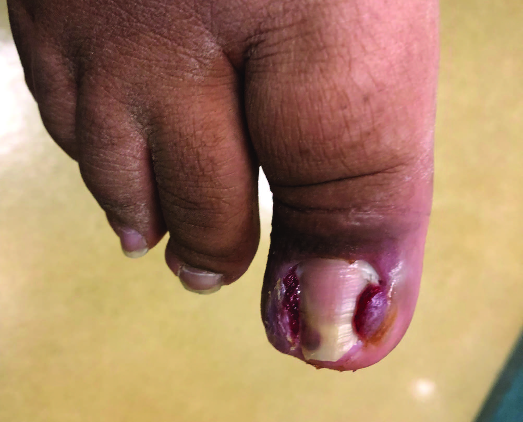 Ingrown Nails: Onychocryptosis and Paronychia
Ingrown toenails often cause significant pain and disability to the patient.2 Great toenails are most affected, but any toenail can become ingrown on either the lateral or medial nail border, or both. They present as painful onychocryptosis (incurvation of the lateral edge of the nail plate) with or without lateral nail fold edema or redness. Ingrown toenails may progress to paronychia with pain, lateral nail fold edema, focal erythema, drainage, granulation tissue, and possible hypertrophy of the periungual tissue. Conservative treatment methods include the “slant back” procedure where one trims the offending lateral nail plate and tapes the lateral nail fold to encourage the skin not to encroach the nail. However, these methods require patience and time on the patient’s part.2
For those patients in whom conservative therapy has failed or whose presentation is too severe for a non-surgical intervention, a partial nail avulsion of the affected side may become indicated. This procedure aims to decrease the width of the nail plate of the offending nail border to relieve pain and pressure. In patients where ingrown nails become a chronic scenario, the procedure can include removal/destruction of the nail matrix, either surgically or chemically, to cause long-term narrowing of the nail plate.2
Onychomycosis
Onychomycosis is a common superficial fungal infection of the nails leading to discoloration, nail plate thickening, and onycholysis. Mycotic nail disease is the most common nail pathology worldwide, reaching all cultures and ethnicities. Onychomycosis is increasing, accounting for up to 90 percent of toenail and at least 50 percent of fingernail infections.3 The most common fungal culprits in the United States are the dermatophytes Trichophyton rubrum and Trichophyton mentagrophytes.4 Non-dermatophyte molds and yeasts also play a role with varying frequency.5,6
Because the initial diagnosis depends on the nail’s appearance and other nail diseases may visually mimic onychomycosis, lab tests such as direct microscopy (potassium hydroxide [KOH]), fungal culture, periodic acid Schiff (PAS) stain, and polymerase chain reaction (PCR) assist in obtaining a definitive diagnosis. Visual nail plate changes help classify onychomycosis, including distal subungual (also known as distal lateral subungual onychomycosis [DLSO], the most common form), proximal subungual, superficial white, and total dystrophic onychomycosis.5,6
Onychomycosis occurs in 10 percent of the general population, 20 percent of individuals 60 years and older, and 50 percent of individuals over 70 years of age.6 Peripheral vascular disease, immunologic disorders, and diabetes mellitus correlate with the increased prevalence in older adults. The risk of onychomycosis is 2.8 times greater in persons with diabetes mellitus, and in patients with HIV infection, prevalence rates range from 15 to 40 percent.6 Other predisposing factors include older age, sex (male more so than female), genetic predisposition, tinea pedis (interdigital or moccasin types), peripheral arterial disease, smoking, nail trauma, inappropriate nail hygiene, and family background of onychomycosis and hyperhidrosis.6
In the last several years, novel treatments of onychomycosis have arisen. FDA-approved medications include oral antifungals (terbinafine and itraconazole), topical antifungals (efinaconazole, tavaborole, and ciclopirox), and laser therapy (approved to temporarily improve the nail’s visual appearance). No matter the treatment, the toenail only grows 1 to 2 mm per month, and treatment can take anywhere from 12 to 18 months due to the slow turnover from the cuticle to the distal tip of the nail.
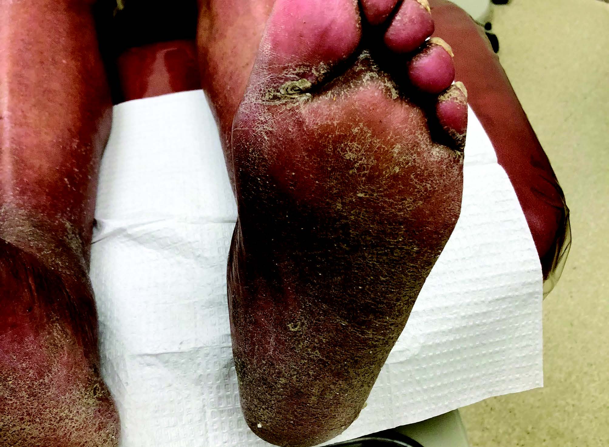 Xerosis
Xerotic and hyperkeratotic disorders result from compromised skin barrier function. The outer layers of the skin need a certain amount of hydration and free fatty acids (that help regulate water loss) to maintain skin integrity. Increased trans-epidermal water loss causes dehydration of the stratum corneum.7 Decrease in sebaceous gland activity with increasing age, frequent bathing, and harsh detergents further reduce the amount of free fatty acids in the skin. The combination of decreased free fatty acids and dehydration causes the cells to shrink due to lack of water and the corneocytes to curl upwards, creating the clinical appearance of scale. Further stressing of the skin can cause fissures and inflammation.7
Xerotic skin has scales present within the skin lines on the plantar foot. Moccasin tinea pedis, on the other hand, usually presents with small, serpiginous, circular plantar scales. Xerosis is more common in the aging population. However, it can also occur due to environmental conditions/climate (low humidity in the wintertime), medications, endocrine disorders (diabetes, thyroid), after infection (post-cellulitic desquamation), and renal dysfunction (especially for patients who are undergoing dialysis). Dry skin management involves using a moisturizer or skin emollient such as CeraVe, Aveeno, Cutemol, or Eucerin. Pruritus can occur with xerosis and will warrant the use of a topical steroid in addition to an emollient or keratolytic.
Hyperkeratotic Lesions
Painful hyperkeratotic lesions, also known as corns and calluses, arise from mechanical stressors and bony deformity, and are extremely common podiatric conditions. Treatments range from conservative debridement, change of shoe gear, orthotics, and topical keratolytics to surgical correction of the underlying deformity.
 Pigmented Lesions
Acquired melanocytic nevi begin to appear after the first six months of life. One may classify these lesions as junctional, compound, or intradermal. Nests of melanocytes may rest at the dermo-epidermal junction as in junctional nevi, or migrate deeper to the dermis as in intradermal nevi. As the melanocytes migrate, the nevi appear elevated and less pigmented clinically.8 Junctional nevi are macular in appearance, but compound are slightly elevated, since they are a combination of the junctional and intradermal types. Intradermal nevi are elevated and often flesh-colored. Overall, these acquired lesions appear symmetrical, evenly pigmented from a deep brown to tan color, and well defined.8 Acquired melanocytic nevi arise from a combination of the environment (sun exposure) and genetics.8
Acral nevi, present on the plantar aspect of the feet, may have pigmented streaks when viewed clinically or with a dermatoscope. This reflects the dermatoglyphics of the plantar skin. Mostly macular, these lesions may also have a slightly raised appearance.
Atypical nevi are benign melanocytic lesions that clinically may overlap with the signs of melanoma (asymmetrical, border notching, color variegation, larger than 6 mm) but are different histologically than melanoma.8 As these lesions begin to appear during puberty, these patients must have periodic total body skin examinations that include a clinical exam, digital photography, and dermoscopy in order to determine which, if any, lesions require skin biopsy.
Skin Rashes
Plantar psoriasis may appear alone, in combination with palmar involvement, or in combination with psoriatic lesions elsewhere on the body. Psoriasis can develop either in childhood or as an adult. Plaque psoriasis, the most common type, presents as an erythematous plaque with a silvery scale. These lesions are geographic, bilateral, and symmetrical, typically occurring on the extensor surfaces.7 The plaques can also be pruritic and affect joints as well as the nails during the progression of the disease. Besides plaque psoriasis, pustular psoriasis appears as sterile pustules on the plantar foot. Plantar plaque and pustular psoriasis are frequently misdiagnosed as either vesicular or moccasin tinea pedis.7
Due to the fissuring that often accompanies psoriatic plaques, one may also misdiagnose it as xerosis. If the patient’s current treatment of either an oral or topical antifungal does not improve the skin condition within the appropriate time frame, a biopsy of the skin may determine the necessity of a topical steroid prescription. Also, if the patient only presents with an onychomycosis-like nail involvement and has failed oral antifungals, a diagnosis of psoriatic nail disease should be a consideration. Patients may also present with the arthritic component of psoriasis, which may manifest in dactylitis of the digits (sausage toes), enthesitis of the Achilles tendon, and distal interphalangeal joint involvement.
In addition to psoriasis and lichen planus, a scaly erythematous rash with fissures could also be an eczematous reaction pattern. Defined plaques may or may not be present, but eczema should be within the differential diagnosis when considering psoriasis. One often-seen eczematous reaction is atopic dermatitis.7 This is usually inherited, as patients will present with a personal or family history of asthma, hay fever, and skin rash appropriate for their age. It is often described as an “itch that gets a rash” and cannot be described as having a primary lesion, as with psoriasis.3
Atopic dermatitis, like the other forms of eczema, can be described as having an acute, sub-acute, and chronic stage of the disease.7 During the acute phase, patients experience intense pruritus with an erythematous scaling and an oozing skin rash. Clinically, this can also appear as dry skin eczema, contact dermatitis, stasis dermatitis, or even a dermatophyte infection. Sub-acute forms of atopic dermatitis present with less pruritus, erythema, scaling, and fissured skin rash. Chronic eczema presents with pruritus, hyper- and hypopigmented plaques of previous inflamed skin, scaly and lichenified skin. Due to the severe skin barrier disruption in all forms of atopic dermatitis, these patients are susceptible to secondary bacterial infections, which should factor into the treatment plan. Overall, the differential diagnosis for atopic dermatitis includes tinea pedis, contact dermatitis, and lichen simplex chronicus (a chronic form of atopic dermatitis).7
Hyperhidrosis
Hyperhidrosis can be a generalized condition with various etiologies, but is usually idiopathic when it is focal, as when it affects the plantar surface of feet.9,10 Hyperhidrosis occurs at locations with a high density of eccrine glands, such as plantar, palmar, and craniofacial surfaces, and also with apocrine glands, such as in the axilla.11,12 In one study, almost 47 percent of patients had primary hyperhidrosis of multiple sites, with plantar hyperhidrosis present in just over 50 percent of cases, making it the most frequently involved anatomic site.11 The age of onset for plantar hyperhidrosis tends to be between 0 and 19 years, with almost 56 percent occurring between 0 and 11 years.13 In an epidemiological study by Lear and colleagues, the plantar surface of the foot showed involvement in almost 46 percent of patients with hyperhidrosis.13
The diagnosis of focal hyperhidrosis is not simply excessive sweating.14 The criteria for diagnosis generally is excessive sweating that lasts for at least 6 months and has at least 2 of the following symptoms: a bilateral and symmetric pattern of sweating that occurs at least once per week; the condition impairs daily activities; started before the age of 25; focal sweating stops during sleep at night, and; family history.14 Treatment includes topical aluminum chloride solution, tap water iontophoresis, onabotulinumtoxinA injections, oral glycopyrrolate, and lastly, lumbar sympathectomy.14
Tinea Pedis
One of the most common infections in the US, tinea pedis is consistently caused by dermatophytes. These are skin, hair, and nail-preferring fungi, of which the top pedal pathogen is Trichophyton rubrum. Dermatophytes are highly contagious and may be transferred between soil, animals, humans, and fomites.7
Wearing shoes, sneakers, and boots creates a warm and moist environment, which is an optimal place for fungus to thrive. Traditionally, tinea pedis occurs in the pedal interdigital areas, where prolonged moisture will cause macerated tissue to occur, but it also presents on the plantar surface of the foot as dry, scaly, and itchy skin known as the moccasin type.7 Populations at risk of developing tinea pedis include those who use communal facilities (pools, dorm showers, gyms), rubber or non-breathable material shoes at work, and those who are obese, have diabetes, are immunocompromised, vascularly compromised, or are unable to perform regular foot hygiene.
Treatment options include prescription and over-the-counter topical medications (such as naftifine, econazole, and ciclopirox), oral medications (off-label uses for terbinafine, itraconazole, and on-label for griseofulvin ultra micro-size), and patient education on proper foot hygiene. Even after educating the patient on the basics of pedal hygiene (drying between toes, changing socks and shoes daily, disinfecting family showering areas, and wearing shower shoes in communal areas), the physician will typically continue to manage the patient for a persistent plantar infection for weeks to months after treating the initial infection.7
Prior to seeking medical attention, the patient will often self-treat with over-the-counter topical preparations that consist of medicated foot powders, sprays and creams, such as Castellani’s paint, gentian violet, undecylenic acid, miconazole, clotrimazole, tolnaftate, butenafine, or terbinafine. After a frustrating trial with many of these products, patients will finally seek professional advice.
Erythrasma/Bacterial Infection
Erythrasma is a superficial infection of the intertriginous areas and on the lower extremities involving the digital interspaces.7 Interdigitally, it presents as a chronic scaly rash that may or may not have maceration or be malodorous. It mostly associates with Corynebacterium minutissium, a member of the normal skin flora that can invade the stratum corneum in a warm, moist environment. Corynebacterium produces porphyrin that fluoresces coral-red under the Wood’s lamp. A Wood’s lamp exam can easily be done in a clinical setting but should optimally be performed in a darkened room. Also, pseudomonas can cause an invasion on the interspaces and will fluoresce green with a Wood’s lamp examination. Differential diagnoses include interdigital tinea pedis, candidal infection, and contact dermatitis. Treatment typically consists of topical or oral erythromycin, clindamycin, or Whitfield’s ointment.7
Final Thoughts
A wide range of skin conditions can present on the lower extremity, from infections to inflammation. Pedal skin is also affected by the shoe gear it is ensconced in and the biomechanical forces it is subject to daily. The unique conditions and the various presentations create a situation where we can be the practitioners with both the knowledge and know-how to assist these patients best.
Dr. Vlahovic is a Clinical Professor in the Department of Podiatric Medicine at the Temple University School of Podiatric Medicine in Philadelphia.
1. Sterling JC, Handfield-Jones S, Hudson PM. Guidelines for management of cutaneous warts. Br J Dermatol. 2001;144(1):4-11.
2. Vlahovic TC. (2020) Permanent ingrown toenails: chemical and surgical procedures. In: Tower DE. (ed) Evidence-Based Podiatry. Springer, Cham. https://doi.org/10.1007/978-3-030-50853-1_1
3. Ghannoum MA, Hajjeh RA, Scher R, et al. A large-scale North American study of fungal isolates from nails: the frequency of onychomycosis, fungal distribution, and antifungal susceptibility patterns. J Am Acad Dermatol. 2000;43(4):641–648.
4. Thomas J, Jacobson GA, Narkowicz CK, et al. Toenail onychomycosis: an important global disease burden. J Clin Pharm Ther. 2010;35(5):497–519.
5. Zaias N. Onychomycosis. Dermatol Clin. 1985;3(3):445–460.
6. Westerberg DP, Yoyack MJ. Onychomycosis: current trends in diagnosis and treatment. Am Fam Physician. 2013;88(11):762–770.
7. Vlahovic TC, Schleicher SM. Skin Disease of the Lower Extremities: A Photographic Guide. HMP Communications; 2012.
8. Wensley KE, Zito PM. Atypical Mole. 202. In: StatPearls [Internet]. Treasure Island (FL): StatPearls Publishing; 2022.
9. Streker M, Tilmann R, Hagen L, Kerscher M. Hyperhidrosis plantaris- a randomized, half-side trial for efficacy and safety of an antiperspirant containing different concentrations of aluminum chloride. J German Soc Dermatol. 2012;10:115-119.
10. Walling HW. Clinical differentiation of primary from secondary hyperhidrosis. J Am Acad Dermatol. 2011;64(4):690-695.
11. Walling HW. Primary hyperhidrosis increases the risk of cutaneous infection: a case-control study of 387 patients. J Am Acad Dermatol. 2009;61(2):242-246.
12. Lima SO, Aragão JF, Machado Neto J, Almeida KB, Menezes LM, Santana VR. Research of primary hyperhidrosis in students of medicine of the State of Sergipe, Brazil. An Bras Dermatol. 2015;90(5):661-665.
13. Lear W, Kessler E, Solish N, Glaser DA. An epidemiological study of hyperhidrosis. Dermatol Surg. 2007;33:S69-S75.
14. Ak M, Dincer D, Haciomeroglu B, Akarsu S, Cinar A, Lapsekili N. Temperament and character properties of primary focal hyperhidrosis patients. Health Qual Life Outcomes. 2013;11:1-5.