Current Insights On The Diagnostic Algorithm For Charcot Neuroarthropathy
Early detection and intervention are critical to maximizing outcomes for patients with Charcot neuroarthropathy. With this in mind, these authors outline key nuances in laboratory evaluation, clinical examination and radiographic parameters for diagnosis along with pertinent aspects of preoperative decision making.
Charcot neuroarthropathy remains a devastating and debilitating complication of diabetes mellitus despite over a century of study and treatment. Charcot neuroarthropathy now most commonly presents in industrialized nations as a sequelae of diabetes (followed by malnutrition and/or alcohol abuse).1 Current population estimates list the development of Charcot neuroarthropathy as between 0.1 and 7.5 percent in the general population, which can escalate to as high as 35 percent in patients with known diabetic peripheral neuropathy (DPN).1
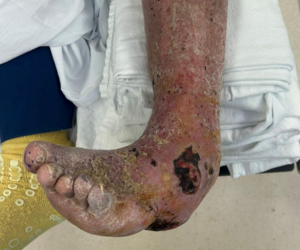
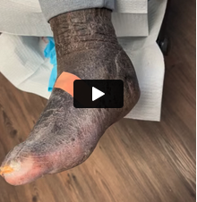
While the diabetic etiology is a mainstay of Charcot neuroarthropathy pathogenesis, systemic contributions include both sensory and autonomic neuropathy, metabolic abnormalities of the bone and trauma. Charcot neuroarthropathy can occur at any weightbearing joint in the body but the repetitive cyclic loading that occurs in gait predisposes the foot and ankle. The condition in this location, also known as Charcot foot, is devastating. Early phase nociceptor activation often subsides, giving way to a relatively painless yet no less destructive deformity. Charcot foot is a source of significant economic burden, long-term morbidity and, in many cases, mortality.2
Acute Charcot is a clinical entity with multiple differential diagnoses. The condition is often missed in early stages due to lack of awareness and nonspecific presentation to the non-limb specialist. As the prevalence of diabetes continues to surge in developed and undeveloped nations alike, early diagnosis of Charcot neuroarthropathy is imperative and interdisciplinary efforts may go a long way toward facilitating early detection of this destructive disease state.3
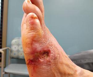
Diagnosis primarily stems from the patient’s history and physical exam. Due to neuropathy and inflammation playing key roles in the early development of Charcot, hallmark findings of the acute disease include new onset of a warm, swollen and erythematous foot, which is infrequently painful. Given the prerequisite neuropathies, patient history regarding recent injury is largely unreliable.4 The initial symptoms are relatively nonspecific and clinicians often misdiagnose the condition as cellulitis, septic arthritis, osteomyelitis, thrombophlebitis or deep venous thrombosis.5
Paradoxically, despite the high frequency of comorbidities and vasculopathies common in patients with longstanding diabetes mellitus, patients with active Charcot neuroarthropathy often have exaggerated or otherwise “bounding” palpable pulses in the foot. For medical providers unfamiliar with the characteristic findings of acute Charcot, these overlapping symptoms may confound appropriate diagnosis, leading to a delay in early intervention. This delay in turn leads to further progression of the destructive osseous process that one can see in the absence of early immobilization and management of the patient with acute Charcot.6
Skin temperature changes in unilateral acute Charcot are common and one may even consider this as a clinical marker for immobilization withdrawal and stage advancement.7 Contralateral skin temperature changes greater than 2oC have been found to be prognostic of inflammatory processes such as Charcot neuroarthropathy and are easy to monitor externally.7 For a review of the general clinical and radiographic guidelines for diagnosing acute Charcot neuroarthropathy, see “An Algorithm Guide For Clinical Diagnosis And Screening Of Acute Charcot Neuroarthropathy” above.
Reviewing Key Imaging Findings For Charcot Neuroarthropathy
Radiographic assessment plays a significant role in the detection and confirmation of Charcot neuroarthropathy. Widely available and inexpensive, simple plain film imaging is highly valuable in portraying osseous density, alignment and structure. Early phase changes are subtle and sometimes non-evident in comparison to the overt abnormalities of late-stage disease. This and a known delay between disease initiation and visible changes lowers the sensitivity of plain films alone, but should not preclude utilization if indicated by physical exam.8
However, one should not use negative findings from this initial radiographic screening to rule out disease. In 1966, Eichenholtz published a staging of Charcot collapse that included stage 1 (development), stage 2 (coalescence) and stage 3 (reconstruction).5 In 1990, Shibata and colleagues added a stage 0 or prodromal stage.9 These stages, marked by radiographic and clinical findings, can drive treatment in a patient with active Charcot neuroarthropathy. Magnetic resonance imaging (MRI) is the optimal second step when a clinician has a high clinical suspicion but plain film radiographs are negative. The detailed soft tissue contrast and multiplane resolution can give early clues to active Charcot in both osseous and soft tissue structures. Subchondral marrow edema with or without microfracture is one of the earliest described changes of stage zero Charcot.10 The diagnostic superiority of MRI is limited to Eichenholtz stages 0 and 1.
In cases in which one suspects overlying osteomyelitis, there may be benefit in more advanced imaging modalities. Triphasic nuclear medicine bone scans are highly sensitive to osseous pathology but are not highly sensitive to Charcot changes. Leukocyte-labeled bone or marrow scans (either Technetium-99 or Indium-111), however, possess improved specificity (69 to 80 percent) for distinguishing infection from an acute Charcot process.11 More recently, there has been a noted increase in the popularity of F-fluorodeoxyglucose positron emission tomography (FDG-PET) for the diagnosis of acute Charcot neutroarthropathy due to its improved sensitivity (greater than 99 percent) and specificity (93.8 percent), high quality images that improve anatomic localization of active process, and more rapid study completion (typically 1.5 to two hours).11
What Lab Tests Can Reveal About Differentiating Between Acute Charcot And Infections
Differentiating between acute Charcot and possible infectious processes is a significant diagnostic challenge. Given the high concomitance of Charcot neuroarthropathy and the presence of diabetic foot ulcerations, it is likely that many patients will experience clinical infections with symptoms (acute edema, erythema, swelling) that mimic those of acute Charcot. However, whereas one normally notes elevations in laboratory values such as white blood cell leukocyte counts, C-reactive protein (CRP) and procalcitonin during infectious processes, these are usually normal to slightly increased in cases of isolated Charcot neuroarthropathy.12 Specifically, CRP is well-established as a sensitive marker of inflammation. Yet multiple studies show that CRP can be normal during active Charcot neuroarthropathy.13
Erythrocyte sedimentation rates (ESRs) may also be mildly elevated during Charcot neuroarthropathy, likely a result of high sensitivity and low specificity.14 Acute Charcot also pushes peripheral monocytes into a proinflammatory phenotype, increasing expression of co-stimulatory surface markers and reducing the secretion of anti-inflammatory cytokines, potentially increasing ESR levels.15 Fortunately, this dissociation between local and systemic inflammatory responses and laboratory data may be useful in differentiating isolated Charcot from active infections, particularly when clinicians consider these findings in combination with clinical progression and radiologic findings.
Emphasizing Early Detection And Effective Patient Education On Offloading
Due to its nonspecific presentation, the acute phase of Charcot neuroarthropathy is often misdiagnosed with delays in intervention rapidly leading to worsening foot deformity, ulceration and possible amputation.16,17 In a 2011 study, Wukich and colleagues in 2011 noted that referring providers correctly identified only one in 22 (4.5 percent) patients as having stage zero Charcot neuroarthropathy.17 An even more concerning finding is that patients in this cohort who progressed to destructive Charcot processes (15 out of 22) had an average of 8.7 weeks pass from incident onset to formal diagnosis and initial treatment whereas patients who were seen an average of 4.6 weeks earlier (7 out of 22) did not progress to the osseous destructive phase. Given that 10 of the 15 patients in the delayed treatment cohort required surgical intervention (an average of 2.9 surgeries per limb), we must raise a great call to arms to increase efforts in early detection, referral and treatment.
The mainstays of treatment in acute Charcot neuroarthropathy remain stabilization, offloading and non-weightbearing.2 It is imperative in this stage to break the cycle of repetitive trauma propagating the acute inflammatory phase and reduce the progression of deformity. Ideally, one should immobilize the extremity in a non-removable total contact cast (TCC) and replace weekly for serial monitoring of Charcot progression. If the patient is unable to tolerate a TCC, a removable cast walker with an offloading insert or modified cast walker such as the “instant” TCC (iTCC) may function as an alternative. However, in a review of 219 cases of patients with Charcot neuroarthropathy, Game and colleagues noted a median time of nine months to achieve resolution in patients whose initial management included non-removable offloading devices in comparison to 12 months for patients who initially wore removable offloading devices.16
Given that adherence with non-weightbearing is associated with positive outcomes in patients with Charcot neuroarthropathy, it is critical that patients understand the severity of this disease and its destructive nature. See “A Pathway For Clinical Management Of Acute Charcot Neuroarthropathy” above.
What You Need To Know Before Surgical Intervention
While a multitude of variables contribute to the overall outcomes of Charcot reconstruction, there is continued debate about the ideal surgical parameters to maximize outcomes and minimize complications. While some authors advocate for an early and aggressive reconstructive timeline, current trends dictate that Charcot surgery should only take place when there is an acute need for intervention. Examples may include active infection recalcitrant to outpatient antibiotics, aggressive skin breakdown, severe dislocation or instability causing vascular compromise.18 In many of these cases, the goals of surgical intervention include stabilization of the extremity (as in cases of acute infection or severe osseous collapse requiring debridement) and allowance for osseous stabilization prior to final reconstructive efforts.
Patients who have definitive Charcot reconstruction typically require medical and surgical optimization to maximize surgical outcomes. The presence of peripheral arterial disease (PAD) in patients with Charcot neuroarthropathy is reportedly as high as 40 percent.19 Therefore, we recommend a thorough review of the patient’s peripheral vascular system by vascular staff prior to reconstruction. We also strongly advocate for a low threshold for noninvasive arterial testing in slow-healing diabetic wounds and any patient with diabetes having a surgical reconstruction procedure.19 In our practice, we customarily refer these patients for lower extremity angiography and they often have vessel disease with treatable lesions.
Poorly controlled diabetes (hemoglobin A1c (HgbA1c) greater than eight percent) is associated with a higher rate of complications. Therefore, we typically delay all surgical reconstructions until the patient is able to sustain an HgbA1c less than eight percent if possible.20 Additionally, patients with longstanding diabetes mellitus are often vitamin D-deficient and will likely require supplementation prior to major reconstructive surgery. Patients may also use this time to prepare their living environment, notify their employer and address other “at-home” concerns before embarking on surgical reconstruction and the extended recovery process.
Final Thoughts
Charcot foot is a source of significant economic burden, long-term morbidity and, in many cases, mortality. Due to the potential for delay in diagnosis, multiple differential diagnoses and the continued surge in diabetes prevalence, early diagnosis is critical and interdisciplinary efforts can help combat this destructive disease state. Early detection and intervention via total contact casting or similar offloading devices are correlated with improved patient outcomes.2,16,17 Therefore, we should strive to improve screening and early referral for patients with Charcot neuroarthropathy.
Dr. Miller is a current Limb Salvage Fellow at MedStar Georgetown University Hospital in Washington, D.C.
Dr. Kennedy is a fellowship-trained foot and ankle surgeon affiliated with the Department of Orthopaedics at McLaren Northern Michigan hospital in Petoskey, Mich.
Dr. Rahnama is a fellowship-trained foot and ankle surgeon and an Assistant Professor at the Georgetown University School of Medicine in Washington, D.C. You can follow him on Instagram @DrAliRahnama for interesting cases and educational material.
1. Rosskopf AB, Loupatatzis C, Pfirrmann CWA, Böni T, Berli MC. The Charcot foot: a pictorial review. Insights Imaging. 2019;10(1);77.
2. Rogers LC, Frykberg RG. The diabetic Charcot foot. In: Veves A, Giurini JM, Guzman RJ, eds. The Diabetic Foot: Medical and Surgical Management. New York: Springer International; 2018:391-413.
3. Stokes A, Preston SH. The contribution of rising adiposity to the increasing prevalence of diabetes in the United States. Prev Med. 2017;101:91-95.
4. Cofield RH, Morrison MJ, Beabout JW. Diabetic neuroarthropathy in the foot: patient characteristics and patterns of radiographic change. Foot Ankle. 1983;4(1):15-22.
5. Rogers LC, Frykberg RG, Armstrong DG, et al. The Charcot foot in diabetes. Diabetes Care. 2011;34(9):2123-2129.
6. Rech ALG, Stüve Y, Toepfer A, Schimke KE. Missing the boat: fatal ending to a missed case of Charcot arthropathy. Endocrinol Diabetes Metab Case Rep. 2020;2020(1):20-0013.
7. Moura-Neto A, Fernandes TD, Zantut-Wittmann DE, et al. Charcot foot: skin temperature as a good clinical parameter for predicting disease outcome. Diabetes Res Clin Pract. 2012;96(2):e11-e14.
8. Morrison WB, Ledermann HP. Work-up of the diabetic foot. Radiol Clin North Am. 2002;40(5):1171-1192.
9. Rosenbaum AJ, DiPreta JA. Classifications in brief: Eichenholtz classification of Charcot arthropathy. Clin Orthop Relat Res. 2015;473(3):1168-1171.
10. Schoots IG, Slim FJ, Busch-Westbroek TE, Maas M. Neuro-osteoarthropathy of the foot—radiologist: friend or foe? Sem Musculoskel Radiol. 2010;14(3):365-375.
11. Milne TE, Rogers JR, Kinnear EM, et al. Developing an evidence-based clinical pathway for the assessment, diagnosis and management of acute Charcot Neuro-Arthropathy: a systematic review. J Foot Ankle Res. 2013;6(1):30.
12. Ertugrul BM, Lipsky BA, Savk O. Osteomyelitis or Charcot neuro-osteoarthropathy? Differentiating these disorders in diabetic patients with a foot problem. Diabet Foot Ankle. 2013;4(1):21855.
13. Jude EB, Selby PL, Mawer EB, Burgess J, Boulton AJM. Inflammatory and bone turnover markers in Charcot arthropathy and osteomyelitis of the feet in diabetic patients. Diabetologia. 2002;45(Suppl 2):A341-A342.
14. Petrova NL, Moniz C, Elias DA, Buxton- Thomas M, Bates M, Edmonds ME. Is there a systemic inflammatory response in the acute Charcot foot? Diabetes Care. 2007;30(4):997- 998.
15. Uccioli L, Sinistro A, Almerighi C, et al. Proinflammatory modulation of the surface and cytokine phenotype of monocytes in patients with acute Charcot foot. Diabetes Care. 2010;33(2):350-355.
16. Game FL, Catlow R, Jones GR, et al. Audit of acute Charcot’s disease in the UK: the CDUK study. Diabetologia. 2012;55(1):32-35.
17. Wukich DK, Sung W, Wipf SA, Armstrong DG. The consequences of complacency: managing the effects of unrecognized Charcot feet. Diabet Med. 2011;28(2):195-198.
18. Ulbrecht JS, Wukich DK. The Charcot foot: Medical and surgical therapy. Curr Diab Reps. 2008;8(6):444-451.
19. Wukich DK, Raspovic KM, Suder NC. Prevalence of peripheral arterial disease in patients with diabetic Charcot neuroarthropathy. J Foot Ankle Surg. 2016;55(4):727-731.
20. Wukich DK, Raspovic KM, Hobizal KB, Sadoskas D. Surgical management of Charcot neuroarthropathy of the ankle and hindfoot in patients with diabetes. Diabetes Metab Res Rev. 2016;32 Suppl 1:292-296.