Emerging Insights On PAD, CLTI And The Gaps In Limb Salvage During The COVID-19 Pandemic
Best medical treatment for peripheral arterial disease (PAD) includes managing hypertension, diabetes, dyslipidemia and associated risk factors like smoking. This reduces morbidity and mortality and can obviate the need for invasive intervention.1 Patients with critical limb-threatening ischemia (CLTI) are at risk of extensive coronary artery disease and cerebrovascular disease with worse outcomes.2 In an observational study, 10 year all-cause mortality was 56 percent for asymptomatic PAD, 63 percent for intermittent claudication and 75 percent for severe limb ischemia. Among all PAD subjects, cardiovascular causes were the most common main cause of death (45 percent) and a cardiovascular event was present as either the main or one of the three most common contributing causes of death in 64 percent of cases.3
Morbidity and mortality associated with PAD significantly increased after the emergence of the COVID-19 pandemic.4 Prior to the pandemic, researchers reported over 150,000 people undergoing lower extremity amputations in the United States yearly, which is directly proportional to rates of peripheral arterial occlusive disease, neuropathy and soft tissue sepsis.5,6 In an Italian observational study, Bellosta and colleagues noted an increased incidence of acute limb ischemia in patients with COVID-19 infections and an almost eight-fold increase in total patients with acute limb ischemia during the peak of the pandemic in comparison with the same period of time in 2019.7
The PARTNERS (Peripheral Arterial Disease Awareness Risk and Treatment: New Resources for Survival) program revealed other important aspects of PAD epidemiology.8 Investigators cited poor recognition of PAD as 44 percent of patients only received a PAD diagnosis after enrollment in the study. Only 49 percent of primary physicians treating patients with a prior diagnosis of PAD actually knew of the diagnosis despite documentation in medical records.
Peripheral arterial disease is very common with an estimated prevalence of 29 percent in high-risk individuals (those greater than 70 years of age without additional risk factors, or those 50 to 69 years of age with a history of cigarette smoking or diabetes).9 Moreover, patients with PAD are generally less intensively managed in comparison to patients with coronary artery disease (CAD).9 Not only is PAD diagnosis frequently overlooked, but cardiovascular risk factors are not treated as appropriately as in patients with CAD.10 Now additional pandemic-related factors like fear of contracting the virus and a surge in demand versus supply of care may lead to even more delayed or severe presentations of PAD.
When A Patient With CLTI Tests Positive For COVID-19
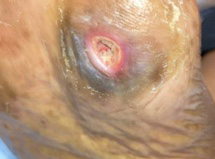
A 73-year-old Caucasian male with a non-healing Wagner grade III wound of the left great toe presented to our hospital in April 2020. His past medical history included type 2 diabetes and hypertension, and he previously had a right partial first ray amputation. The skin around the left hallux ulcer was pale and bluish with hair loss and purulent drainage. His feet were cold to the touch, with bilateral extremity discoloration and diminished distal pulses.
Clinically, the patient exhibited Rutherford category V PAD. He admitted self-wound care at home for about 10 days prior to hospital presentation. We felt his delay in seeking care was possibly due to fear of contracting COVID-19 and/or a lack of knowledge about the severity of PAD.
Arterial doppler revealed an occluded left superficial femoral artery with a reconstituted collateral flow due to a Trans-Atlantic Inter-Society Consensus (TASC) D aortoiliac lesion.
There was routine hospital COVID-19 testing at this time and the patient tested positive for COVID-19. During this time, our state had 2,500 hospital admissions daily for COVID-19 and overall confirmed cases in our state had surged up to 108,998, including 8,196 deaths.11 As a result, hospitals implemented strict triage protocols, even for cases involving acute stroke and myocardial infarction. This patient’s pathology involved a chronic lesion so due to his COVID-19 status and intensive triage, we had to pursue hospital committee approval to surgically address his condition. This took crucial time for a patient at risk of permanent disability and limb loss. In the meantime, we monitored the patient daily and conservatively managed him with thromboprophylactic drugs.
After the rigorous maintenance of his condition and subsequent committee approval for surgical treatment, we performed peripheral angiography, which confirmed 100 percent occlusion of the left superficial femoral artery with residual disease on the right side as well. We recommended urgent angioplasty due to the CLTI. These interventions pose multiple challenges due to the presence of widespread multilevel disease, long and complex occlusive lesions and involvement of the tibial vessels, which itself is a major interventional challenge. In this case, the threat was even greater due to his COVID-19 hypercoagulable state, his late presentation and delayed intervention due to quarantine procedures.
We followed a conventional antegrade approach but it failed because there was a flush occlusion. The superficial femoral artery was completely blocked from the ostium so there was not enough support to cross the lesion. In order to establish access in a case like this, multiple trials of contrast for road mapping and radiation administration are necessary, which sometimes causes significant blood loss. Most of the time, this results in aborting the procedure completely and eventual amputation of the limb.
What You Should Know About The Retrograde Access Approach
In accordance with the recent standard change in cases of failed antegrade approach and prevention of foot amputation, we used a pedal access (retrograde) approach.12 This is a two-step procedure.
Step 1: Establishing pedal access. This requires a great amount of time along with expert knowledge of foot anatomy and ultrasound to maintain awareness of the blood flow and calcium deposition along the walls of vessels. Even with significant experience, vessel access alone can take anywhere from 45 minutes to an hour, especially in cases of severely diseased vessels. This requires a peripheral interventionist expert to modify the failed outcome of the conventional method.
Three vessels are usable for pedal access. They include the dorsalis pedis/anterior tibial artery, which is the easiest of the three, the peroneal artery or the posterior tibial artery.
We locally anesthetize the puncture site, avoiding compression of the intended access vessel. These vessels usually have a very low perfusion pressure so extrinsic compression by local anesthetic volume can further interfere with successful access. One can use fluoroscopy to confirm access, especially in heavily calcified vessels. Another guiding measure is roadmapping, which uses antegrade angiography from the femoral access site to identify the pedal/tibial target vessel.
In our procedure, we used duplex-guided access. This involves identifying the vessel with the ultrasound probe in either a longitudinal or transverse position relative to the vessel. One can use Doppler color flow to identify the target artery. Through grayscale mode, the needle accesses the anterior wall of the vessel under duplex guidance. Echogenic-appearing micropuncture needles are best as it can be difficult to see the tip of a regular needle using the duplex ultrasound probe.
Once we establish access, evidenced by back bleeding, we pass the micro-puncture access wire (0.018 inch) through the needle into the vessel under fluoroscopic guidance. After removing the needle, one can pass a micro-puncture 4-French sheath over the wire and secure access. Sometimes we use only the dilator of the sheath without the sheath itself to secure access. Once the retrograde introducer is in place, we fully heparinize the patient in the usual fashion to avoid any thrombosis in the tibial vessels during the intervention.
Step 2: Retrograde occlusion crossing with atherectomy and angioplasty. The appropriate endovascular tool depends on the type of lesion. For a retrograde approach, the modalities used to cross and treat stenoses and occlusions include:
1. Wire/catheter technique;
2. Chronic total occlusion (CTO) crossing devices (TruePath™ (Boston Scientific); TurboElite Laser™ (Spectranetics) or Crosser™ CTO recanalization catheters (Bard));
3. Atherectomy devices such as the Diamondback™ 360 coronary orbital atherectomy system (Cardiovascular Systems, Inc.) and;
4. Balloon angioplasty and self-expanding stents for superficial femoral and popliteal lesions.
Working Through The Challenges Of Crossing The Lesion In A Patient With CLTI
We first obtained access in the anterior tibial artery with the foot in slight plantarflexion but we could not cross the lesion. This illustrates another challenge. Achieving access doesn’t guarantee one can cross the lesion.
With our second try, we accessed the posterior tibial vein. The blood flow was very sluggish and other nearby vessels did not allow us to differentiate between the posterior tibial artery and vein. Therefore, our second attempt also failed.
We finally succeeded with our third attempt via the posterior tibial artery with the foot dorsiflexed and laterally rotated, and the knee slightly flexed. This was risky because the posterior tibial artery is very prone to vasospasm. Sometimes, there is a complete arterial spasm, resulting in absolute blood supply cessation. In these cases, the access site then poses another threat of non-healing if the spasm does not abate or if the procedure is aborted. In these cases, we inject vasodilators.
Once we established posterior tibial access, we were finally able to cross the lesion and snared the wire from the top. We then created the floss, a wire going from the left groin all the way to the left foot, forming a nice rail over which we performed atherectomy and angioplasty.
The blood vessels opened up nicely. The access itself took over an hour and the entire procedure took four hours. We potentially saved this patient’s foot, which may have subsequently improved his limb and life longevity.
Final Thoughts
The COVID-19 pandemic continues to pose challenges to adequate and timely PAD treatment. Peripheral arterial disease poses an equivalent risk to that of coronary heart disease.13 This case emphasizes lack of awareness of a “foot attack,” clearly apparent with this patient’s delay in seeking treatment. If this patient sustained a heart attack, treatment delay may be less likely.
There are nearly two million people living with limb loss in the United States. Approximately 185,000 amputations occur in the United States each year.14 When it comes to people with diabetes who have a lower extremity amputation, up to 55 percent will require a contralateral amputation within two to three years.15 Peripheral arterial disease is a significant contributor to amputation, which has physical and possible mental health ramifications.16
Timely treatment of PAD can prevent amputation. Despite the challenges presented by treating patients during the COVID-19 pandemic, it is vital for patients and hospitals alike to be aware of the risks associated with PAD and CLTI, and the gravity of a “foot attack.”
Dr. Singh is a graduate of Smolensk State Medical University in Russia and works as a Junior Doctor in India. She is a prospective internal medicine residency applicant for the 2021-2022 match.
Dr. Shah is an interventional cardiologist and endovascular specialist. He is the founder and director of Apex Heart and Vascular Care in Passaic, N.J.
1. Burns P, Gough S, Bradbury AW. Management of peripheral arterial disease in primary care. BMJ. 2003;326(7389):584-588.
2. Cotter G, Cannon CP, McCabe CH, et al. Prior peripheral arterial disease and cerebrovascular disease are independent predictors of adverse outcome in patients with acute coronary syndromes: are we doing enough? Results from the Orbofiban in patients with unstable coronary syndromes-thrombolysis in myocardial infarction (OPUS-TIMI) 16 study. Am Heart J. 2003;145(4):622-627.
3. Sartipy F, Sigvant B, Lundin F, Wahlberg E. Ten year mortality in different peripheral arterial disease stages: a population based observational study on outcome. Eur J Vasc Endovasc Surg. 2018;55(4):529-536.
4. Scheinfeld MH, Ye K, Goldman IA. Lower-extremity arterial thrombosis associated with COVID-19 is characterized by greater thrombus burden and increased rate of amputation and death. Radiology. 2020;297(2):E263-E269.
5. Dillingham TR, Pezzin LE, Shore AD. Reamputation, mortality, and health care costs among persons with dysvascular lower-limb amputations. Arch Phys Med Rehabil. 2005 Mar;86(3):480-486.
6. Beckman JA, Creager MA, Libby P. Diabetes and atherosclerosis: epidemiology, pathophysiology, and management. JAMA. 2002;287(19):2570-2581.
7. Bellosta R, Luzzani L, Natalini G. Acute limb ischemia in patients with COVID-19 pneumonia. J Vasc Surg. 2020;72(6):1864-1872.
8. Hirsch AT, Criqui MH, Treat-Jacobson D, et al. Peripheral arterial disease detection, awareness, and treatment in primary care. JAMA. 2001;286:1317–1324.
9. Hiatt WR, Fowkes FG, Heizer G, et al. Ticagrelor versus clopidogrel in symptomatic peripheral artery disease. N Engl J Med. 2017;376:32–40.
10. McDermott MM, Mehta S, Ahn H, Greenland P. Atherosclerotic risk factors are less intensively treated in patients with peripheral arterial disease than in patients with coronary artery disease. J Gen Intern Med. 1997;12(4):209-215.
11. Worldometer. New Jersey Coronavirus Cases. Available at: https://www.worldometers.info/ coronavirus/usa/new-jersey/ . Accessed March 2, 2021.
12. Goltz JP, Planert M, Horn M, et al. Retrograde transpedal access for revascularization of below-the-knee arteries in patients with critical limb ischemia after an unsuccessful antegrade transfemoral approach. Rofo. 2016;188(10):940-948.
13. Subherwal S, Patel MR, Kober L, et al. Peripheral artery disease is a coronary heart disease risk equivalent among both men and women: results from a nationwide study. Eur J Prev Cardiol. 2015 Mar;22(3):317-25.
14. Ziegler-Graham K, MacKenzie EJ, Ephraim PL, Travison TG, Brookmeyer R. Estimating the prevalence of limb loss in the United States: 2005 to 2050. Arch Phys Med Rehabil. 2008;89(3):422-429.
15. Pandian G, Hamid F, Hammond M. Rehabilitation of the patient with peripheral vascular disease and diabetic foot problems. In: DeLisa JA, Gans BM (eds). Rehabilitation Medicine: Principles And Practice. Philadelphia:Lippincott‐Raven;1998:1517-1544.
16. Kashani JH, Frank RG, Kashani SR, Wonderlich SA, Reid JC. Depression among amputees. J Clin Psychiatry. 1983;44(7):256-258.