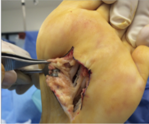
Study Assesses One-Year Radiographs After Triplane Tarsometatarsal Arthrodesis
In a recent poster presentation at the American College of Foot and Ankle Surgeons (ACFAS) 2020 Scientific Conference, Dayton and colleagues compared radiographic measurements before and after triplane tarsometatarsal arthrodesis for hallux valgus. Analyzing records of 108 patients, the authors found that this procedure provided reliable correction with low recurrence and complications at one year.
J.P. McAleer, DPM, FACFAS uses a similar technique for treating hallux valgus. Lawrence DiDomenico, DPM, FACFAS shares he also uses a dorsal medial approach to tarsometatarsal arthrodesis and considers fusing the intercuneiform joint as well. Bob Baravarian, DPM, FACFAS prefers a staple and plate fixation system with constant compression across the staple and no screws crossing the joint.
The study authors acknowledge that one should recognize the existence of radiographic reporting bias.
“There will always be user bias and user error,” agrees Dr. DiDomenico, the Director of Podiatric Residency Training at St. Elizabeth Health Center in Youngstown, Ohio. “One must consider the technical component of the radiograph, such as the position of the beam to the foot (and the position of the foot itself).”
Four weightbearing views of the foot with evaluation in proper angle and base of gait are essential to avoid misdiagnosis preoperatively, and for confirming deformity reduction intraoperatively, says Dr. McAleer, who is in private practice in Jefferson City, Mo. Dr. Baravarian cautions surgeons to not completely rely on the anatomic axis measurements in procedure selection and also consider joint angles along with joint instability.
The study authors question the relevance of the distal metatarsal articular angle due to poor agreement in post-reduction measurements of this angle. Dr. Baravarian feels this assertion is overstated but Drs. DiDomenico and McAleer agree this is a relevant consideration.
“The distal metatarsal articular angle is a perfect example of how (surgeons evaluate) a three-dimensional problem like hallux valgus in two dimensions, resulting in a lack of complete analysis of the complex nature of first ray alignment and mechanics,” says Dr. McAleer.
“(First tarsometatarsal arthrodesis) is a great procedure for the right patient but is not for every patient,” says Dr. Baravarian, an Assistant Clinical Professor at the UCLA School of Medicine. He stresses that everyday patient life and surgical recovery are also important factors to consider when planning hallux valgus correction.
Dr. McAleer points out that while radiographs are useful in initial evaluation of first ray deformity, angular relationships are only one aspect of that evaluation. He adds that the health of the joints and the presence of concomitant deformities such as metatarsus adducts are also important to gauge from these initial radiographs.
Emphasizing that the triplane tarsometatarsal arthrodesis is predictable and allows for complete reduction of deformity, Dr. DiDomenico feels this study supports three-dimensional correction in a straightforward fashion.
Should Surgeons Delay Arthrodesis Procedures Due To Low Vitamin D Levels?
By Jennifer Spector, DPM, FACFAS, Associate Editor
A new study suggests that with appropriate perioperative supplementation, those with insufficient serum Vitamin D levels have similar fusion rates for common pedal arthrodesis procedures as patients with sufficient Vitamin D levels.
In the recent award-winning poster presentation at the aforementioned ACFAS conference, study authors assessed fusion rates of various pedal arthrodeses (including first MPJ, first tarsometatarsal and subtalar joint fusions) and the relationship with preoperative vitamin D levels with or without perioperative supplementation. In the study involving 100 patients, researchers drew serum 25-hydroxyvitamin D levels within three months of surgery.
Those study patients with sufficient (>30 ng/mL) levels did not receive any perioperative supplementation. Those with insufficient levels (20-29 ng/mL) received advice to supplement with 2,000 IU of vitamin D3 daily over the counter. Finally, patients with deficient lab levels (10-19 ng/mL) took 50,000 IU of vitamin D2 daily for 12 weeks plus 2,000 IU of vitamin D3 daily.
“The main conclusion of the study is that adult patients undergoing pedal arthrodesis procedures, with appropriate supplementation, did not have a significant difference in fusion rates when comparing various levels of serum vitamin D,” says Troy J. Boffeli, DPM, FACFAS, the lead author of the study.
Abimbola O. Johnson, DPM, a study co-author, shares that the study patients (including those who initially had sufficient, insufficient or deficient levels of vitamin D) overall achieved successful fusion at a rate of 92 percent or better at 10 weeks post-op, and 98 percent or better at 14 weeks post-op. Interestingly, Drs. Boffeli and Johnson point out that 35 percent of the study population had some degree of hypovitaminosis D.
The co-authors point out that while the lab test for serum 25-hydroxyvitamin D is relatively inexpensive, physicians commonly order it as part of a workup for a non-union.
“At this stage, the cost of a non-union will greatly surpass the cost of lab work,” shares Dr. Johnson, a second-year resident at HealthPartners Institute/Regions Hospital in St. Paul, Minn.
Dr. Boffeli adds that some surgeons may obtain this lab work preoperatively but then cancel the procedure upon identification of hypovitaminosis D.
“This study lays the groundwork to dispute (this notion) as all patients with low vitamin D (in this study) received supplementation perioperatively and went on to heal successfully,” relates Dr. Boffeli, the Director of the Foot and Ankle Surgery Residency Training Program at HealthPartners/ Regions Hospital in St. Paul, Minn.
Drs. Boffeli and Johnson both say the particular procedures (first MPJ, first tarsometatarsal and subtalar joint fusions) noted in the study have strong union rates in the general population, and that the results may not extend to all fusion procedures. Additionally, they excluded patients with vitamin D levels under 10 ng/mL as the team did delay surgery in those cases. Overall, they share that further research may identify fusion rates in patients with hypovitaminosis D who did not receive supplementation.
Endoscopic Versus Open Gastrocnemius Recession: Which Procedure Leads To More Complications?
By Jennifer Spector, DPM, FACFAS, Associate Editor
Open gastrocnemius recessions reportedly lead to twice as many postoperative complications than endoscopic gastrocnemius recessions.
That was a key finding from James and colleagues in a recent retrospective meta-analysis presented at the aforementioned ACFAS conference. Infection and dehiscence occurred nine times more often with open gastrocnemius recessions. Additionally, the researchers found that complex regional pain syndrome (CRPS) and deep vein thrombosis/pulmonary embolism (DVT/PE) complications only occurred after open gastrocnemius recession procedures. True nerve injuries occurred slightly less often in the endoscopic gastrocnemius recession group but the study authors noted a 30 percent higher resolution rate of these injuries in this group.
John Grady, DPM, FASPS, a co-author of the study, notes the findings on postoperative complications do reflect his experience in practice as well. He adds that a recent technique modification has also helped reduce nerve-related post-op complications.
“I now isolate the sural nerve endoscopically and put it on the opposite side of my cannula prior to cutting the gastrocnemius, which decreases neural complications,” notes Dr. Grady, Chair of the Podiatric Section of the Department of Orthopedics at Advocate Christ Medical Center and Advocate Children’s Hospital.
Priya Parthasarathy, DPM, DABPM, feels the results of this study do not correlate with her experience.
“I do a fair number of open gastroc recessions and have not had a wound or nerve complication that I know of,” says Dr. Parthasarathy, who is in private practice in Silver Spring, Md.
Jashan Valjee, DPM, FACFAS, points out that patients with increased morbidities will do better with the endoscopic procedure as wound healing complications are much less.
“If the patient has a high body mass index (BMI), the endoscopic gastrocnemius recession would be preferable as it can lessen the struggle against soft issue dissecting down to the gastrocnemius aponeurosis,” explains Dr. Valjee, who is in private practice in Glen Burnie and Hanover, Md.
Dr. Grady conversely feels that an obese patient may pose challenges in endoscopic dissection to the point where an open procedure might be preferable.
Dr. Parthasarathy agrees that if mobility or other procedures cause intraoperative positioning issues, one can perform the endoscopic gastrocnemius recession with the patient completely supine. However, she adds that open gastrocnemius recession is achievable through a fairly small incision and can better guarantee adequate lengthening through direct visualization.
“I usually do an open procedure on the more moderate to severe equinus patients who are still in the category of gastroc recession,” says Dr. Parthasarathy.
“There seems to be less strength loss seems with the endoscopic gastrocnemius recession and cosmetically, there is no comparison,” says Dr. Grady, who is in private practice in Oak Lawn, Ill.
Dr. Valjee adds that this study focuses on complication rates and not necessarily the success of either procedure
“It would be interesting to see a comparison of patient satisfaction scores and goals met between the two procedures,” notes Dr. Valjee.