


Pertinent Surgical Pearls on Management of Skew Foot Deformity in the Young Adult
Skew foot deformity is a rare complex congenital condition that involves the subtalar joint, midtarsal joint, and Lisfranc articulations. Referred to as a Z- or serpentine foot, it affects formation and growth of the osseous pedal structures and is a rigid deformity. The skew foot deformity presents with severe talonavicular joint and subtalar joint valgus instability with a large talar head uncoverage angle (oblique talus). The midfoot is severely abducted on the rearfoot and the metatarsals at the Lisfranc joint are severely adducted. These two opposite forces lead to hypoplastic development of the cuneiforms.
Furthermore, the ankle is in external rotation relative to the talus giving a “flat top” appearance. As lines are drawn through the rearfoot (talar neck bisection) and midfoot (to the intermediate cuneiform) and then back through the second metatarsal bisection, the “classic” Z-foot is depicted (as shown in Figure 1A). One can observe this Z configuration radiologically and clinically (Figures 1C and 1D).1
In 1949, McCormick and Blount originally coined the deformity skew or S(serpentine)-foot.2 Prior to this, in 1933, Peabody and Muro labeled this foot structure congenital metatarsus varus to differentiate it from benign congenital metatarsus adductus.3 This referred to the distinct mechanical advantage the tibialis anterior muscle has in both metatarsus adductus, but more predominately in a skew foot, in creating forefoot varus and extreme metatarsus primus elevatus. A review of the literature demonstrates that the natural history of untreated skew foot is undocumented.4
Authors advise that nonoperative treatment for those with skew foot is unsuccessful and surgical correction is necessary, although there are only limited studies addressing surgical intervention and outcome results in young adults.4,5 Correction of this deformity can be very challenging; however, surgeons must be able to notice the multifaceted biomechanical components and apply appropriate surgical principles to guide repair. This article aims to augment the current literature by presenting a case study illustrating successful repair of a skew foot.
When a Young Adult Male Presents With Bilateral Congenital Deformities
A 24-year-old male with a longstanding history of severe bilateral foot deformity presented with restrictions in shoe wear and chronic pain. The deformity was never recognized or addressed in infancy or childhood. He states he sought help at 12 years of age from pediatric orthopedists, who had no treatment options to offer. He has no prior treatment and was walking in sandals.
On clinical examination, the patient demonstrated severe metatarsus adductovarus, which was nonreducible and rigid in nature. The rearfoot was also in valgus and nonreducible. Ankle joint dorsiflexion was adequate at 5 degrees and 30 degrees of external rotation was noted. Lateral view radiographs revealed severe metatarsus primus elevatus with hallux flexus and a talonavicular break (Figure 1B).
The entire forefoot was rotated with a fixed varus. Note visualization of each respective metatarsal. The dorsoplantar radiographic view notes 50 percent talar uncoverage, and a severe abducted malposition of the navicular and cuneiforms. Engel’s angle was near 90 degrees (Figure 1B). Also the posterior position of the fibula relative to the tibia was noted.
The patient was only able to wear open-toed sandals. His deformity was so severe that traditional shoes could not be worn. The patient was limited due to pain and was working in primarily a sitting cashier’s job. Through his youth he was unable to do any form of athletic activities.
Surgical Considerations for Correction of Skew Foot Deformity
The senior author, Dr. Visser’s, approach begins with planning to address the deformity at all levels including the ankle joint, rearfoot, midfoot and forefoot. When approaching the ankle joint, one must evaluate for equinus and the degree of external tibial fibular rotation. In addressing the rearfoot, one should reduce talonavicular uncoverage and address heel valgus. At the midfoot, the senior author encourages surgeons to consider a large and extensive closing osteotomy across the cuboid and cuneiforms. Lastly, at the forefoot, one can address severe elevatus of the first metatarsal with a tibialis anterior tendon transfer and also included is a first metatarsal-medial cuneiform-navicular arthrodesis.
For this particular patient, the surgical approach included the following steps:
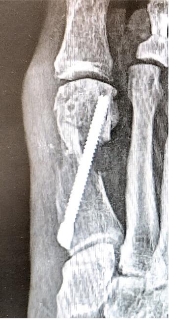
Step one: Extensive medial column arthrodesis involving (1) subtalar joint capsulotomy, (2) talonavicular, (3) navicular-cuneiform, and (4) first metatarsal-medial cuneiform (Figure 2)
Step two: Plantar fasciotomy
Step three: Transfer of tibialis anterior tendon to the cuboid
Step four: Large cuneocuboid wedge osteotomy involving the intermediate cuneiform, lateral cuneiform and cuboid (Figures 3A, B, C)
An Architectural Approach to Skew Foot Reconstruction of Pedal Foot Deformity
It is important that the surgeon consider the biomechanical aspects of surgical correction of the skew foot, including calcaneovalgo, cuneocuboid abducto, and metatarsus adducto varus components.
In the case discussed above, upon completion of the described procedures, a plantigrade foot was accomplished and a stable forefoot to rearfoot alignment was established (Figure 4). Below are some descriptions, in the senior author’s experience, of the biomechanical points of view of the key interventions involved in correction:
Correction of proximal line of the Z
- Subtalar joint capsulotomy: restores the calcaneus in a neutral position.
- Talonavicular arthrodesis: establishes talar convergence and with subtalar joint mobility (from subtalar joint capsulotomy) creates closed kinetic chain supination of the rearfoot correcting heel valgus. This also reduces a portion of the external rotation.
Correction of digital Z-line
- First metatarsal-medial cuneiform-navicular arthrodesis: corrects first ray metatarsus primus elevatus and adductus. Also corrects hallux flexus and forefoot varus.
- Transfer of the tibialis anterior to the cuboid eliminates the supination force on the first ray and forefoot varus.
- Straightening of the Z
- Plantar fascial release: Removes traction force between forefoot and rearfoot.
Straightens the center of the Z-line
- Cuneocuboid wedge osteotomy: Corrects forefoot adduction and varus deformity proximally. Aligns the forefoot, midfoot and the rearfoot.
In Conclusion
Skew foot deformity poses great challenges to patients and foot and ankle specialists alike. Its association with concomitant deformities and worsening of the deformity in the untreated young adult foot poses significant adversity in surgical planning and ultimately, procedure selection. By adhering to sound surgical principles and remaining fluid in approach, surgeons can obtain excellent long-standing clinical results.
Dr. Visser is the Director of the Foot and Ankle Surgery Residency program at SSM Health DePaul Hospital in St. Louis. He is a Fellow of the American College of Foot and Ankle Surgeons.
Dr. Smith is a second-year podiatric resident at SSM Health DePaul Hospital.
References
1. Domínguez G, Munuera PV. Metatarsus adductus angle in male and female feet: normal values with two measurement techniques. J Am Podiatr Med Assoc. 2008: 98(5):364–369. https://doi.org/10.7547/0980364
2. McCormick DW, Blount WP. Metatarsus adductovarus: skewfoot. J Am Med Assoc. 1949;141(7):449–453. https://doi.org/10.1001/jama.1949.02910070021005
3. Peabody CW, Muro F. Congenital metatarsus varus. J Bone Joint Surg. 1933;15(1):171-189.
4. Peterson HA. Skewfoot (forefoot adduction with heel valgus). J Pediatric Orthop. 1986;6(1):24–30. https://doi.org/10.1097/01241398-198601000-00005
5. Hamel J. Skew foot/serpentine foot. In: Foot and Ankle Surgery in Children and Adolescents. Springer;2021:79-90. https://doi.org/10.1007/978-3-030-58108-4_2


