Earlobe correction is a common aesthetic surgical procedure that dermatologists can offer their patients. It is an optimal solution for an earlobe that is mishapened from many years of wear and tear. In most cases, this outpatient procedure is straightforward and rarely presents any complications. The most common technique consists of excising around the previous fistula with a primary closure of the anterior and posterior defect. If the defect involves the edge of the lobule, the sides of the cleft are excised and a suture is placed connecting the tips, usually vertical mattress sutures are used for eversion.1
Hematoma, which is a collection of blood under a surgical dead space, is an infrequent complication in cutaneous surgery. Studies report a frequency between 0.1% and 0.6% for Mohs surgery2,3 and up to 15% for face lifts.4 Although Mohs surgery has low relative incidence of hematoma, it could have severe consequences and any dermatologic surgeon must be confident with its management. The majority of hematomas occur within 24 hours of the surgery from the combination of the unstable clots along with the disappearance of the epinephrine effect that contribute to bleeding vessels. Postoperative pressure dressings are essential to avoid this complication. Among the risks factors described for hematoma are gender, type of anesthesia, hypertension, patient’s activity and anxiety after the surgery, abnormal coagulation studies, and medications that may affect clotting.5-10
In this article, we report a case of bilateral hematomas occurring 48 hours following earlobe correction along with a simple technique for its management.
Case
A 45-year-old woman presents to the clinic inquiring about earlobe correction surgery. Her past medical history was relevant for hypothyroidism controlled with levothyroxine and regular intake of prophylactic aspirin 81 mg. Upon physical examination, the patient presented with earlobes that had an expanded orifice in the center with preservation of the inferior border (Figures 1A-B). The patient was offered, and agreed to with an informed consent, a bilateral earlobe correction surgery. She took her last aspirin 5 days prior to the surgery. Conventional earlobe correction was done bilaterally, excising around the fistula, and closing the defect anteriorly and posteriorly, without complications. Routine pressure bandages were applied.
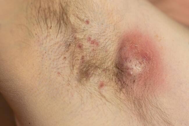
Two days later, the patient presented oozing blood from both wounds, soaking through her bandage gauze. The only symptom she reported was slight pain upon palpation of the area. On physical examination, she had bilateral hematomas on both earlobes with evident blood through intact suture lines. There were no firm or fluctuant masses upon palpation. There was no erythema or warmth at the surgical sites and no signs of possible infection. A 18-gauge needle attached to a syringe was introduced through the suture line to dislodge the clots that were present on the edges, less than 0.2 cc was aspirated. Subsequently, we were able to squeeze less than 1 cc of blood through the suture line. She was bandaged again with another set of pressure dressings. The procedure was well tolerated. Topical and systemic antibiotics were given. Upon follow-up, the hematomas were completely resolved. Ten weeks later, she returned for repiercing of her bilateral earlobes and she tolerated the procedure well (Figures 2A-B and Figures 3A-3B).
Discussion
Earlobe surgery is typically a simple and straightforward procedure that dermatologists can offer to their patients. Usually it is done in the outpatient setting and very rarely leads to any complications.1,11,12 Earlobe tears can be classified as partial, complete, or multiple.12 Depending on the type, the surgeon will decide on which procedure will be better for the repair of the defect.
Local anesthetic is administered to the area followed by stabilization of the earlobe with chalazion clamps, tongue depressors, or skin hooks. Chalazion clamps are preferred because they provide the additional benefit of hemostasis. If the defect is within the upper two-thirds of the lobe, the fistula can be excised and closed anteriorly and posteriorly. In contrast, if the defect is in the lower one-third of the lobe, it is better to extend the defect to a full tear and close it with subcutaneous and epidermal stitches. In the case of a complete tear, the borders that form the cleft are excised with a V- or C-type incision and then borders are reapproximated.1,12
Hematoma, which can be major or minor, is an infrequent complication of cutaneous surgery and to our knowledge it has not been previously described in earlobe repair surgery. Studies have shown that it is an independent factor for infection.13-15 A major hematoma is a rapid accumulation of blood that requires open evacuation and coagulation of the bleeding vessels. These hematomas can cause tissue ischemia, necrosis, and infection. Minor hematomas are smaller collections of blood that can be aspirated through the suture line or through a small slit on the affected area. They can cause seromas, tissue irregularities, and hyperpigmentation.4
Our patient presented with bilateral minor hematomas 2 days after earlobe repair, which were easily managed with the technique described above without the need to remove the sutures. Even though our patient denies any clotting or bleeding problem, she admits taking aspirin 81 mg 5 days before the surgery. We think this could have been the possible culprit of this complication. This case illustrates that in these situations early intervention is key, resulting in a good cosmesis. As dermatologic surgeons, it is important that we are aware of the possibility of this occurrence, even in low risk surgeries.
Dr Correa-Selm a is micrographic surgery and procedural dermatology fellow at Affiliated Dermatologists and Dermatologic Surgeons in Morristown, NJ.
Dr Lee is director of the ACGME-approved Micrographic Surgery and Procedural Dermatology Fellowship and a practicing dermatologist at Affiliated Dermatologists and Dermatologic Surgeons in Morristown, NJ.
Disclosure: The authors report no relevant financial relationships.
References
1. Alves Ribeiro A, de Matos Lurenco L, Cardoso de Barros Matsuda T, Ferrari NM. Split earlobe repair: literature review and new technique proposal. Surg Cosmetic Dermatol. 2009;1(3):141-144.
2. Alam M, Ibrahim O, Nodzenski M, et al. Adverse events associated with Mohs micrographic surgery: multicenter prospective cohort study of 20,821 cases at 23 centers. JAMA Dermatol. 2013;149(12):1378-1385.
3. Cook JL, Perone JB. A prospective evaluation of the incidence of complications associated with Mohs micrographic surgery. Arch Dermatol. 2003;139(2):143-152.
4. Niamtu J 3rd. Expanding hematoma in face-lift surgery: literature review, case presentations, and caveats. Dermatol Surg. 2005;31(9 Pt 1):1134-1144.
5. Lawson W, Naidu RK. The male facelift. An analysis of 115 cases. Arch Otolaryngol Head Neck Surg. 1993;119(5):535-539.
6. Baker DL, Aston SJ, Guy CL, Rees TD. The male rhytidectomy. Plast Reconstr Surg. 1977;60(4):514-522.
7. Baker DC. Complications of cervical rhytidectomy. Clin Plast Surg.1983;10(3):543-562.
8. Rees TD, Lee YC, Couburn RJ. Expanding hematoma after rhytidectomy: a retrospective study. Plast Reconstr Surg. 1973;51(2):149-153.
9. Kamer FM, Kushnick SD. The effect of propofol on hematoma formation in rhytidectomy. Arch Otolaryngol Head Neck Surg. 1995;121(6):658-661.
10. Berner RE, Morain WD, Noe JM. Postoperative hypertension as an etiological factor in hematoma after rhytidectomy: prevention with chlorpromazine. Plast Reconstr Surg. 1976;57(3):314-319.
11. Xu JH, Shen H, Hong XY. The aesthetic repair of complete traumatic cleft earlobe with a three-flap method. Ann Plast Surg. 2010;65(3):318-320.
12. Niamtu J 3rd. Eleven pearls for cosmetic earlobe repair. Dermatol Surg. 2002;28(2):180-185.
13. Rogers HD, Desciak EB, Marcus RP, Wang S, MacKay-Wiggan J, Eliezri YD. Prospective study of wound infections in Mohs micrographic surgery using clean surgical technique in the absence of prophylactic antibiotics. J Am Acad Dermatol. 2010;63(5):842-851.
14. Rogues AM, Lasheras A, Amici JM, et al. Infection control practices and infectious complications in dermatological surgery. J Hosp Infect. 2007;65(3):258-263.
15. Amici JM, Rogues AM, Lasheras A, et al. A prospective study of the incidence of complications associated with dermatological surgery. Br J Dermatol. 2005;153(5):967-971.
 Earlobe correction is a common aesthetic surgical procedure that dermatologists can offer their patients. It is an optimal solution for an earlobe that is mishapened from many years of wear and tear. In most cases, this outpatient procedure is straightforward and rarely presents any complications. The most common technique consists of excising around the previous fistula with a primary closure of the anterior and posterior defect. If the defect involves the edge of the lobule, the sides of the cleft are excised and a suture is placed connecting the tips, usually vertical mattress sutures are used for eversion.1
Hematoma, which is a collection of blood under a surgical dead space, is an infrequent complication in cutaneous surgery. Studies report a frequency between 0.1% and 0.6% for Mohs surgery2,3 and up to 15% for face lifts.4 Although Mohs surgery has low relative incidence of hematoma, it could have severe consequences and any dermatologic surgeon must be confident with its management. The majority of hematomas occur within 24 hours of the surgery from the combination of the unstable clots along with the disappearance of the epinephrine effect that contribute to bleeding vessels. Postoperative pressure dressings are essential to avoid this complication. Among the risks factors described for hematoma are gender, type of anesthesia, hypertension, patient’s activity and anxiety after the surgery, abnormal coagulation studies, and medications that may affect clotting.5-10
In this article, we report a case of bilateral hematomas occurring 48 hours following earlobe correction along with a simple technique for its management.
Case
A 45-year-old woman presents to the clinic inquiring about earlobe correction surgery. Her past medical history was relevant for hypothyroidism controlled with levothyroxine and regular intake of prophylactic aspirin 81 mg. Upon physical examination, the patient presented with earlobes that had an expanded orifice in the center with preservation of the inferior border (Figures 1A-B). The patient was offered, and agreed to with an informed consent, a bilateral earlobe correction surgery. She took her last aspirin 5 days prior to the surgery. Conventional earlobe correction was done bilaterally, excising around the fistula, and closing the defect anteriorly and posteriorly, without complications. Routine pressure bandages were applied.
Two days later, the patient presented oozing blood from both wounds, soaking through her bandage gauze. The only symptom she reported was slight pain upon palpation of the area. On physical examination, she had bilateral hematomas on both earlobes with evident blood through intact suture lines. There were no firm or fluctuant masses upon palpation. There was no erythema or warmth at the surgical sites and no signs of possible infection. A 18-gauge needle attached to a syringe was introduced through the suture line to dislodge the clots that were present on the edges, less than 0.2 cc was aspirated. Subsequently, we were able to squeeze less than 1 cc of blood through the suture line. She was bandaged again with another set of pressure dressings. The procedure was well tolerated. Topical and systemic antibiotics were given. Upon follow-up, the hematomas were completely resolved. Ten weeks later, she returned for repiercing of her bilateral earlobes and she tolerated the procedure well (Figures 2A-B and Figures 3A-3B).
Discussion
Earlobe surgery is typically a simple and straightforward procedure that dermatologists can offer to their patients. Usually it is done in the outpatient setting and very rarely leads to any complications.1,11,12 Earlobe tears can be classified as partial, complete, or multiple.12 Depending on the type, the surgeon will decide on which procedure will be better for the repair of the defect.
Local anesthetic is administered to the area followed by stabilization of the earlobe with chalazion clamps, tongue depressors, or skin hooks. Chalazion clamps are preferred because they provide the additional benefit of hemostasis. If the defect is within the upper two-thirds of the lobe, the fistula can be excised and closed anteriorly and posteriorly. In contrast, if the defect is in the lower one-third of the lobe, it is better to extend the defect to a full tear and close it with subcutaneous and epidermal stitches. In the case of a complete tear, the borders that form the cleft are excised with a V- or C-type incision and then borders are reapproximated.1,12
Hematoma, which can be major or minor, is an infrequent complication of cutaneous surgery and to our knowledge it has not been previously described in earlobe repair surgery. Studies have shown that it is an independent factor for infection.13-15 A major hematoma is a rapid accumulation of blood that requires open evacuation and coagulation of the bleeding vessels. These hematomas can cause tissue ischemia, necrosis, and infection. Minor hematomas are smaller collections of blood that can be aspirated through the suture line or through a small slit on the affected area. They can cause seromas, tissue irregularities, and hyperpigmentation.4
Our patient presented with bilateral minor hematomas 2 days after earlobe repair, which were easily managed with the technique described above without the need to remove the sutures. Even though our patient denies any clotting or bleeding problem, she admits taking aspirin 81 mg 5 days before the surgery. We think this could have been the possible culprit of this complication. This case illustrates that in these situations early intervention is key, resulting in a good cosmesis. As dermatologic surgeons, it is important that we are aware of the possibility of this occurrence, even in low risk surgeries.
Dr Correa-Selm a is micrographic surgery and procedural dermatology fellow at Affiliated Dermatologists and Dermatologic Surgeons in Morristown, NJ.
Dr Lee is director of the ACGME-approved Micrographic Surgery and Procedural Dermatology Fellowship and a practicing dermatologist at Affiliated Dermatologists and Dermatologic Surgeons in Morristown, NJ.
Disclosure: The authors report no relevant financial relationships.
References
1. Alves Ribeiro A, de Matos Lurenco L, Cardoso de Barros Matsuda T, Ferrari NM. Split earlobe repair: literature review and new technique proposal. Surg Cosmetic Dermatol. 2009;1(3):141-144.
2. Alam M, Ibrahim O, Nodzenski M, et al. Adverse events associated with Mohs micrographic surgery: multicenter prospective cohort study of 20,821 cases at 23 centers. JAMA Dermatol. 2013;149(12):1378-1385.
3. Cook JL, Perone JB. A prospective evaluation of the incidence of complications associated with Mohs micrographic surgery. Arch Dermatol. 2003;139(2):143-152.
4. Niamtu J 3rd. Expanding hematoma in face-lift surgery: literature review, case presentations, and caveats. Dermatol Surg. 2005;31(9 Pt 1):1134-1144.
5. Lawson W, Naidu RK. The male facelift. An analysis of 115 cases. Arch Otolaryngol Head Neck Surg. 1993;119(5):535-539.
6. Baker DL, Aston SJ, Guy CL, Rees TD. The male rhytidectomy. Plast Reconstr Surg. 1977;60(4):514-522.
7. Baker DC. Complications of cervical rhytidectomy. Clin Plast Surg.1983;10(3):543-562.
8. Rees TD, Lee YC, Couburn RJ. Expanding hematoma after rhytidectomy: a retrospective study. Plast Reconstr Surg. 1973;51(2):149-153.
9. Kamer FM, Kushnick SD. The effect of propofol on hematoma formation in rhytidectomy. Arch Otolaryngol Head Neck Surg. 1995;121(6):658-661.
10. Berner RE, Morain WD, Noe JM. Postoperative hypertension as an etiological factor in hematoma after rhytidectomy: prevention with chlorpromazine. Plast Reconstr Surg. 1976;57(3):314-319.
11. Xu JH, Shen H, Hong XY. The aesthetic repair of complete traumatic cleft earlobe with a three-flap method. Ann Plast Surg. 2010;65(3):318-320.
12. Niamtu J 3rd. Eleven pearls for cosmetic earlobe repair. Dermatol Surg. 2002;28(2):180-185.
13. Rogers HD, Desciak EB, Marcus RP, Wang S, MacKay-Wiggan J, Eliezri YD. Prospective study of wound infections in Mohs micrographic surgery using clean surgical technique in the absence of prophylactic antibiotics. J Am Acad Dermatol. 2010;63(5):842-851.
14. Rogues AM, Lasheras A, Amici JM, et al. Infection control practices and infectious complications in dermatological surgery. J Hosp Infect. 2007;65(3):258-263.
15. Amici JM, Rogues AM, Lasheras A, et al. A prospective study of the incidence of complications associated with dermatological surgery. Br J Dermatol. 2005;153(5):967-971.
Earlobe correction is a common aesthetic surgical procedure that dermatologists can offer their patients. It is an optimal solution for an earlobe that is mishapened from many years of wear and tear. In most cases, this outpatient procedure is straightforward and rarely presents any complications. The most common technique consists of excising around the previous fistula with a primary closure of the anterior and posterior defect. If the defect involves the edge of the lobule, the sides of the cleft are excised and a suture is placed connecting the tips, usually vertical mattress sutures are used for eversion.1
Hematoma, which is a collection of blood under a surgical dead space, is an infrequent complication in cutaneous surgery. Studies report a frequency between 0.1% and 0.6% for Mohs surgery2,3 and up to 15% for face lifts.4 Although Mohs surgery has low relative incidence of hematoma, it could have severe consequences and any dermatologic surgeon must be confident with its management. The majority of hematomas occur within 24 hours of the surgery from the combination of the unstable clots along with the disappearance of the epinephrine effect that contribute to bleeding vessels. Postoperative pressure dressings are essential to avoid this complication. Among the risks factors described for hematoma are gender, type of anesthesia, hypertension, patient’s activity and anxiety after the surgery, abnormal coagulation studies, and medications that may affect clotting.5-10
In this article, we report a case of bilateral hematomas occurring 48 hours following earlobe correction along with a simple technique for its management.
Case
A 45-year-old woman presents to the clinic inquiring about earlobe correction surgery. Her past medical history was relevant for hypothyroidism controlled with levothyroxine and regular intake of prophylactic aspirin 81 mg. Upon physical examination, the patient presented with earlobes that had an expanded orifice in the center with preservation of the inferior border (Figures 1A-B). The patient was offered, and agreed to with an informed consent, a bilateral earlobe correction surgery. She took her last aspirin 5 days prior to the surgery. Conventional earlobe correction was done bilaterally, excising around the fistula, and closing the defect anteriorly and posteriorly, without complications. Routine pressure bandages were applied.
Two days later, the patient presented oozing blood from both wounds, soaking through her bandage gauze. The only symptom she reported was slight pain upon palpation of the area. On physical examination, she had bilateral hematomas on both earlobes with evident blood through intact suture lines. There were no firm or fluctuant masses upon palpation. There was no erythema or warmth at the surgical sites and no signs of possible infection. A 18-gauge needle attached to a syringe was introduced through the suture line to dislodge the clots that were present on the edges, less than 0.2 cc was aspirated. Subsequently, we were able to squeeze less than 1 cc of blood through the suture line. She was bandaged again with another set of pressure dressings. The procedure was well tolerated. Topical and systemic antibiotics were given. Upon follow-up, the hematomas were completely resolved. Ten weeks later, she returned for repiercing of her bilateral earlobes and she tolerated the procedure well (Figures 2A-B and Figures 3A-3B).
Discussion
Earlobe surgery is typically a simple and straightforward procedure that dermatologists can offer to their patients. Usually it is done in the outpatient setting and very rarely leads to any complications.1,11,12 Earlobe tears can be classified as partial, complete, or multiple.12 Depending on the type, the surgeon will decide on which procedure will be better for the repair of the defect.
Local anesthetic is administered to the area followed by stabilization of the earlobe with chalazion clamps, tongue depressors, or skin hooks. Chalazion clamps are preferred because they provide the additional benefit of hemostasis. If the defect is within the upper two-thirds of the lobe, the fistula can be excised and closed anteriorly and posteriorly. In contrast, if the defect is in the lower one-third of the lobe, it is better to extend the defect to a full tear and close it with subcutaneous and epidermal stitches. In the case of a complete tear, the borders that form the cleft are excised with a V- or C-type incision and then borders are reapproximated.1,12
Hematoma, which can be major or minor, is an infrequent complication of cutaneous surgery and to our knowledge it has not been previously described in earlobe repair surgery. Studies have shown that it is an independent factor for infection.13-15 A major hematoma is a rapid accumulation of blood that requires open evacuation and coagulation of the bleeding vessels. These hematomas can cause tissue ischemia, necrosis, and infection. Minor hematomas are smaller collections of blood that can be aspirated through the suture line or through a small slit on the affected area. They can cause seromas, tissue irregularities, and hyperpigmentation.4
Our patient presented with bilateral minor hematomas 2 days after earlobe repair, which were easily managed with the technique described above without the need to remove the sutures. Even though our patient denies any clotting or bleeding problem, she admits taking aspirin 81 mg 5 days before the surgery. We think this could have been the possible culprit of this complication. This case illustrates that in these situations early intervention is key, resulting in a good cosmesis. As dermatologic surgeons, it is important that we are aware of the possibility of this occurrence, even in low risk surgeries.
Dr Correa-Selm a is micrographic surgery and procedural dermatology fellow at Affiliated Dermatologists and Dermatologic Surgeons in Morristown, NJ.
Dr Lee is director of the ACGME-approved Micrographic Surgery and Procedural Dermatology Fellowship and a practicing dermatologist at Affiliated Dermatologists and Dermatologic Surgeons in Morristown, NJ.
Disclosure: The authors report no relevant financial relationships.
References
1. Alves Ribeiro A, de Matos Lurenco L, Cardoso de Barros Matsuda T, Ferrari NM. Split earlobe repair: literature review and new technique proposal. Surg Cosmetic Dermatol. 2009;1(3):141-144.
2. Alam M, Ibrahim O, Nodzenski M, et al. Adverse events associated with Mohs micrographic surgery: multicenter prospective cohort study of 20,821 cases at 23 centers. JAMA Dermatol. 2013;149(12):1378-1385.
3. Cook JL, Perone JB. A prospective evaluation of the incidence of complications associated with Mohs micrographic surgery. Arch Dermatol. 2003;139(2):143-152.
4. Niamtu J 3rd. Expanding hematoma in face-lift surgery: literature review, case presentations, and caveats. Dermatol Surg. 2005;31(9 Pt 1):1134-1144.
5. Lawson W, Naidu RK. The male facelift. An analysis of 115 cases. Arch Otolaryngol Head Neck Surg. 1993;119(5):535-539.
6. Baker DL, Aston SJ, Guy CL, Rees TD. The male rhytidectomy. Plast Reconstr Surg. 1977;60(4):514-522.
7. Baker DC. Complications of cervical rhytidectomy. Clin Plast Surg.1983;10(3):543-562.
8. Rees TD, Lee YC, Couburn RJ. Expanding hematoma after rhytidectomy: a retrospective study. Plast Reconstr Surg. 1973;51(2):149-153.
9. Kamer FM, Kushnick SD. The effect of propofol on hematoma formation in rhytidectomy. Arch Otolaryngol Head Neck Surg. 1995;121(6):658-661.
10. Berner RE, Morain WD, Noe JM. Postoperative hypertension as an etiological factor in hematoma after rhytidectomy: prevention with chlorpromazine. Plast Reconstr Surg. 1976;57(3):314-319.
11. Xu JH, Shen H, Hong XY. The aesthetic repair of complete traumatic cleft earlobe with a three-flap method. Ann Plast Surg. 2010;65(3):318-320.
12. Niamtu J 3rd. Eleven pearls for cosmetic earlobe repair. Dermatol Surg. 2002;28(2):180-185.
13. Rogers HD, Desciak EB, Marcus RP, Wang S, MacKay-Wiggan J, Eliezri YD. Prospective study of wound infections in Mohs micrographic surgery using clean surgical technique in the absence of prophylactic antibiotics. J Am Acad Dermatol. 2010;63(5):842-851.
14. Rogues AM, Lasheras A, Amici JM, et al. Infection control practices and infectious complications in dermatological surgery. J Hosp Infect. 2007;65(3):258-263.
15. Amici JM, Rogues AM, Lasheras A, et al. A prospective study of the incidence of complications associated with dermatological surgery. Br J Dermatol. 2005;153(5):967-971.