What Are These Yellow-Brown Papules on This Infant’s Trunk?
Case Report
A 14-month-old infant boy was referred to dermatology for evaluation of a rash initially noted at 4 weeks of age. The patient’s mother reported a pruritic, raised, and dark rash with stable individual lesions. The patient had no known allergies or medication allergies. Past medical history, including birth, development, nutritional, and surgical history, were noncontributory. There was no family history of atopy.
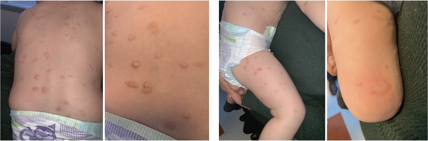
Skin examination revealed an eruption of edematous skin-colored to yellow-brown papules and plaques with surrounding erythema involving the face, chest, abdomen, back, and bilateral upper and lower extremities (Figure 1). Some lesions demonstrated development of wheals upon stroking (Figure 2). A punch biopsy was obtained.
What Is The Diagnosis?
Check your answer below.
Diagnosis
Urticaria Pigmentosa (Maculopapular Cutaneous Mastocytosis)
Urticaria pigmentosa (UP) is the most common cutaneous presentation of mastocytosis, a condition characterized by a pathologic expansion of clonal mast cells.1 Mastocytosis is divided into systemic mastocytosis and cutaneous mastocytosis, both of which may present with skin findings. Age of onset confers varying clinical presentation and prognosis.1 The World Health Organization classification of cutaneous mastocytosis includes diff use cutaneous mastocytosis, mastocytoma, or UP, also known as maculopapular cutaneous mastocytosis (MPCM).1,2 Systemic mastocytosis most often involves the bone marrow, but can involve the liver, spleen, and lymph nodes.
Mast cells develop and proliferate in response to activation of proto oncogene c-KIT (CD117), a transmembrane tyrosine kinase receptor, by KIT ligand (stem cell factor).3,4 Both adult mastocytosis and pediatric mastocytosis are associated with activating c-KIT mutations, but diff er with frequency of anaphylaxis, chronicity of disease, and cutaneous fi ndings.1,4,5 In pediatric-onset mastocytosis, risk of anaphylaxis is less than 10% compared with approximately 50% in adults.1,6 Furthermore, in pediatric patients, mastocytosis is more likely to resolve in adolescence, whereas mastocytosis in adults tends to be chronic.1,7-9 In adult-onset mastocytosis, there is increased risk of systemic disease and mast cell leukemia.10
Clinical Presentation
In UP, there are 2 main variants seen in different age groups. Adults typically display a monomorphic variant with small monomorphic hyperpigmented macules and papules, whereas pediatric patients typically display the polymorphic variant with larger tanto-brown polymorphic plaques or nodules. Lesions typically spare the face, palms, and soles.11 Spontaneous blistering can be seen.1 In the pediatric-onset population, most cases occur between birth and age 2 years.1
Lesions can be provoked by physical stimuli (temperature and pressure), emotional stress, and medications.1,12,13 The development of a wheal and erythema upon physical stroking is eponymously known as Darier sign and is thought to be due to release of histamine, leukotrienes, and prostaglandins from cutaneous mast cells.1,12,13
Laboratory Findings
An elevated serum tryptase level is associated with systemic involvement, and measurement can be used as a prognostic biomarker of disease course.14 Given that systemic mastocytosis is not common in pediatric populations, tryptase levels are typically less than 20 µg/L.1,14,15 Systemic symptoms include diarrhea and abdominal pain.11 Although internal organ involvement is not common in UP, clinicians should assess for possible extracutaneous involvement with physical exam or radiographic evaluation. Patients found to have organomegaly, tryptase greater than 20 µg/L, severe symptoms, or hematologic abnormalities should be referred to hematology for consideration of bone marrow biopsy.15
Histopathology
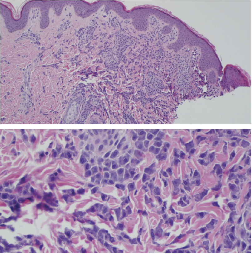
In lesions of UP, biopsies demonstrate diff use mast cells in the dermis, on average 4- to 8-fold higher than uninvolved skin.1 Polymorphic MPCM tends to have higher mast cell numbers compared with monomorphic MPCM. Detecting and quantifying mast cells can be aided by use of immunohistochemical stains with antibodies against tryptase or CD117, or histochemical stains such as Giemsa or toluidine blue.1,16 In patients with polymorphic MPCM, mast cells tend to be spherical with a fried egg appearance, while white cells in monomorphic MPCM tend to be spindle shaped.1 There may be purple granules within mast cells.17
Differential Diagnosis
Rarely, blisters may develop on UP lesions, raising clinical suspicion of bullous arthropod bite reaction and bullous impetigo. Obtaining a social history can clarify risk factors, such as exposure to arthropods or sick contacts, respectively. The wheals noted on patients with UP may suggest a physical urticaria, including dermographism; however, in UP the wheal is limited to the lesion. Nodular scabies can mimic the pruritic erythematous nodules of UP and can be differentiated by histologic examination of biopsied nodules showing primarily eosinophilic or mast cell infiltrates.18 Juvenile xanthogranuloma has a similar clinical appearance, but a positive Darier sign suggests UP.
Treatment
The primary objective of treatment is to decrease mast cell degranulation by avoiding triggers.12 Clinicians should educate patients, parents, and caregivers about avoidance of possible mast cell degranulating agents, including opioids such as morphine and codeine, muscle relaxants such as succinylcholine, and analgesics such as nefopam, which may directly or indirectly induce mast cell activation.19 Adverse events are rare in pediatric populations, but risks can be managed by incremental medication administration.20 Clinicians should also encourage use of medical alert bracelets.
Topical treatments include cromolyn sodium, topical steroids, and topical calcineurin inhibitors.11,12 Systemic therapy includes H1 receptor antagonists. H2 receptor antagonists can be used to manage gastric hypersecretion due to mastocytosis. There is limited evidence that systemic cromolyn sodium may be useful for pruritus.12
In patients with anaphylactic episodes, auto-injectable epinephrine pens should be available. Counseling should be provided about perioperative risks, especially because certain medications could trigger anaphylaxis. Depending on concern for systemic mastocytosis, a referral to hematology may be needed.
Our Patient
A punch biopsy was obtained, which showed diff use aggregates of mast cells in the upper dermis (Figure 3) consistent with mastocytosis. After the initial visit, the patient was lost to follow up despite numerous phone calls to the patient’s parents.
Conclusion
UP is a common presentation of mastocytosis, a rare disease. Diagnosis and management require a combination of history, physical examination, histologic examination, and laboratory testing, all of which can guide prognosis and determine the risk of systemic manifestations. Clinicians should be vigilant for signs of systemic mastocytosis due to increased risks of anaphylaxis. Avoidance of triggers and management of symptoms are mainstays of UP treatment.
References
1. Hartmann K, Escribano L, Grattan C, et al. Cutaneous manifestations in patients with mastocytosis: consensus report of the European Competence Network on Mastocytosis; the American Academy of Allergy, Asthma & Immunology; and the European Academy of Allergology and Clinical Immunology. J Allergy Clin Immunol. 2016;137(1):35-45. doi:10.1016/j.jaci.2015.08.034
2. Valent P, Akin C, Metcalfe DD. Mastocytosis: 2016 updated WHO classification and novel emerging treatment concepts. Blood. 2017;129(11):1420-1427. doi:10.1182/blood-2016-09-731893
3. Theoharides TC, Valent P, Akin C. Mast cells, mastocytosis, and related disorders. New Engl J Med. 2015;373(2):163-172. doi:10.1056/nejmra1409760
4. Longley BJ, Tyrrell L, Lu SZ, et al. Somatic c-KIT activating mutation in urticaria pigmentosa and aggressive mastocytosis: establishment of clonality in a human mast cell neoplasm. Nat Genet. 1996;12(3):312-314. doi:10.1038/ng0396-312
5. Bodemer C, Hermine O, Palmérini F, et al. Pediatric mastocytosis is a clonal disease associated with D 816 v and other activating c-KIT mutations. J Invest Dermatol. 2010;130(3):804-815. doi:10.1038/jid.2009.281
6. Brockow K, Jofer C, Behrendt H, Ring J. Anaphylaxis in patients with mastocytosis: a study on history, clinical features and risk factors in 120 patients. Allergy. 2008;63(2):226-232. doi:10.1111/j.1398-9995.2007.01569.x
7. Caplan RM. The natural course of urticaria pigmentosa: analysis and follow-up of 112 cases. Arch Dermatol. 1963;87(2):146-157. doi:10.1001/archderm.1963.01590140008002
8. Heinze A, Kuemmet TJ, Chiu YE, Galbraith SS. Longitudinal study of pediatric urticaria pigmentosa. Pediatr Dermatol. 2017;34(2):144-149. doi:10.1111/pde.13066
9. Ben-Amitai D, Metzker A, Cohen HA. Pediatric cutaneous mastocytosis: a review of 180 patients. Isr Med Assoc J. 2005;7(5):320-322.
10. Carter MC, Metcalfe DD, Komarow HD. Mastocytosis. Immunol Allergy Clin North Am. 2014;34(1):181-196. doi:10.1016/j.iac.2013.09.001
11. Macri A, Cook C. Urticaria pigmentosa. In: StatPearls [internet]. StatPearls Publishing; 2021. Accessed February 9, 2023. https://www.ncbi.nlm.nih.gov/ books/NBK482503
12. Castells M, Metcalfe DD, Escribano L. Diagnosis and treatment of cutaneous mastocytosis in children: practical recommendations. Am J Clin Dermatol. 2011;12(4):259-270. doi:10.2165/11588890-000000000-00000
13. Surjushe A, Jindal S, Gote P, Saple D. Darier’s sign. Indian J Dermatol Venereol Leprol. 2007;73(5):363-364. doi:10.4103/0378-6323.35751
14. Brockow K, Akin C, Huber M, Metcalfe DD. Assessment of the extent of cutaneous involvement in children and adults with mastocytosis: relationship to symptomatology, tryptase levels, and bone marrow pathology. J Am Acad Dermatol. 2003;48(4):508-516. doi:10.1067/mjd.2003.98
15. Carter MC, Clayton ST, Komarow HD, et al. Assessment of clinical findings, tryptase levels, and bone marrow histopathology in the management of pediatric mastocytosis. J Allergy Clin Immunol. 2015;136(6):1673-1679.e3. doi:10.1016/j. jaci.2015.04.024
16. Wolff K, Komar M, Petzelbauer P. Clinical and histopathological aspects of cutaneous mastocytosis. Leuk Res. 2001;25(7):519-528. doi:10.1016/S0145- 2126(01)00044-3
17. Rapini RP. Myeloproliferative disorders. In: Rapini RP, ed. Practical Dermatopathology. 3rd ed. Elsevier; 2021:341-364.
18. Mauleón-Fernandez C, Sáez-de-Ocariz M, Rodríguez-Jurado R, Durán- McKinster C, Orozco-Covarrubias L, Ruiz-Maldonado R. Nodular scabies mimicking urticaria pigmentosa in an infant. Clin Exp Dermatol. 2005;30(5): 595-596. doi:10.1111/j.1365-2230.2005.01832.x
19. Czarny J, Lange M, Ługowska-Umer H, Nowicki RJ. Cutaneous mastocytosis treatment: strategies, limitations and perspectives. Postepy Dermatol Alergol. 2018;35(6):541-545. doi:10.5114/ada.2018.77605
20. Carter MC, Uzzaman A, Scott LM, Metcalfe DD, Quezado Z. Pediatric mastocytosis: routine anesthetic management for a complex disease. Anesth Analg. 2008;107(2):422-427. doi:10.1213/ane.0b013e31817e6d7