What Caused These Yellowish, Thick Plaques?
© 2023 HMP Global. All Rights Reserved.
Any views and opinions expressed are those of the author(s) and/or participants and do not necessarily reflect the views, policy, or position of The Dermatologist or HMP Global, their employees, and affiliates.
Case Report
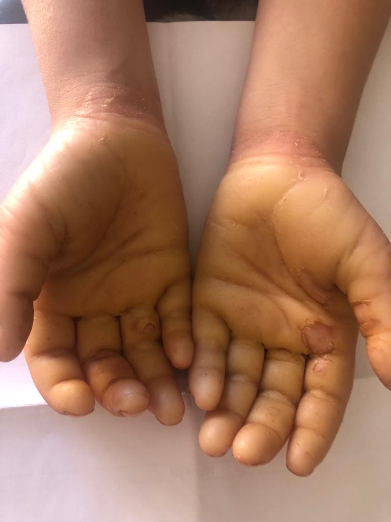
A 6-year-old female patient presented with recurrent, yellowish, thickened plaques on her bilateral palms and soles. Her parents noted that the plaques were visible immediately upon birth and initially limited to the palms and soles; however, they had recently begun to involve the dorsal aspects of the hands and feet, along with mild hyperhidrosis. The patient was born from a second-degree consanguineous marriage and her family history was positive for similar but milder manifestations in a younger sister. On physical examination, there were diffuse bilateral, palmoplantar hyperkeratotic plaques and erythema primarily on the ventral aspects of the palms and soles but extending partially onto the dorsal aspects (Figure). Significant malodor was appreciated. A punch biopsy from the left palm was performed.
What Is The Diagnosis?
Check your answer below!
Diagnosis
Mal de Meleda (MdM)
MdM, or keratosis palmoplantaris transgrediens, is a rare, hereditary dermatologic disease inherited in an autosomal recessive manner. This disease has an estimated worldwide prevalence of 1:100 000, although it may be underdiagnosed due to lack of availability of genetic testing. MdM was initially named after the island of Mljet (Meleda) in Croatia, where multiple cases were described in 1826.1 However, in recent decades, cases have been reported in a wide range of countries.1 Although over 26 gene mutations have been implicated in the pathogenesis of MdM, the main culprit mutation encodes a short nonglycosylated peptide named secreted Ly-6/uPar related protein-1 (SLURP1) located on the long arm of chromosome 8.1,2 This peptide has an immunomodulatory effect, suppressing tumor necrosis factor alpha and neutrophil activity.3
Clinical Presentation
MdM classically presents at birth or in early infancy with extensive erythema and exfoliating hyperkeratosis of the palms and soles. Plaques may extend to the dorsal surface of the feet and hands in a “gloves and stocking” mold, known as the transgrediens pattern. MdM can also exhibit increasingly severe lesions with age, which is known as the progrediens pattern.2
Nail abnormalities, such as onycholysis, koilonychia, and hyperkeratosis, are the most common associated features. Hyperhidrosis is also common, which often causes interdigital maceration along with an unpleasant fetid odor that can be psychologically debilitating while also increasing the risk of fungal infections.4
Affected fingers may also exhibit particular patterns, including fifth finger dysplasia, knuckle pads, constriction bands, flexion contractures (pseudoainhum), and spontaneous amputations.5 Oral manifestations of the disease, such as perioral erythema and lower lip angular cheilitis, have also been reported.2,6 There have also been cases of malignant melanoma arising within long-standing hyperkeratotic lesions.7,8
Differential Diagnosis
The identification of MdM may be challenging not only due to considerable phenotypic diversity, but also because of the clinical and genetic overlap with other members of the heterogenous palmoplantar keratoderma (PPK) group of diseases (Table 1). There is no unanimously accepted classification of hereditary PPK, but these conditions can be generally divided into 4 main categories. MdM is typically described as a diffuse, nonepidermolytic PPK.
Table 1. Major Categories of Hereditary PPK
| Type of PPK | Definition |
|---|---|
| Diffuse | Hyperkeratotic plaques that include the entire surface of palms and soles, which can be further subdivided into nontransgrediens pattern (sharp demarcation at the palmar or plantar surfaces) and transgrediens pattern (more extensive lesions involving other areas, such as the dorsal surfaces of the hands and feet, Achilles tendon area, elbows, and knees) |
| Focal | Painful hyperkeratotic plaques typically found on weight-bearing areas |
| Striate | Linear, hyperkeratotic lesions typically found along the flexor aspects of fingers and toes |
| Punctate | Small, circular hyperkeratotic papules usually found on the palms and soles, often described as a “raindrop” pattern |
In general, the combination of extensive erythema and exfoliating hyperkeratosis of the palms and soles, nail involvement, and hyperhidrosis should raise clinical suspicion for MdM. Histologically, the disease exhibits nonspecific findings, such as hyperkeratosis and acanthosis, with some cases exhibiting parakeratosis. Furthermore, perivascular lymphocytic dermal infiltrate has been reported as stigmata of upper dermis involvement. However, given that epidermolysis has not been described in this condition, the absence of epidermolysis can help distinguish MdM from epidermolytic subtypes of PPK.
There are several diffuse PPK syndromes that present with similar phenotypes and genetic mutations to MdM and are all inherited in an autosomal recessive manner (Table 2). Genetic testing combined with clinical findings are therefore necessary for precise diagnosis.2
Table 2. Focused Differential Diagnosis of MdM
| Syndrome Name | Features |
|---|---|
| Gamborg-Nielsen type PPK9 | The only hereditary PPK sharing the same genetic mutation (SLURP1) with MdM; however, it lacks nail involvement and any lesions outside of the palms and soles. |
| Nagashima type PPK10 | Hyperhidrosis is a common presenting finding; however, this syndrome has a mild and nonprogressive hyperkeratosis without extracutaneous manifestations and the causative gene (SERPINB7) is distinct from MdM. |
| SERPINA12 type PPK11,12 | Similar to Nagashima type; however, may have mutilating plaques with associated erythema, as well as tendon involvement. The causative gene is distinct from MdM. |
Management and Treatment
Once the diagnosis of MdM is established, therapeutic goals are based around symptom relief and quality of life maximization.13
Topical keratolytics, such salicylic acid and urea, are the mainstays of treatment. They can be applied under occlusion and combined with mechanical keratolysis, such as pumice stones or blades, to increase absorption.14 If these keratolytics are insufficient, topical retinoids can also be used. Topical steroids can be added to reduce associated erythema, dryness, or skin irritation.13
Systemic treatment with oral retinoids has been shown to be effective in the treatment of MdM. Multiple studies suggest that systemic retinoids (acitretin, isotretinoin, and alitretinoin) in doses of 0.5 to 1 mg/kg may result in a significant recovery from symptoms after 3 to 4 months of therapy.2,15,16 Combination of oral retinoids and topical therapy such as calcipotriol-betamethasone can also help improve side effect tolerance and compliance.16 The main adverse side effect associated with oral retinoids is skin dryness, which can be managed symptomatically. Oral retinoids are contraindicated in patients who are pregnant or considering pregnancy.
Other nonspecific treatments for MdM include symptomatic management such as prophylactic topical antifungals, orthotics to limit mechanical pressure, and surgery to prevent spontaneous amputation.13 In a recent study, nonepidermolytic PPK was treated effectively with blue LED light.17
Clinicians should carefully monitor patients for involvement of tendons and joints, and escalate therapy appropriately to avoid debilitating contractures.
Patients considering family planning may likely benefit from genetic counseling, and any first-degree relatives with appropriate syndromes can be referred for genetic testing. The symptoms of MdM can significantly affect a patient's quality of life, often resulting in serious psychological disability. Therefore, patients may also benefit from referral to mental health specialists.
Our Patient
Diagnosis was confirmed via clinical history and genetic testing. Given the relatively mild nature of disease, she was treated with topical keratolytics, as well as emollients. After 2 months, the erythema and hyperkeratosis improved significantly.
Conclusion
MdM is a rare, nonepidermolytic, diffuse PPK caused by SLURP1 mutations and inherited in an autosomal recessive fashion. It is characterized mainly by symmetric palmoplantar hyperkeratosis that progressively extends to the dorsal surfaces of the hands and feet, and associated hyperhidrosis and malodor that may worsen with age. Diagnosis involves a combination of clinical findings and genetic testing. There is no cure, and management focuses on symptomatic management and quality of life improvement. Topical emollients and keratolytics can be used in mild cases; however, patients with refractory or debilitating disease may need systemic therapy with oral retinoids or surgical intervention to avoid debilitating contractures.
References
1. Bakija-Konsuo A. Mal de Meleda—through history and today. Acta Dermatovenerol Croat. 2014;22(2):79-84.
2. Perez C, Khachemoune A. Mal de Meleda: a focused review. Am J Clin Dermatol. 2016;17(1):63-70. doi:10.1007/s40257-015-0157-1
3. Swamynathan S, Tiwari A, Loughner CL, et al. The secreted Ly6/uPAR-related protein-1 suppresses neutrophil binding, chemotaxis, and transmigration through human umbilical vein endothelial cells. Sci Rep. 2019;9(1):5898. doi:10.1038/s41598-019-42437-x
4. Marrakchi Z, Marrachi S, Meziou TJ, Turki H, Zahaf A. [Mal de Meleda. 16 cases]. Tunis Med. 2006;84(7):423-426.
5. Chhabra G, Verma P, Sharma S. Mal de Meleda palmoplantar keratoderma with pseudoainhum. Skinmed. 2021;19(5):383-384.
6. Nath AK, Chaudhuri S, Thappa DM. Mal de Meleda with lip involvement: a report of two cases. Indian J Dermatol. 2012;57(5):390-393. doi:10.4103/0019-5154.100497
7. Arousse A, Mokni S, H’mida Ben Brahim D, et al. Amelanotic melanoma arising in an area of SLURP-1 mutated Mal de Meleda. Int J Dermatol. 2019;58(8):966-968. doi:10.1111/ijd.14231
8. Mozzillo N, Nunziata CA, Caracò C, Fazioli F, Botti G, Melanoma Cooperative Group. Malignant melanoma developing in an area of hereditary palmoplantar keratoderma (Mal de Meleda). J Surg Oncol. 2003;84(4):229-233. doi:10.1002/jso.10317
9. Zhao L, Vahlquist A, Virtanen M, et al. Palmoplantar keratoderma of the Gamborg-Nielsen type is caused by mutations in the SLURP1 gene and represents a variant of Mal de Meleda. Acta Derm Venereol. 2014;94(6):707-710. doi:10.2340/00015555-1840
10. Huang C, Yang Y, Huang X, Zhou Z. Nagashima-type palmoplantar keratosis: clinical characteristics, genetic characterization, and clinical management. Biomed Res Int. 2021;2021:8841994. doi:10.1155/2021/8841994
11. Mohamad J, Sarig O, Malki L, et al. Loss-of-function variants in SERPINA12 underlie autosomal recessive palmoplantar keratoderma. J Invest Dermatol. 2020;140(11):2178-2187. doi:10.1016/j.jid.2020.02.030
12. Steele L, Tawfik SS, O’Toole EA. The proteolytic network in palmoplantar keratoderma: SERPINA12 joins the family. J Invest Dermatol. 2020;140(11):2111-2113. doi:10.1016/j.jid.2020.06.031
13. Bodemer C, Steijlen P, Mazereeuw-Hautier J, O’Toole EA. Treatment of hereditary palmoplantar keratoderma: a review by analysis of the literature. Br J Dermatol. 2021;184(3):393-400. doi:10.1111/bjd.19144
14. Schiller S, Seebode C, Hennies HC, Giehl K, Emmert S. Palmoplantar keratoderma (PPK): acquired and genetic causes of a not so rare disease. J Dtsch Dermatol Ges. 2014;12(9):781-788. doi:10.1111/ddg.12418
15. van de Kerkhof PC, van Dooren-Greebe RJ, Steijlen PM. Acitretin in the treatment of Mal de Meleda. Br J Dermatol. 1992;127(2):191-192. doi:10.1111/j.1365-2133.1992.tb08060.x
16. Pospischil I, Enzelsberger K, Gross S, Hoetzenecker W, Fischer TW. Mal de Meleda: diagnostic work-up and therapy with low-dose acitretin. Acta Derm Venereol. 2022;102:adv00758. doi:10.2340/actadv.v102.995
17. Lodi G, Sannino M, Cannarozzo G, Bennardo L, Nisticò SP. Non-epidermolytic palmoplantar keratoderma treated with blue LED light. Photodermatol Photoimmunol Photomed. 2022;38(6):614-615. doi:10.1111/phpp.12791