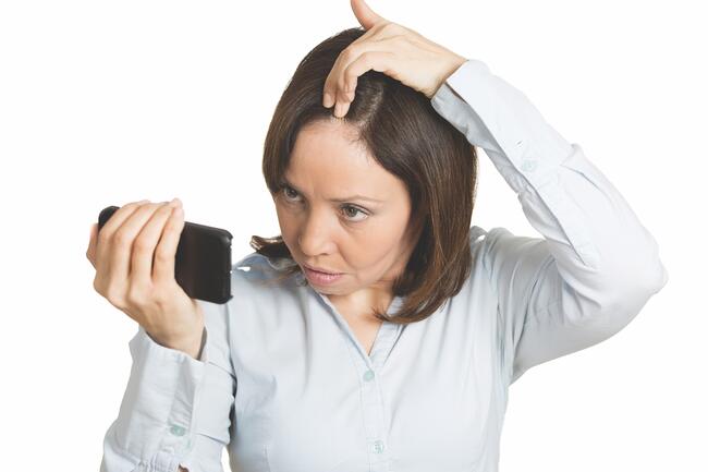
Reflectance Confocal Microscopy and Dermoscopy: Insights Into Linear Alopecia in Pediatric Patients
According to a recent study published in Skin Research and Technology, reflectance confocal microscopy (RCM) combined with dermoscopy can differentiate between alopecia areata (AA), trichotillomania (TM), nevus sebaceous (NS), and linear scleroderma en coup de sabre (LSCS).
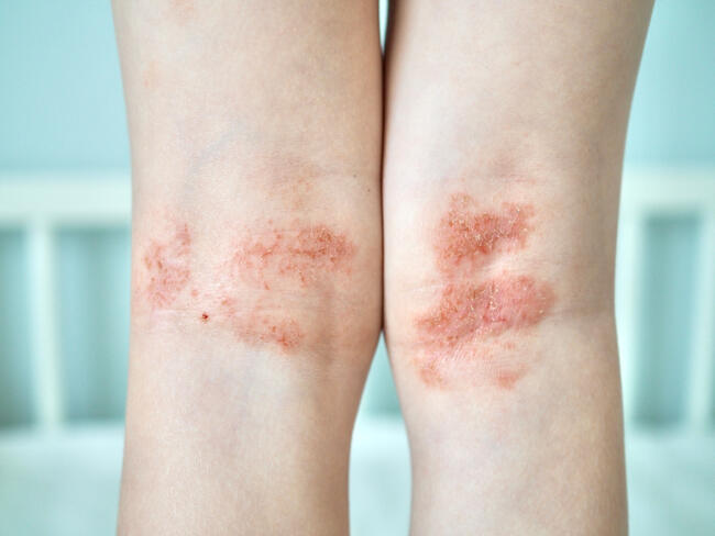
Researchers aimed to enhance the diagnostic accuracy of AA, TM, NS, and LSCS, all of which can manifest as linear alopecia, posing challenges for accurate diagnosis. They enrolled a total of 36 pediatric patients with linear alopecia, with 14 having AA, 7 with TM, 9 with NS, and 6 with LSCS. The research utilized RCM in conjunction with dermoscopy to evaluate and distinguish the characteristics of these conditions.
The key to differential diagnosis was found in various features observed through RCM and dermoscopy. For AA, a decreased hair follicle density in the dermis was identified, whereas TM exhibited normal-sized and dense follicular openings. NS presented with distinctive petal-like and frogspawn-like structures, and LSCS showed missing dermal papillary rings, sebaceous glands, and follicles, accompanied by abundant fibrous material in the dermis. Dermoscopic findings across the conditions included alopecia and reduced hair density. Patients with AA displayed yellow dots, black dots, and exclamation mark hairs, whereas patients with TM showed irregularly broken hairs and blood spots. Patients with NS and LSCS both exhibited an absence of follicular openings, with NS presenting whitish and yellowish round structures, and LSCS showcasing an atrophic area with white patches, linear vessels, and an absence of yellow or black dots.
“RCM combined with dermoscopy can provide additional information on disease states and differentiate between AA, TM, NS, and LSCS,” the authors concluded.
Reference
Wang Y, Chen L, Qin B, Li Q. Linear alopecia in pediatrics: RCM and dermoscopy reveal diagnostic cues. Skin Res Technol. 2023;29(11):e13523. doi:10.1111/srt.13523