


Descriptive Study of Ulcers Treated in a Diabetic Foot Unit
Abstract
Diabetic foot (DF) is one of the most prevalent complications in diabetic patients and an important cause of morbidity, mortality, and health costs. Multidisciplinary diabetic foot units (DFU) aim to improve the management of DF and reduce its complications. The objective of this report is to describe the type of patient received by our DFU, location and severity of their pathology and the main results of treatment. Material and Methods. This is a descriptive study of a cohort of patients treated at the DFU of the Vascular Department of the Hospital Universitario Fundación Alcorcón (HUFA) between November 2009 and September 2012. Every patient had a complete clinical evaluation that included vascular, neurological, and biomechanical evaluation. The number and location of the ulcer, the presence of ischemia or neuropathy, the TEXAS classification and the clinical course were prospectively recorded. Statistical analysis using the Chi square and ANOVA tests were used. Results. 288 patients were included, 239 of whom had a total of 404 ulcers. Of them, 58.4% were digital ulcers, 20.5% metatarsal, 5.4% midfoot and 15.6% heel ulcers. Regarding Texas Classification, 18.2% of the lesions were group A, 9.7% group B, 52.2% group C, and 20% group D. At follow-up, the healing rate was 77.2% and re-ulceration occurred in 35.1% instances. The minor amputation rate was 36.1%, major amputation 6.3%, and mortality 5.9%. Conclusion. Our experience in our DFU suggests that this multidisciplinary team can improve the results of DF ulcer management, reduce major amputation rates, and increase survival of our patients. Conclusion: Our experience in our DFU suggests that this multidisciplinary team can improve the results of DF ulcer management, reduce major amputation rates, and increase survival of our patients.
VASCULAR DISEASE MANAGEMENT 2021;18(5):E77-E86
Key words: diabetic foot, toe and flow, ulcer, diabetes mellitus
One of the most important complications of diabetes is diabetic foot (DF). Worldwide, the prevalence of DF is estimated between 1.3% and 4.8% of all patients diagnosed with Diabetes.1 During their lifetime, between 15% and 25% of diabetics will develop a DF ulcer, with an annual risk of ulceration in developed countries of 5% (25 times higher than the non-diabetic population).2 Once the ulcer heals, it is estimated that after 1, 3, and 5 years, 34%, 61% and 70% of the patients, respectively, will develop a new ulcer.3 A significant part of DF patients will require major or minor amputation (7% - 20%).4 Survival after having suffered a major amputation at 3 and 5 years is about 50%5 and 32%-40%, respectively.5,6 Furthermore, 5 years after a minor amputation, about 15% will undergo a major amputation of the same limb and, at least half of the patients, of the contralateral limb.7,8
This evidence underlines the importance of aggressive management of DF patients, aimed at reducing the incidence of ulcers and the prompt healing of existing ones, as well as reducing the rates of re-ulceration and amputation. The objective of this paper is to present the clinical characteristics of the patients received by our DFU in a period of 3 years, the techniques used to study these patients in our center, and the main results obtained at follow-up.
Materials and Methods
At the end of 2009, a diabetic foot unit (DFU) based on the “Toe and Flow” model was created at the HUFA.9 It was the first such unit in the whole region of Madrid, and the first in Spain that had the active participation of a podiatrist specialized in DF working as “gatekeeper”, included in our Vascular Surgery Service. The “Toe and Flow” model implies an intimate functional relationship between the podiatrist (Toe) and the vascular surgeon (Flow), and with whom other specialists can interact. Our DFU has 7 vascular surgeons, 1 podiatrist specialized in DF, and 2 nurses. Our DFU has a close collaboration with other specialists such as, the Internal Medicine Service, with its Surgical Patient Support Unit, Infectious Diseases Unit (with the antimicrobial treatment optimization program (PROA)), and the Endocrinology Unit. Each of them has a designated doctor that works actively with us, sharing some days of office work.
Study Design: This is a descriptive study of the activity and results of the DFU at the HUFA. We present a retrospective descriptive study on a prospectively maintained database on the evolution of ulcers treated in the DFU.
Inclusion Criteria: Patient with diabetes mellitus, with or without the presence of a DF ulcer, treated at the DFU of the HUFA during the three-year period between November 2009 and September 2012.
Data Collection and Variables: The data obtained in every visit was stored in a database designed for this purpose (MicrosoftTM AccessTM). On patient reception a complete medical history, including clinical, vascular and epidemiological data, diabetic course, and glycemic control over time and data on the ulcer (location, duration, complications, etc) were recorded in the database.
Ulcers were classified according to the Texas Classification, noting the presence or absence of probe to bone test and the presence of clinical signs of infection.10-12 We also recorded the results of microbiological cultures and antibiogram, antibiotic treatment, and type of wound therapy (including the use of negative pressure therapy and ozone therapy), and the reason for stopping the follow-up of every ulcer (major or minor amputation, or death). In the event that there was more than one ulcer at the same location, the one with the worst prognosis was recorded. We studied every new ulcer or re-ulceration that could develop during follow-up in the same way. The presence of deformities and previous amputations in the foot were recorded too.
Every patient had a neurologic and vascular screening. We evaluated the deep sensation using an ultraneurobiotensiometer (NBT) (NovalabIbérica SAL, Alcalá de Henares, Madrid, Spain) and superficial sensation using Semmes-Weinstein monofilament (MSW 5.07 10g) (NovalabIbérica SAL, Alcalá de Henares, Madrid, Spain). The presence of a Charcot foot was also noted.Vascular status was assessed by peripheral pulses palpation (pedal and posterior tibial) and ankle/brachial index (Super Dopplex II, Huntleigh healthcare Ltd., Cardiff, UK).
A complete biomechanical study consisting of joint biomechanical assessment, radiological study, and a static and dynamic computerized study of the gait (F-ScanTM (RsscanInternationalTM, Belgium) was performed in the first visit, in non-ulcerated patients, or once healed, in those with foot ulceration. According to the protocol, after the ulcer healed and the biomechanical study had been carried out, an individualized orthopedic treatment was established using footwear therapy, customized insole, and/or silicone orthosis, if indicated. Progress was monitored through protocolized biomechanical reviews. The patients received specific diabetes education with special emphasis on the complications that could occur in the foot, associated with adequate preventive measures.
Patients with ischemic or neuroischemic limbs after basic vascular screening, underwent a complete vascular examination with of Eco-Doppler (Envisoror iE33 Philips, Bothel WA, USA). If necessary, open or endovascular revascularization was attempted.
Statistical analysis: Categorical variables were described as percentages and compared with the Chi-Squared test, while for continuous variables we used the mean and standard deviation and comparison of means with the ANOVA test, respectively. The Bonferroni method was used for adjusting of the multiple comparisons.
Results
During the study period, 288 patients were treated in the DFU. Most of them were men (73.5%) with a mean age of 68.0 ± 12.4 years. Time from diagnosis of diabetes was 16.6 ± 12.1 years and the mean HbA1c values at the first visit was 7.5% ± 1.5%, reflecting poor glycemic control. Mean follow-up was of 509 ± 309 days and during this period patients were seen a mean of 18.1 +/- 19.9 times, for a total of 5205 visits. We performed 124 revascularization procedures (23.4% open, 71.8% endovascular, and 4.8% hybrid), 18 major amputations in 18 (6.3%) patients, 157 minor amputations in 104 (36.1%) patients and 17 patients (5.9%) died. Most of these minor amputations (80.3%) were at the digital level and 19.1% at the metatarsal level. During the study period, 83.0% of the patients (239) had at least one ulcer, of whom 217 had it on first visit while 22 developed it during the follow-up period, for a total of 404 ulcers (1.69 ± 0.98 ulcers per ulcerated patient) (Table 1). In 84 of the 239 patients with healed ulcers (35.1%) re-ulceration occurred during follow-up. Of the 404 ulcers, 154 (38.1%) were re-ulcerated, the talar location being significantly lower than the others (p<0.001). We had 154 re-ulcerations (154/404, 38.1%) distributed as follows: 65.6% digital, 22.1% metatarsal, 17.0% at the midfoot, and 4.5% at the heel (Table 1).
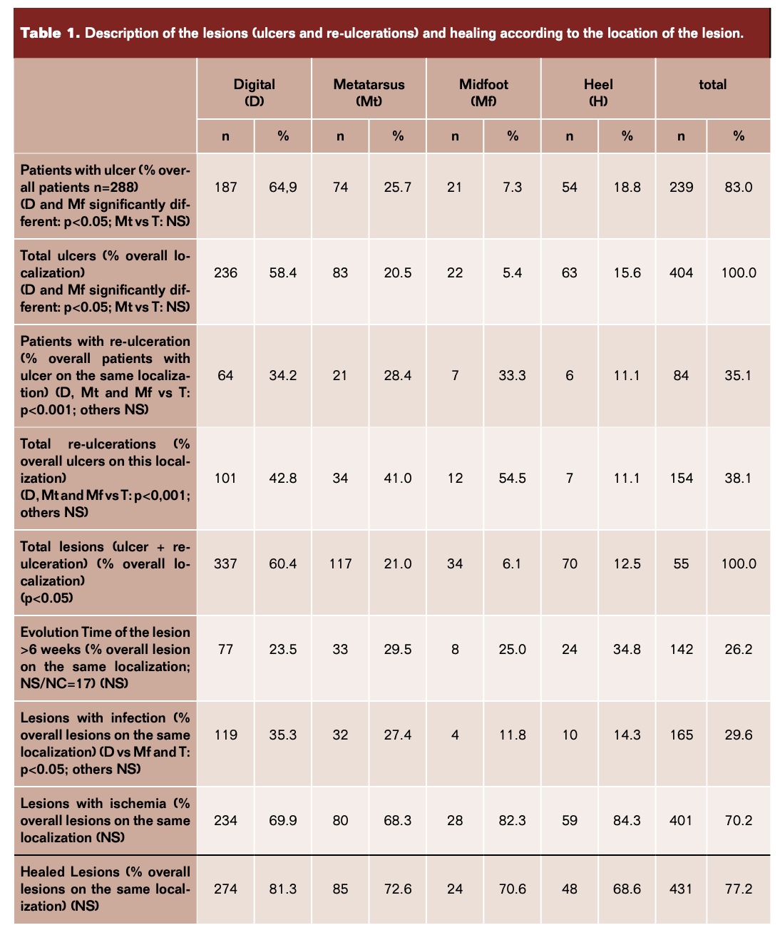
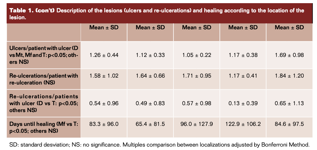
Table 1 shows the main characteristics of the ulcers (n=558, when joining initial ulcers and re-ulcerations): 60.4% were digital, 21% metatarsal, 6.1% midfoot, and 12.5% heel ulcers. When they were first received at the DFU, 26.2% had been present for more than 6 weeks, irrespective of the location in the foot; 29.6% had clinical signs of infection, more prevalent at the digital level, p<0.05; and 70.2% had data of ischemia, without significant differences between groups. During follow-up, 77.2% of the ulcers healed in 84.6 +/- 97.5 days, again without differences between groups.
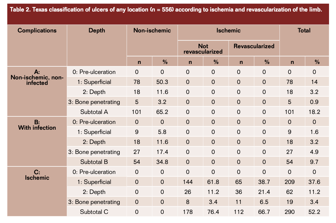
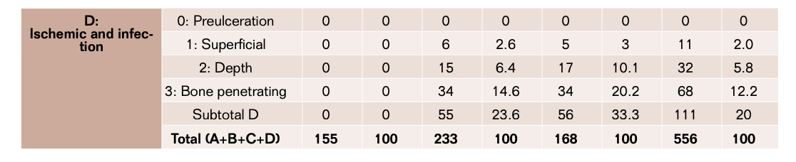
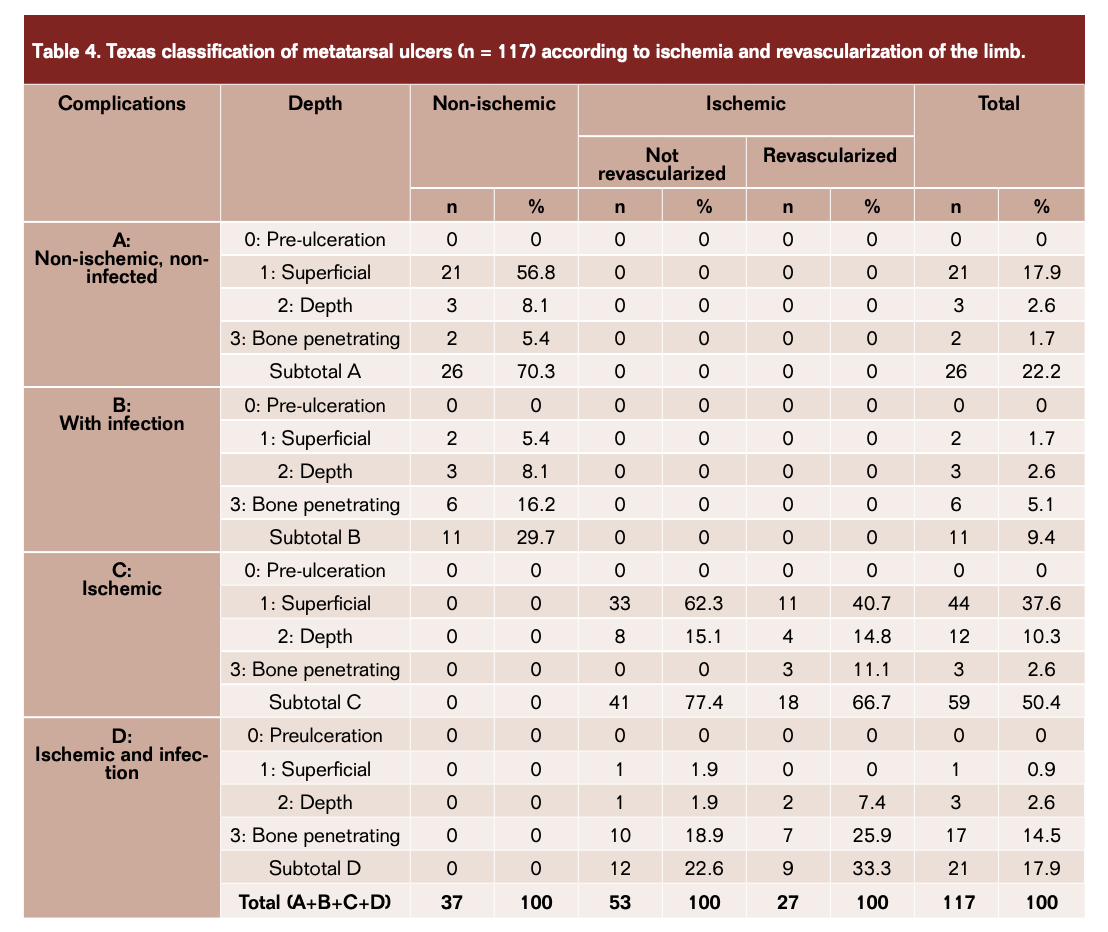
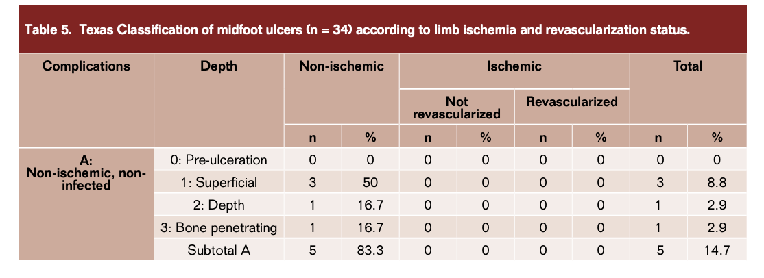
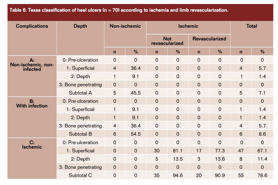
In Table 2 we show the distribution of ulcers according to the Texas Classification: 18.2% Texas A, 9.7% Texas B, 52.2% Texas C, and 20% Texas D. Besides, it shows the distribution of the ulcers in the absence or presence of ischemia and, in the latter, if they were revascularized or not. Tables 3 to 6 show these same data when the ulcers were present at the digital, metatarsal, midfoot, or heel level. We can see that digital and metatarsal ulcers have a similar Texas classification that it is very different from that at the midfoot and heel location, and that these last ones, in turn, are very similar between them.
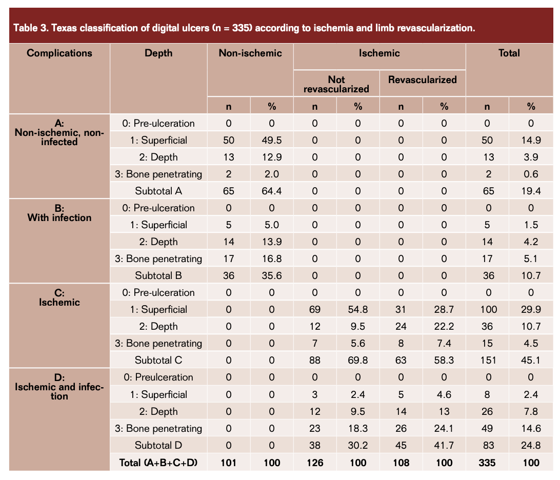
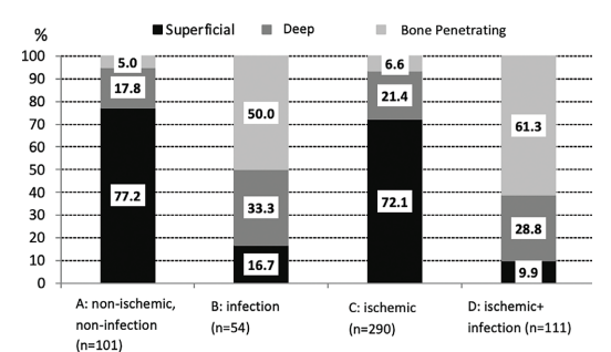
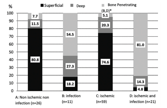
In Figure 1, we can see the distribution of the ulcers according to their depth and tissue involvement (A to D). We can observe that bone involvement is far more frequent in the presence of infection (50% in B and 61.3% in D, vs 5% in group A and 6.6% in group C) and that, when it is not present, the percentage of superficial ulcers is similar between groups, with or without ischemia (77.2% vs 72.1%). The depth of the ulcers is statistically similar in groups A and C and in groups B and D.
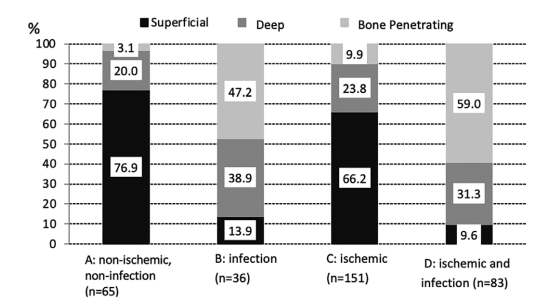
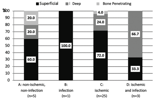
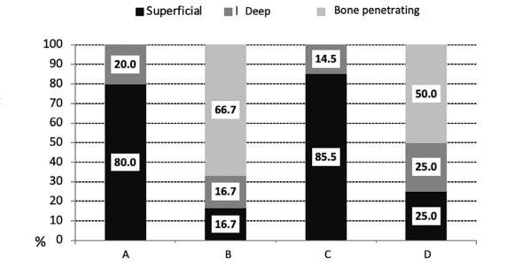
Figures 2 to 5 show that these data do not change significantly with the location of the ulcers and that, again, in each level, bone involvement is more frequent in the presence of infection.
Discussion
The prevalence of diabetes worldwide is estimated at 300 million, and by 2030 it is projected to increase dramatically to 425 million.1 Foot ulceration will develop in up to 25% of the patients with diabetes during their lifetime.13 Diabetic foot ulcer is a heterogeneous disease that continues to be a major problem both for the patient and for health care systems.14 As much as 85% of amputations are preceded by a foot ulcer, which creates a gateway for microorganisms and leads to infection.15 On many occasions, there is a late referral of these patients to specialized care, further worsening the prognosis of their disease.16-18 In addition to the risk of complications, such as infection or amputation, a diabetic foot ulcer has an important effect on the economic and social life of patients.1,19 The survival of patients who have undergone a major amputation is diminished when compared with patients in whom it has been possible to save the limb.20 Therefore, not only productivity and quality of life decrease, but life expectancy is also reduced after a major amputation of the lower limbs.21
In recent years, many studies have been published on the management of DFU. The increased prevalence of neuroischemic ulcers and their difficult treatment, have changed the management of this disease. Vartanian et al, published in 2015 a retrospective follow-up of 89 patients with neuroischemic wounds, with specific differences with our study, but comparable: it was a shorter follow-up time (16 months vs mean 509 ± 309 days), more young people (60.4 years vs mean age 68.4 ± 12.4 years), more chronic wounds (>6 weeks: 67% vs 26.2%), more endovascular procedures in our series (50% vs 71.8%) and different wound location (41% vs 78.9% toes and forefoot and 24% vs 15.6% hindfoot).22 The aggressive treatment of peripheral arterial disease (PAD), within he so-called “Foot Attack” protocol, has been one of the milestones of this preventive management.23 Successful revascularization has been shown to reduce the rate of major amputation in diabetic patients with critical limb ischemia.24, 25 Faglia et al25 published in 2007 data from a cohort study of 564 consecutive diabetic patients from January 1999 to December 2003; the mean age and diabetes duration was similar to our study (67.3 ± 10.1 and 17.0 ± 11.1 years, respectively), endovascular procedures were performed in 74.5% patients, similar in our study too (71.8%), but the major amputations were 9.8% vs 6.3% in our study. Alexandrescu et al have shown a decreased amputation rate in their unit using a percutaneous revascularization strategy.26 However, there is continued risk of amputation even in successfully revascularized patients, both in the short and long term during follow-up, for several reasons: 1) incomplete revascularization that does not obtain direct blood flow to the foot27 2) established gangrene that may require removal of a large part of the foot that make rehabilitation impossible28 3) the presence of untreatable restenosis in patients undergoing endovascular treatment 4) bypass occlusion in cases of open surgery.29
t
The location of the wound stratifies the chances of successful healing too. Pickwell et al30 have shown, related to the results of the Eurodiale Study, that the healing time of diabetic foot ulcers is stratified according to the location of the ulcer: those located at the digital and forefoot level have a greater healing probability, and wounds susceptible to partial amputation offer the faster healing rate once it is performed. Comparing both studies, the complete healing achieved was 59% at 12 weeks vs 77.2% in an average time of 84.6 ± 97.5 days in our study.
The estimated cost of foot ulcers and amputations to the US health care system was about 29 billion dollars in 2007.31 In a descriptive analysis of the relationship between regional on vascular care and the rate of amputation,32 it was shown that the costs of hospital care in the year prior to amputation exceeded $20,000 per patient for hospital care alone. But after the amputation, the costs do not stop.33 Several studies suggest that enhanced vascular care, measured by any type of diagnostic or therapeutic vascular procedure in the year prior to amputation, is related to lower risks of amputation and diminished the future in-patient time.34,35 A team of professionals who are super-specialized in the management of the diabetic foot is required to prevent the amputation of the lower extremities in people with diabetes. The Diabetic Rapid Response Acute Foot Team (DRRAFT) guidelines suggest that the vascular surgeon and the diabetic podiatrist constitute a minimum in a diabetic foot team.36 A single-center hospital in the US reduced major amputations by 72% after implementing a process of care focused on podiatry and vascular services.32 Other previous experiences have shown that the implementation of multidisciplinary teams has reduced amputations in the treatment of DF, reaching a reduction of amputations of more than 50% in hospitals that have introduced multidisciplinary teams for the care of the feet.37 Furthermore, by reducing costly amputations, such equipment can save more than four times its cost.38 The results obtained by our DFU compare favorably with those published in the international literature, despite the fact that in our series the patients seem to have a more marked vascular compromise than in others. Aggressive revascularization therapy may justify these very encouraging results.
Conclusion
Our initial experience with a multidisciplinary team for the preservation of extremities in the diabetic patient (DFU) suggests that wound healing and improvement in quality of life is possible and can be achieved in the majority of patients with high-risk diabetic foot. The care required for these patients often requires multiple invasive procedures, hospital care, and outpatient visits. Our retrospective study on a prospectively maintained data base, and several reports by others, have shown reductions in major amputations and foot complications with multidisciplinary cooperation, suggesting that better communication and organization with significant enthusiasm makes a difference. Our initial experience reflects that of others, in that the amputation prevention model is not only clinically effective, but also financially viable.
Acknowledgment
Abbott Vascular initially funded the creation of the Diabetic Foot Unit at Vascular Surgery Department, University Hospital Foundation Alcorcón.
Corresponding Author: Sandra Vicente Jiménez; Email: Svicente@fhalcorcon.com
REFERENCES
1. Boulton AJM, Vileikyte L, Ragnarson-Tennvall G, Apelqvist J. The global burden of diabetic foot disease. Lancet. 2005;12;366(9498):1719-1724.
2. Reiber Ge, LeMaster JW. Epidemiology and Economic impact of foot ulcers and amputations in people with diabetes. In: Bowker JH, Pfeifer MA, eds. Levin and O’Neal’s The Diabetic Foot. Philadelphia. Mosby, Elsevier. 2008: 3-22.
3. Glover JL, Weingarten MS, Buchbinder DS, Poucher RL, Deitrick GA, 3rd, Fylling CP. A 4-year outcome-based retrospective study of wound healing and limb salvage in patients with chronic wounds. Adv Wound Care. 1997;10(1):33-38.
4. Larsson J, Agardh CD, Apelqvist J, Stenström A. Long-term prognosis after healed amputation in patients with diabetes. Clin Orthop Relat Res. 1998;(350):149-158.
5. Sumpio BE, Armstrong DG, Lavery LA, Andros G, SVS/APMA writing group. The role of interdisciplinary team approach in the management of the diabetic foot: a joint statement from the Society for Vascular Surgery and the American Podiatric Medical Association. J Vasc Surg. 2010;51(6):1504-1506.
6. Levin ME. Preventing amputation in the patient with diabetes. Diabetes Care. 1995;18(10):1383-1394.
7. Izumi Y, Satterfield K, Lee S, Harkless LB. Risk of reamputation in diabetic patients stratified by limb and level of amputation: a 10-year observation. Diabetes Care. 2006;29(3):566-570.
8. Reiber GE, Smith DG. Lower extremity foot ulcers and amputations in diabetes. In: Harris MI CC, Stern MP. Diabetes in America1995. p. 409-27.
9. Rogers LC, Andros G, Caporusso J, et al. Toe and flow essential components and structure of the amputation prevention team. J Am Podiatr Med Assoc. 100(5):342-348.
10. Armstrong DG, Lavery LA, Harkless LB. Validation of a diabetic wound classification system. The contribution of depth, infection, and ischemia to risk of amputation. Diabetes Care. 1998;21(5):855-859.
11. Grayson ML, Gibbons GW, Balogh K, Levin E, Karchmer AW, et al. Probing to bone in infected pedal ulcers. A clinical sign of underlying osteomyelitis in diabetic patients. JAMA. 1995;273(9):721-723.
12. Lipsky BA, Berndt AR, Cornia PB, et al. Infectious Diseases Society of America clinical practice guideline for the diagnosis and treatment of diabetic foot infections. Clin Infect Dis. 2012;54(12):e132-173.
13. Singh N, Armstrong DG, Lipsky BA. Preventing foot ulcers in patients with diabetes. JAMA. 2005;293(2):217-228.
14. Elgzyri T, Larsson J, Thörne J, Eriksson K-F, Apelqvist J. Outcome of ischemic foot ulcer in diabetic patients who had no invasive vascular intervention. Eur J Vasc Endovasc Surg. 2013;46(1):110-117.
15. Pecoraro RE, Reiber GE, Burgess EM. Pathways to diabetic limb amputation. Basis for prevention. Diabetes Care. 1990;13(5):513-521.
16. Manu C, Lacopi E, Bouillet B, et al. Delayed referral of patients with diabetic foot ulcers across Europe: patterns between primary care and specialised units. J Wound Care. 2018;27(3):186-192.
17. García-Klepzig JL, Sánchez-Ríos JP, Manu C, et al. Perception of diabetic foot ulcers among general practitioners in four European countries: knowledge, skills and urgency. J Wound Care. 2018;27(5):310-319.
18. Sánchez-Ríos JP, García-Klepzig JL, Manu C, et al. Referral of patients with diabetic foot ulcers in four European countries: patient follow-up after first GP visit. J Wound Care. 2019;28(Sup8):S4-S14.
19. Bus SA, Lavery LA, Monteiro-Soares M, et al. Guidelines on the prevention of foot ulcers in persons with diabetes (IWGDF 2019 update). Diabetes Metab Res Rev. 2020;36 Suppl 1:e3269.
20. Larsson J, Agardh CD, Apelqvist J, Stenström A. Long-term prognosis after healed amputation in patients with diabetes. Clin Orthop Relat Res. 1998;350:149-158.
21. Wu SC, Driver VR, Wrobel JS, Armstrong DG. Foot ulcers in the diabetic patient, prevention and treatment. Vasc Health Risk Manag. 2007;3(1):65-76.
22. Vartanian SM, Robinson KD, Ofili K, et al. Outcomes of neuroischemic wounds treated by a multidisciplinary amputation prevention service. Ann Vasc Surg. 2015;29(3):534-542.
23. Hinchliffe et al. Guideline on diagnosis, prognosis and management of peripheral artery disease among people with diabetes (IWGDF 2019 update). DiabMetab Res Rev. 2020. e3276
24. Faglia E, Clerici G, Caminiti M, Quarantiello A, Curci V, Morabito A. Predictive values of transcutaneous oxygen tension for above-the-ankle amputation in diabetic patients with critical limb ischemia. Eur J Vasc Endovasc. 2007;33(6):731-736.
25. LoGerfo FW, Gibbons GW, Pomposelli FB, et al. Trends in the care of the diabetic foot. Expanded role of arterial reconstruction. Arch Surg. 1992;127(5):617-621.
26. Alexandrescu V, Hubermont G, Coessens V, et al. Why a multidisciplinary team may represent a key factor for lowering the inferior limb loss rate in diabetic neuro-ischaemic wounds: application in a departmental institution. Acta Chir Belg. 2009;109(6):694-700.
27. Pound N, Chipchase S, Treece K, Game F, Jeffcoate W. Ulcer-free survival following management of foot ulcers in diabetes. Diabet Med. 2005;22(10):1306-1309.
28. Ryer EJ, Trocciola SM, DeRubertis B, et al. Analysis of outcomes following failed endovascular treatment of chronic limb ischemia. Ann Vasc Surg. 2006;20(4):440-446.
29. Nowygrod R, Egorova N, Greco G, et al. Trends, complications, and mortality in peripheral vascular surgery. J Vasc Surg. 2006;43(2):205-216.
30. Pickwell KM, Siersma VD, Kars M, Holstein PE, Schaper NC. Diabetic foot disease: impact of ulcer location on ulcer healing. Diabetes Metab Res Rev. 2013;29(5):377-383.
31. Goodney PP, Travis LL, Brooke BS, et al. Relationship between regional spending on vascular care and amputation rate. JAMA Surg. 2014 January; 149(1): 34–42
32. Philip P. Goodney, MD, MS, Kerianne Holman, et al.Regional intensity of vascular care and lower extremity amputation rates. J Vasc Surg. 2013 June; 57 (6): 1471-80. E3.
33. Holman KH, Henke PK, Dimick JB, Birkmeyer JD. Racial disparities in the use of revascularization before leg amputation in medicare patients. J Vasc Surg. 2011; 54:420–426. 426, e421.
34. Ho V, Wirthlin D, Yun H, Allison J. Physician supply, treatment, and amputation rates for peripheral arterial disease. J Vasc Surg. 2005; 42:81–87.
35. Van Gils CC, Wheeler LA, Mellstrom M, Brinton EA, Mason S, Wheeler CG. Amputation prevention by vascular surgery and podiatry collaboration in high-risk diabetic and nondiabetic patients. The operation desert foot experience. Diabetes Care. 1999; 22:678–683.
36. Fitzgerald, RH;Mills JL, et al. The Diabetic Rapid Response Acute Foot Team:Essential Skills for Targeted Limb Salvage. E Plasty. May 5, 2009. Vol 9.
37. Edmonds et al. Improved Survival of the Diabetic Foot The Role of a Specialised Foot Clinic –Quarterly Journal of Medicine, New Series 60, No. 232, pp. 763-771, 1
38. M. Kerr, G. Rayman, W. J. Jeffcoate. Research: Health Economics Cost of diabetic foot disease to the National Health Service in England. Diabet Med 31, 1498-1504 (2014)
