


Management of Thrombosed Contralateral Common Iliac Vein Secondary to Wallstent Extension Into the Inferior Vena Cava
VASCULAR DISEASE MANAGEMENT 2020;17(4):E63-E66.
Abstract
Jailing of the contralateral iliac vein may occur as a result of the iliac vein stent protruding into the inferior vena cava (IVC). Endothelialization and fibrotic tissue growth over the stent can lead to obstruction of flow to the contralateral iliac vein and subsequent development of acute deep venous thrombosis (DVT). Management of such a complication is a necessary skill for the endovascular operator. The advent of new, dedicated venous stents with more accurate deployment at the confluence is likely to reduce such a complication.
Introduction
Managing venous disease has evolved over the past two decades and catheter-based therapy is now the dominant method of treatment in these patients.1 Stenting of the common iliac vein (CIV) is considered the primary method of treatment in symptomatic patients.2 New, dedicated stents have been recently approved. Venous stents are made of nitinol, providing good radial strength and more accurate deployment at the confluence.3,4
The Wallstent (Boston Scientific) has been the main stent used off label to treat iliac vein compression. These stents are difficult to deploy accurately at the intended target, have a lower radial force, and carry the risk of migration. Quite often, when these stents are deployed, they are unintentionally released far into the IVC and therefore cross over the origin of the contralateral common iliac vein. Previously, extending the stent to the IVC wall to prevent its migration has been recommended.5 Although the majority of the time this does not have significant consequences, we present a case of contralateral iliac vein thrombosis occurring as a result of extending the Wallstent far into the IVC across the origin of the CIV.
Case Report
A 37-year-old female presented with swelling involving the right lower extremity for almost 5 days that was progressively getting worse, and was associated with numbness and pain. She had a history of left CIV DVT 2 years prior to her presentation and had a 16 mm Wallstent placed. She was placed on oral anticoagulation. The latter was discontinued 1 year after a back surgery (L4/L5 fusion) and was never resumed. She had a positive family history of DVT, was an active smoker, and had no recent surgery or travel. Upon presentation, Doppler ultrasound (DUS) showed extensive DVT involving the right lower extremity, with an occlusion extending from the popliteal vein to the CIV.
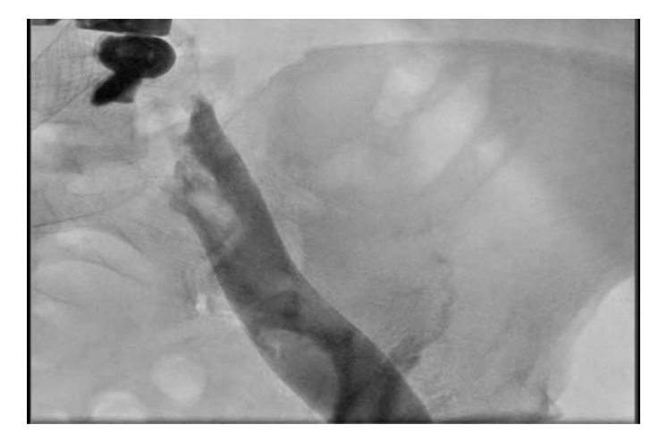
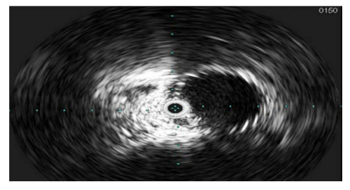
The patient was placed in a prone position and access via the ipsilateral right popliteal vein was obtained. A Destination sheath (Terumo) was advanced over a Wholey wire (Medtronic) to the area of the thrombus (Figure 1). The wire was advanced through the occluded thrombus in the right CIV, with care taken in order to avoid entering through the contralateral stent struts that extended into the IVC. Intravascular ultrasound (IVUS) was used to confirm the location of the wire between the wall of the IVC and the Wallstent (Figure 2).
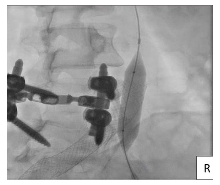
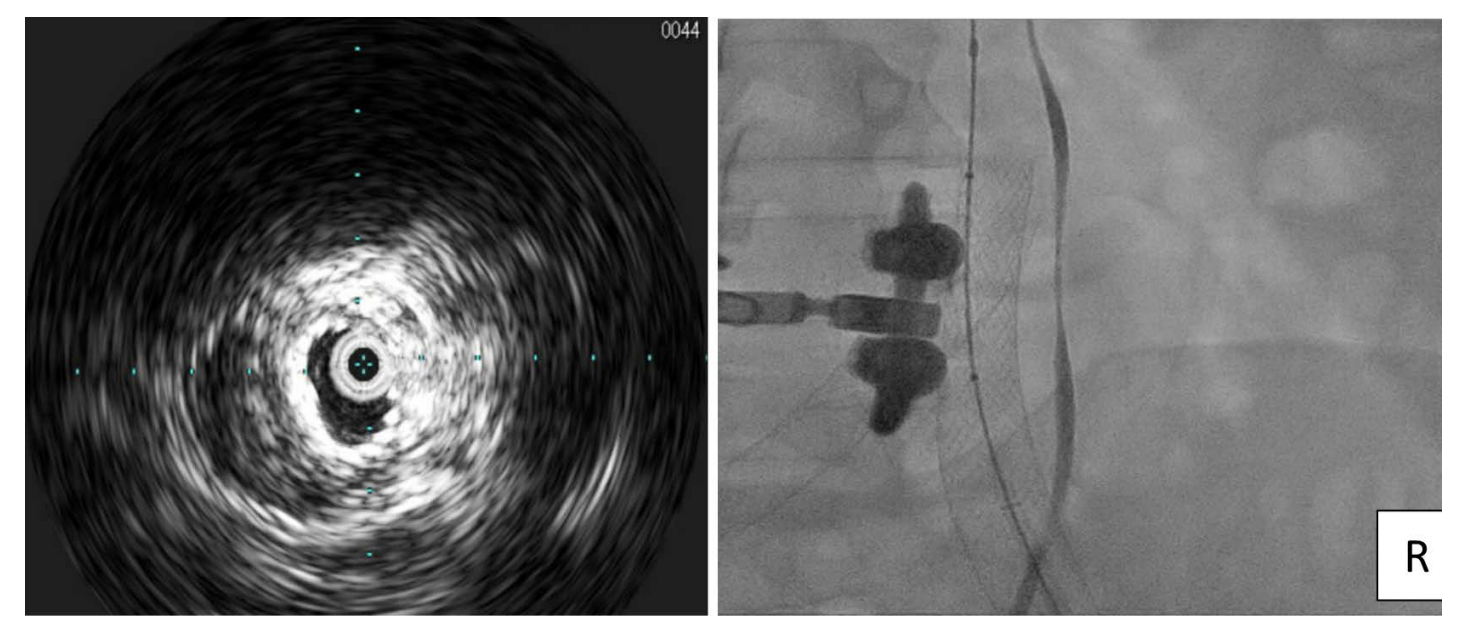
The extensive thrombus in the right CIV was treated with embolectomy using an AngioJet ZelanteDVT thrombectomy catheter (Boston Scientific) after power-pulse spray with tissue plasminogen activator (tPA). Post embolectomy, dilation with an 8 mm Atlas Gold Balloon (BD) at 5 atmospheres (atm) was followed by dilation with a 12 mm Atlas Gold balloon at 4 atm (Figure 3). A 20 x 80 mm WallStent was advanced into the ipsilateral iliac vein and into the IVC adjacent to the previously placed left CIV stent, then post dilated with the 14 mm balloon. The 20 mm diameter of the Wallstent was chosen because the contralateral stent placed in left CIV was measured at 16 mm. IVUS images showed stents adjacent to one another (Figure 4). Popliteal and femoral vein pressure dropped from 48 mmHg at baseline to 18 mm post stenting. Angiography confirmed excellent flow in both the left and right CIV stents (Video 1). The patient was discharged on aspirin and apixaban. At a one-month clinic visit, resolution of pain and swelling had already occurred.
Discussion
Contralateral DVT after common iliac vein (CIV) stent placement has been reported in 1 to 9.7% of patients.6,7 Extension of stenting far into the IVC appears to be a risk factor.6,7 Khairy et al identified factors associated with contralateral DVT after iliac vein stenting and reported that noncompliance with anticoagulation, presence of IVC filter, history of DVT, and acuity of the thrombosis are predictors of the occurrence of DVT.8 Murphy et al have reported a comparison between the Z-stent (Cook Medical) extension technique and extending the Wallstent in the IVC, with higher contralateral DVT noted with the Wallstent. The Z-stent has wider open cells, which may explain the difference between the two stents.9
The following are important learning points in managing a thrombotic occlusion of a CIV secondary to a protruding, contralateral Wallstent into the IVC.
- IVUS is important to ascertain that the wire crossing the occlusion is outside the contralateral stent struts and in-between the IVC and the stent itself.
- Reducing clot burden before definitive treatment is important and often requires the use of a lytic agent.
- The choice of stent in these scenarios is limited to non-nitinol stents in order to avoid use of the new venous stents that carry a high radial force and theoretically may compromise the contralateral old stent.
- Use of the Atlas Gold balloon for Wallstent dilation is recommended, as its noncompliant characteristics allow dilatation at high pressure without overstretching the vessel and its short shoulders minimize dilation beyond the stent edges.10
- Use of IVUS post stent placement is important to ensure adequate lumen gain, which correlates with stent patency.
- Ensuring patient compliance with anticoagulation in a thrombotic occlusion even after stent placement is key to preventing future thrombotic events.
Using the new, dedicated venous stents will likely lead to high patency rates and more importantly, provide the ability to position these stents more accurately at the confluence, avoiding excessive protrusion into the IVC.11 Given the unpredictability in accurately deploying the Wallstent, it is anticipated that dedicated venous stents will take over the treatment of iliac vein compression. However, sizing constraints of some of the new nitinol stents may preclude their use in large-diameter vessels.
Disclosure: Drs Radaideh and Robken report no conflicts of interest regarding the content herein. Dr Shammas reports that he receives research and educational grants from Boston Scientific, Intact Vascular, Philips, VMG, and BD.
Manuscript submitted December 27, 2019, final version accepted February 21, 2020.
Address for correspondence: Qais Radaideh, MD, Midwest Cardiovascular Research Foundation, 1622 E. Lombard Street, Davenport, Iowa 52803. Tel. +1.563.324.2828. Email: qaisrad89@gmail.com
REFERENCES
1. Radaideh Q, Patel NM, Shammas NW. Iliac vein compression: epidemiology, diagnosis and treatment. Vasc Health Risk Manage. 2019;15:115-122.
2. Rossi FH, Kambara AM, Izukawa NM, Rodrigues TO, Rossi CB, Sousa AG, et al. Randomized double-blinded study comparing medical treatment versus iliac vein stenting in chronic venous disease. J Vasc Surg Venous Lymphat Disord. 2018;6(2):183-191.
3. Rizvi SA, Ascher E, Hingorani A, Marks N. Stent patency in patients with advanced chronic venous disease and nonthrombotic iliac vein lesions. J Vasc Surg Venous Lymphat Disord. 2018;6(4):457-463.
4. Abou Ali AN, Avgerinos ED, Chaer RA. Role of venous senting for iliofemoral and vena cava venous obstruction. Surg Clin North Am. 2018;98(2):361-371.
5. Neglen P, Raju S. Balloon dilation and stenting of chronic iliac vein obstruction: technical aspects and early clinical outcome. J Endovasc Ther. 2000;7(2):79-91.
6. Le TB, Lee TK, Park KM, Jeon YS, Hong KC, Cho SG. Contralateral deep vein thrombosis after iliac vein stent placement in patients with May-Thurner syndrome. J Vasc Interv Radiol. 2018;29(6):774-780.
7. Mandel JE, Ostrozhynskyy Y, Hingorani A, Marks N, Ascher E. Underexpansion of Wallstents(R) in the treatment of nonthrombotic iliac vein lesions. Ann Vasc Surg. 2018;52:163-167.
8. Khairy SA, Neves RJ, Hartung O, O'Sullivan GJ. Factors associated with contralateral deep venous thrombosis after iliocaval venous stenting. Eur J Vasc Endovasc Surg. 2017;54(6):745-751.
9. Murphy EH, Johns B, Varney E, et al. Deep venous thrombosis associated with caval extension of iliac stents. J Vasc Surg Venous Lymphat Disord. 2017;5(1):8-17.
10. Shammas NW, Shammas GA, Jones-Miller S, Radaideh Q. Safety of the Atlas Gold balloon in treating iliofemoral veins: experience from a single center. J Invasive Cardiol. 2018 Nov;30(11):401-405.
11. Lichtenberg MKW, de Graaf R, Stahlhoff WF, Ozkapi A, Rassaf T, Breuckmann F. Venovo venous stent in the treatment of non-thrombotic or post-thrombotic iliac vein lesions - short-term results from the Arnsberg venous registry. Vasa. 2019;48(2):175-180.
