


Multidisciplinary Management of Cervical Ectopic Pregnancy: A Report of Two Cases
VASCULAR DISEASE MANAGEMENT 2023;20(5):E83-E87
Abstract
Cervical ectopic pregnancy (CP) is a rare but potentially life-threatening cause of obstetric-related hemorrhage. Medical and surgical treatment options are available; however, due to the inability of the cervical stroma to contract, there remains a risk for significant bleeding. Classically, CP has been managed by obstetricians alone, but there is a growing role for vascular interventional radiology in minimizing blood loss while preserving fertility in these patients. While uterine artery embolization has been used in various cases of peripartum hemorrhage, lone use of intra-arterial occlusion balloons is rarely described in the literature. Two cases are presented of patients who had minimal intraoperative blood loss secondary to interventional radiology placement of intra-arterial occlusion balloons prior to evacuation of CP. Standardized protocols for multidisciplinary management of CPs can lead to efficient treatment and improve patient outcomes.
Introduction
Cervical ectopic pregnancies (CPs) occur when the blastocyst implants abnormally along the endocervical canal and make up approximately 0.1% of all ectopic gestations.1 Most commonly, patients present with painless bleeding in the late first trimester or early second trimester.2,3 Hemorrhage is difficult to control as the cervical stroma does not contract in the same manner as the uterine myometrium and is therefore unresponsive to uterotonics.4 Mortality associated with hemorrhage from CPs has declined, largely due to advances in sonography allowing earlier diagnosis and advancement of treatment options; however, there can still be significant morbidity from blood loss.4,5 Presented here are 2 cases of cervical ectopic pregnancy that were jointly managed by obstetrics and interventional radiology (IR) in our institution.
Case Report 1
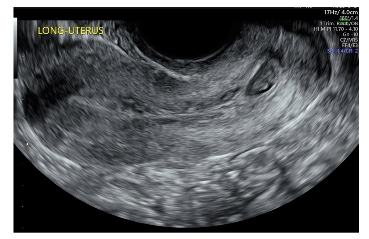
A 29-year-old woman (gravida 3, para 0020) presented to her outpatient obstetrician/gynecologist with heavy vaginal bleeding following a positive home urine pregnancy test. Beta human chorionic gonadotropin (β-hGC) measured 1269.7 mIU/mL. An ultrasound was performed and demonstrated CP measuring approximately 6 weeks’ gestational age with cardiac activity present (Figure 1). Her prior pregnancies included a biochemical pregnancy followed by complete molar gestation, treated with dilation and curettage (D&C). She was subsequently sent to the hospital for intervention. The patient was offered both D&C with vascular IR (VIR) support as well as systemic methotrexate with intra-sac potassium chloride injection, from which she chose the former option.
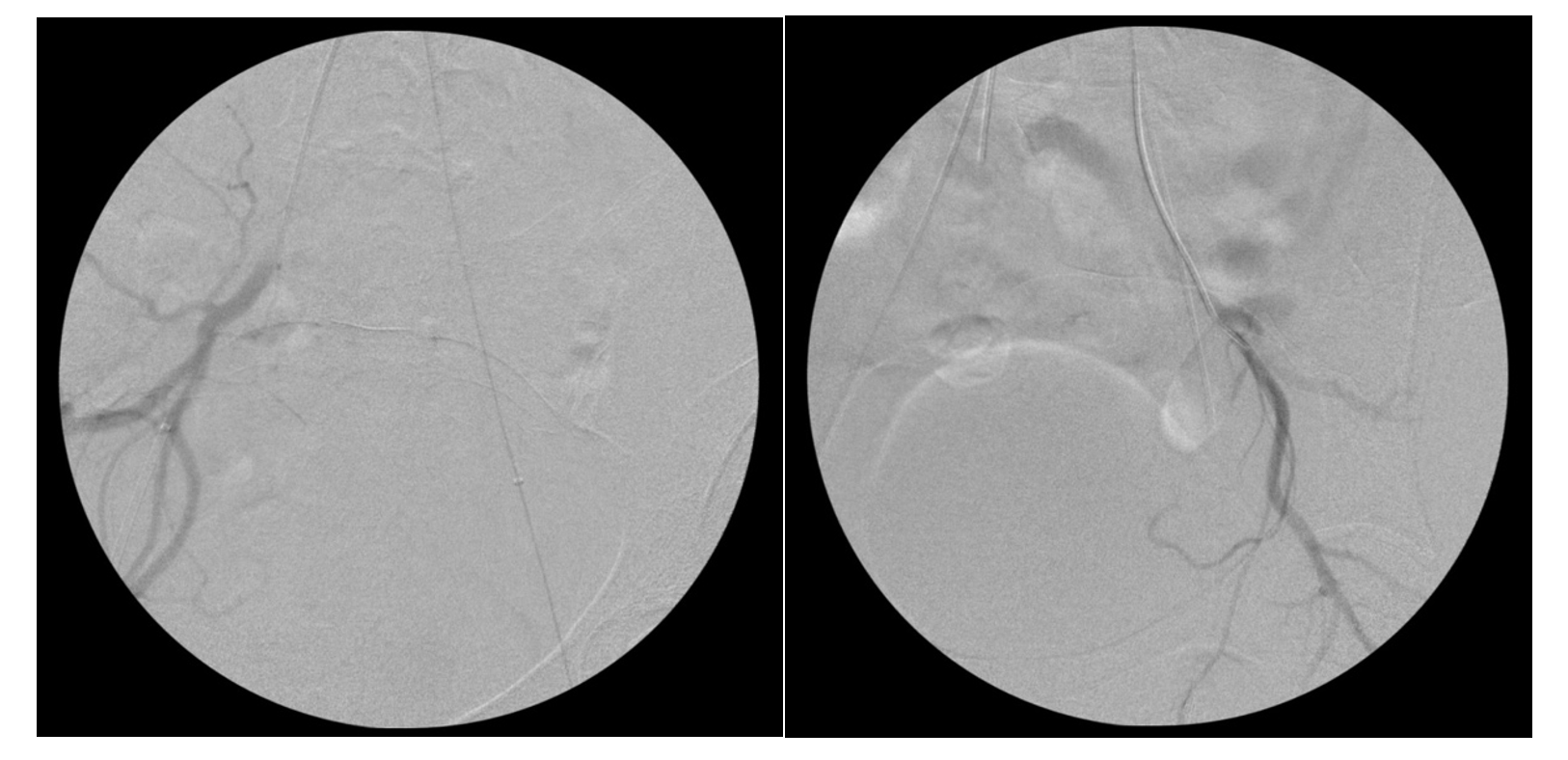
The procedure was performed in the operating room. Initially, the patient was placed supine. With the use of an ultrasound and C-arm for fluoroscopy, bilateral femoral arterial access was obtained, and 6F sheaths were placed. A 5F Cobra 2 catheter was used to cross the aortic bifurcation to the contralateral internal iliac artery (IIA). The catheters were then exchanged over a regular 0.035" Glidewire (Terumo) for 5.5F Fogarty balloons (Edwards Lifesciences), positioned in the proximal IIAs (Figure 2 and Figure 3). Test inflation of the balloons was performed under fluoroscopy to determine balloon volume required for vessel occlusion. The balloons were deflated, and the catheters and sheaths were well-secured. The patient was then shifted into lithotomy position and uterine vacuum aspiration was performed by the gynecology team. Following evacuation, bilateral angiograms were performed and demonstrated no active bleeding. Total estimated blood loss for the combined procedure was 20 mL. The patient remained in the hospital overnight for pain control and was discharged the next day. At postop day 5, a telephone follow-up was completed and the patient reported she was doing well.
Case Report 2
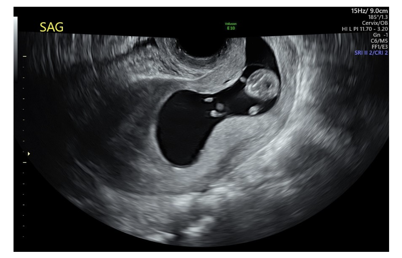
A 35-year-old woman (gravida 1, para 0) was found during an initial first trimester ultrasound to have a CP at approximately 10 weeks’ gestation (Figure 4) with fetal cardiac activity. She had no bleeding or pain at the time of presentation and β-hCG was 80,931.3 mIU/mL. Management options were discussed with the patient, including D&C with VIR support, systemic methotrexate, intra-sac methotrexate, and gravid hysterectomy. The patient elected to proceed with D&C with VIR support as she desired future fertility.
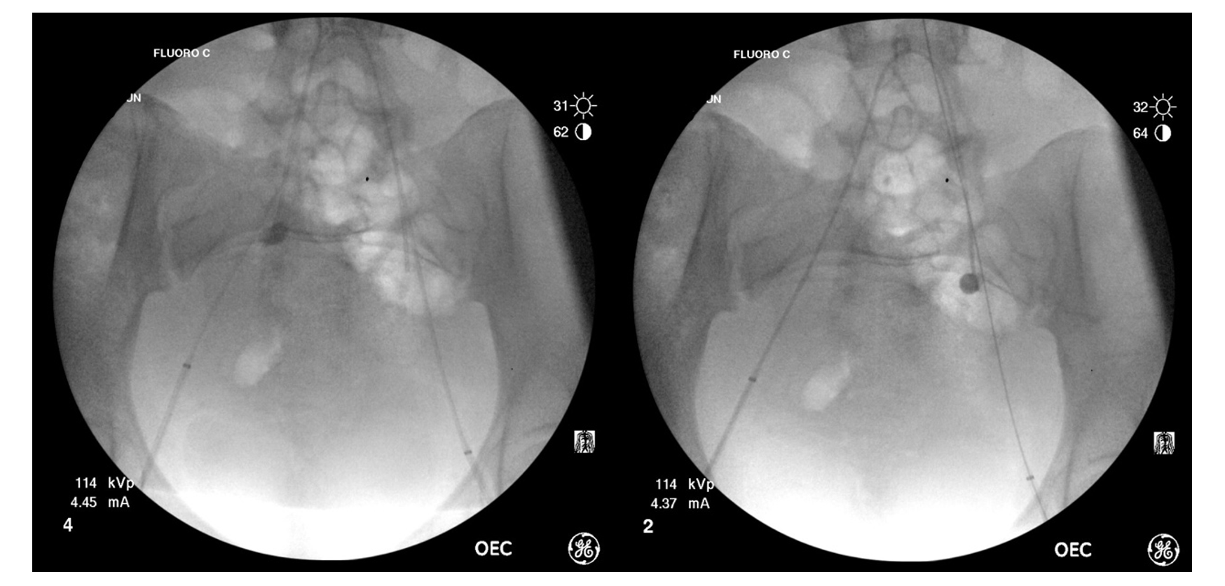
The procedure was performed in the operating room. Initially, the patient was placed supine and bilateral femoral arterial access was obtained with placement of 6F sheaths. A 5F Cobra 2 catheter was used to cross the aortic bifurcation to the contralateral IIA. The catheters were then exchanged over a regular 0.035" Glidewire for 5.5F Fogarty balloons, positioned in the proximal IIAs. Test inflation of the balloons was performed under fluoroscopy to determine balloon volume required for adequate vessel occlusion (Figure 5 and Figure 6). The balloons were deflated, and catheters and sheaths were well-secured. The patient was then shifted into lithotomy position. Due to the size, the CP was seen protruding through a dilated cervix. Uterine vacuum aspiration was subsequently performed by the gynecology team. Following evacuation, the balloons were deflated, and no further bleeding was noted on physical inspection. Total estimated blood loss for the combined procedure was 32 mL. The patient was monitored in recovery for 4 hours postprocedure (as bilateral groin hemostasis was achieved by manual compression and no closure device was used). She was discharged home the same day. On postprocedural telephone follow-up after 2 days, the patient reported some cramping and bleeding and passage of a small amount of clot that was decreasing overall. She otherwise felt well.
Discussion
CP is rare and a potential cause of significant hemorrhage leading to maternal morbidity and mortality. Risk factors include prior cesarean section, curettage, or other cervical/uterine surgery, and in vitro fertilization.1,5,6 Previously, diagnosis was difficult and often delayed; however, this has improved significantly with advances in ultrasound imaging, and patients with cervical implantation are identified earlier in their pregnancies.4,5 To date, there is no standardized algorithm for the management of CP and treatment options are offered based on the experience of the obstetrician, available options in the institution, and gestational age at presentation.6,7 These are typically broken into conservative and surgical options, both of which have their own risks and benefits.
Medical treatments may be better suited for patients in whom the ectopic gestation is discovered earlier. Weight-based dosing of systemic methotrexate (MTX) is most often used, with a success rate of more than 90%.6 In the presence of cardiac activity, targeted intra-amniotic injection of potassium chloride is sometimes performed, but it carries a high risk of hemorrhage and requires a skilled operator.6 Factors that may impact the success of medical management alone include gestational age, β-hCG level, and the presence of fetal cardiac activity.3 The main benefit of medical treatments is that they are fertility-sparing, and thus preferred in patients who wish to conceive again.
Surgical options also exist for the management of CP. Hysterectomy was historically the mainstay of treatment for CP as treatment options were limited. Today, hysterectomy is a last resort in cases of intractable bleeding, greater gestational age of the pregnancy, or lack of alternatives.4,8 However, it is the least preferred of the treatment options due to the loss of the ability to conceive and the additional morbidity and mortality associated with emergent surgery. Most patients are younger in age and desire to maintain their ability to conceive. Therefore, D&C or dilation and evacuation are often performed. These are shorter procedures with low risk to future fertility, though complications such as cervical trauma, uterine perforation, Asherman syndrome, severe bleeding, retained products, and infection can occur.9 The main risk in dealing with CP is trauma to the cervix itself, which is mitigated by using misoprostol or other cervical preparation before beginning manual dilation.10 Additional hemostatic agents may also be used throughout the procedure, including local injection of vasopressin into the cervix and/or intravenous administration of tranexamic acid.11,12
Cervical ectopic gestation is generally discussed in the literature in case reports due to the low overall incidence. Most reported cases discuss a combined treatment approach to increase the likelihood of success in removing the ectopic tissue as well as preserving fertility.1,4,8 In our institution, we have worked diligently in collaboration with our obstetrician colleagues to decrease bleeding risk in peripartum patients. These include not only CP, but also cases of placenta accreta spectrum as well as postpartum hemorrhage. The specific intervention varies case-by-case and includes both uterine artery embolization (UAE) and temporary endovascular arterial occlusion balloon (AOB) placement.
UAE is an excellent option for decreasing bleeding from cervical ectopic gestation as it is commonly used for other types of peripartum hemorrhage.1,7 A case report was published by Majumdar et al of a patient who had incomplete termination of a 6-week CP following systemic (intramuscular) administration of MTX. She subsequently presented with vaginal bleeding and UAE was performed prior to vacuum aspiration of retained products.8 UAE can be performed primarily or in conjunction with occlusion balloon placement. The theoretical potential for decreased future fertility with UAE has been discussed in the literature, with concerns primarily involving the potential for endometrial necrosis due to embolization and for ovarian dysfunction and premature menopause if the ovarian arteries require embolization.1 A recent large systematic review of the literature suggests that pregnancy rates after UAE are comparable to age-adjusted rates in the general population;13 however, there are no randomized controlled study data to confirm these findings.
Temporary AOB placement is another choice for decreasing blood loss while preserving fertility. Historically, AOBs have been placed for temporary control of pelvic bleeding during surgery, particularly in the setting of trauma.14 Their use has been expanded in recent years to cases of obstetric hemorrhage, including abnormal placentation and ectopic pregnancies, though the data is divided on their efficacy.15-17 One case report described the use of an AOB during surgical wedge resection of an intramural ectopic pregnancy.18 Another study reported a multidisciplinary approach to controlling peripartum hemorrhage in placenta accreta spectrum patients by use of resuscitative endovascular balloon occlusion of the aorta (REBOA).19 Intra-arterial occlusion balloons are helpful for decreasing blood flow to and reducing the amount of hemorrhage from spontaneously bleeding CPs, including during their operative management. The benefit of AOB placement is that the blood flow to the uterus is only temporarily disrupted, minimizing the risk of long-term damage and preserving future fertility. More importantly, hemostasis can be evaluated dynamically, as the balloons can be deflated and the cervix can be directly monitored for signs of bleeding. In the event there is persistent bleeding, the balloons can immediately be reinflated for temporary hemostasis until additional measures are taken.
There are very few reports in the literature regarding use of occlusion balloons alone during surgical management of cervical ectopics, as UAE is performed more often. The first report in 2006 by Yang et al describes a process similar to the one used in our cases, but with balloon placement in the bilateral common iliac arteries rather than the IIAs.20 We have found hypogastric AOB placement to require minimal additional effort on the part of the operator to obtain more localized pelvic hemostasis without affecting the lower extremities. Additionally, the balloons require less volume to be occlusive as the vessels are smaller in caliber. There are risks of inadequate occlusion or arterial rupture, but these are lowered as the balloon placement and test inflation are performed under fluoroscopy.
Conclusions
A multidisciplinary approach to treatment of CP can help decrease blood loss during operative management. While there is a slight additional risk due to increased procedural/sedation time, the benefits of decreased bleeding, shorter length of stay, and preserved fertility are higher when our teams work together. There can be some logistical complexities when coordination between departments is needed, but development of a standardized protocol for these situations can lead to efficient management and improved outcomes in patients with CP. Additionally, the sole use of AOBs can be easily applied to a wider range of situations of peripartum hemorrhage, including other ectopic gestations of the uterus. n
The authors have completed and returned the ICMJE Form for Disclosure of Potential Conflicts of Interest. The authors report no financial relationships or conflicts of interest regarding the content herein.
Manuscript accepted April 13, 2023.
Address for correspondence: Priya Mody, MD, Department of Radiology, Community Division, University of North Carolina at Chapel Hill, 101 Manning Dr., Chapel Hill, NC 27514. Email: priya_mody@med.unc.edu
REFERENCES
1. Fowler ML, Wang D, Chia V, et al. Management of cervical ectopic pregnancies: a scoping review. Obstet Gynecol. 2021;138(1):33-41. doi:10.1097/AOG.0000000000004423
2. Samal SK, Rathod S. Cervical ectopic pregnancy. J Nat Sci Biol Med. 2015;6(1):257-260. doi:10.4103/0976-9668.149221
3. Albahlol IA. Cervical pregnancy management: an updated stepwise approach and algorithm. J Obstet Gynaecol Res. 2021;47(2):469-475. doi:10.1111/jog.14617
4. Pandya MR, Modesara JA. A rare case of cervical ectopic pregnancy. Indian J Obstet Gynecol Res. 2022;9(2):312-316. doi:10.18231/j.ijogr.2022.060
5. Hirakawa M, Tajima T, Yoshimitsu K, et al. Uterine artery embolization along with the administration of methotrexate for cervical ectopic pregnancy: technical and clinical outcomes. Am J Roentgenol. 2009;192(6):1601-1607. doi:10.2214/ajr.08.1921
6. Singh S. Diagnosis and management of cervical ectopic pregnancy. J Hum Reprod Sci. 2013;6(4):273-276. doi:10.4103/0974-1208.126312
7. Ben Farhat L, Ben Salah Y, Askri A, Dali N, Hendaoui L. Conservative treatment of a twin cervical pregnancy with uterine artery embolization. Diagn Interv Radiol. 20010;16(3):248-250. doi:10.4261/1305-3825.dir.2073-08.1
8. Majumdar A, Shekhar B, Satwik A. Uterine artery embolisation: a rescuer in cervical ectopic pregnancy. BMJ Case Rep. 2021;14(9):e244623. doi:10.1136/bcr-2021-244623
9. Kakinuma T, Kakinuma K, Sakamoto Y, et al. Safety and efficacy of manual vacuum suction compared with conventional dilatation and sharp curettage and electric vacuum aspiration in surgical treatment of miscarriage: a randomized controlled trial. BMC Pregnancy Childbirth. 2020;20(1):695. doi:10.1186/s12884-020-03362-4
10. Meirik O, Huong NTM, Piaggio G, Bergel E, von Hertzen H, WHO Research Group on Postovulatory Methods of Fertility Regulation. Complications of first-trimester abortion by vacuum aspiration after cervical preparation with and without misoprostol: a multicentre randomised trial. Lancet. 2012;379(9828):1817-1824. doi:10.1016/s0140-6736(11)61937-5
11. Ishikawa H, Unno Y, Omoto A, Shozu M. Local injection of diluted vasopressin followed by suction curettage for cervical ectopic pregnancy. Eur J Obstet Gynecol Reprod Biol. 2016;207:173-177. doi:10.1016/j.ejogrb.2016.11.004
12. Zakhari A, Sanders AP, Solnik MJ. Tranexamic acid in gynecologic surgery. Curr Med Res Opin. 2020;36(3):513-520. doi:10.1080/03007995.2019.1708533
13. Mohan PP, Hamblin MH, Vogelzang RL. Uterine artery embolization and its effect on fertility. J Vasc Interv Radiol. 2013;24(7):925-930. doi:10.1016/j.jvir.2013.03.014
14. Gamberini E, Coccolini F, Tamagnini B, et al. Resuscitative endovascular balloon occlusion of the aorta in trauma: a systematic review of the literature. World J Emerg Surg. 2017;12:42. doi:10.1186/s13017-017-0153-2
15. Picel AC, Wolford B, Cochran RL, Ramos GA, Roberts AC. Prophylactic internal iliac artery occlusion balloon placement to reduce operative blood loss in patients with invasive placenta. J Vasc Interv Radiol. 2018;29(2):219-224. doi:10.1016/j.jvir.2017.08.015
16. Chen D, Xu J, Tian Y, et al. Effect of prophylactic balloon occlusion of internal iliac artery in pregnancies complicated by placenta previa and accreta. BMC Pregnancy Childbirth. 2021;21(1):640. doi:10.1186/s12884-021-04103-x
17. Knuttinen MG, Jani A, Gaba RC, Bui JT, Carrillo TC. Balloon occlusion of the hypogastric arteries in the management of placenta accreta: a case report and review of the literature. Semin Intervent Radiol. 2012;29(3):161-168. doi:10.1055/s-0032-1326924
18. Li S, Liu H, Li X, Liu Z, Li Y, Li N. Transfemoral temporary aortic balloon occlusion in surgical treatment of second trimester intramural ectopic pregnancy. J Obstet Gynaecol Res. 2016;42(6):716-718. doi:10.1111/jog.12968
19. Russo RM, Carver A, Clifford C, Rolston A, Uppal S, Napolitano LM. A team approach to peripartum hemorrhage control incorporating resuscitative endovascular balloon occlusion of the aorta. J Trauma Acute Care Surg. 2022;93(2):e89-e94. doi:10.1097/TA.0000000000003612
20. Yang JH, Shih JC, Liu KL, Yang YS. Combined treatment with temporary intraoperative balloon occlusion of common iliac arteries and hysteroscopic endocervical resection with postoperative cervical balloon for intractable cervical pregnancy in an infertile woman. Fertil Steril. 2007;88(5):1438.e11-3. doi:10.1016/j.fertnstert.2007.01.033
