


Tension Hemothorax in a Neurofibromatosis-related Angiopathy: A Comprehensive Analysis
© 2024 HMP Global. All Rights Reserved.
Any views and opinions expressed are those of the author(s) and/or participants and do not necessarily reflect the views, policy, or position of Vascular Disease Management or HMP Global, their employees, and affiliates.
VASCULAR DISEASE MANAGEMENT. 2024;21(9):E74-E83
Abstract
Objectives: This literature review investigates the incidence, etiology, and treatment strategies for spontaneous hemothorax in patients with neurofibromatosis type 1 (NF1). Methods: A comprehensive literature review was performed to identify articles on spontaneous hemothorax in patients with NF1. Relevant articles were selected and data on demographics, clinical presentation, sources of bleeding, treatment modalities, and outcomes were extracted and analyzed. Results: The review identified 47 relevant articles detailing 52 cases of spontaneous hemothorax in patients with NF1, with a mean age of 44 years and a male-to-female ratio of 27:25. The predominant etiology was arterial aneurysms or pseudoaneurysms. Common treatment interventions included chest tube insertion, thoracoscopy, angiography with endovascular embolization, and open thoracotomy. Cardiac arrest occurred in 16 patients, and rebleeding was observed in 12 cases within 7 days post-surgery. No significant differences in treatment outcomes were found among endovascular treatment, thoracotomy, and thoracoscopy groups. Conclusion: Spontaneous hemothorax in patients with NF1 is a rare but serious condition requiring prompt diagnosis and treatment. The findings highlight the importance of recognizing arterial aneurysms or pseudoaneurysms as a primary source of bleeding and suggest that a multidisciplinary approach is crucial for optimizing patient outcomes. Further research is needed to develop standardized treatment protocols and improve long-term prognosis in this patient population.
Introduction
Neurofibromatosis type 1 (NF1), also known as von Recklinghausen's disease, is the most common type of phacomatosis.1 NF1 is characterized by polymorphic clinical manifestations, of which skin and neurological symptoms are the most prevalent, along with skeletal and vascular system involvement.
Vascular changes occur in only 1.0% to 3.6% of patients with NF1.2,3 Most commonly affected are medium- and large-caliber vessels, with disruptions attributed to weakness in the vessel wall leading to the formation of aneurysmal dilations and stenoses.
The motivation for conducting this analysis stemmed from encountering similar complications twice in our practice; both patients with NF1 developed tension hemothorax following posterior spinal surgery, without any vertebrectomies.
The main questions of this study are the following: What is the incidence and etiology of tension hemothorax in patients with NF1, particularly following spinal surgery or other intervention on the thorax and thoracic spine? What is the optimal approach to treating this condition?
We hypothesize that tension hemothorax occurrence in patients with NF1 is a rare but potentially fatal complication, primarily attributed to vascular weakness related to NF1-associated vasculopathy.
Methods
The initial literature review was conducted independently by 2 physicians. The search encompassed several databases, including PubMed (National Institutes of Health), Google Scholar (Alphabet, Inc.), and eLibrary (U.S. General Services Administration). Search terms included neurofibromatosis type 1, hemothorax, scoliosis, and surgical treatment, covering the period from 2003 to 2023 to include recent publications. Following the primary selection, a secondary screening was performed. Relevant articles describing clinical cases or case series involving patients with NF1 and either spontaneous or postoperative tension hemothorax were identified and retrieved for analysis. Articles meeting the inclusion criteria were then selected for further analysis, which included the presence of patients diagnosed with NF1; occurrence of either spontaneous or postoperative tension hemothorax; and clinical cases or case series detailing the condition, its treatment, and follow-up. Duplicates were removed from the dataset.
Relevant data encompassing patient demographics, clinical presentation, diagnostic findings, treatment modalities, and outcomes were extracted from the selected articles. Analysis of the extracted data aimed to identify common patterns, trends, and factors associated with tension hemothorax occurrence in patients with NF1, particularly following spinal surgery.
Statistical analyses utilized Microsoft Excel 2019, employing analysis of variance to assess intergroup differences.
Results
Eighty-five articles were identified in the initial analysis, and 47 were deemed relevant upon application of the inclusion criteria. These publications collectively detailed 52 cases of patients with NF1 who experienced tension hemothorax4 (Table 1). Among these cases, 52% were men (n = 27) and 48% were women (n = 25). The average age of the patients was 44 years, with ages ranging from 14 to 72 years. Among the 52 patients, 20 (38%) were diagnosed with scoliosis.
In the majority of instances (n = 44, 85%), no specific predisposing factors were discerned in the development of hemothorax, and there was no associated injury in any case. However, in 2 cases, hemothorax manifested shortly after surgical procedures: once following thoracic-level vertebrectomy,4 and the second following excision of a mediastinal tumor.5 In 2 cases, the onset of hemothorax followed a bout of coughing.6,7 Two patients presented with hemothorax alongside arterial hypertension stemming from chronic hypertension.8,9 One pregnant woman experienced gestational hypertension,10 while another woman was within 4 days postpartum.11 Additionally, 2 patients exhibited hemothorax alongside chronic kidney disease,8,12 and 3 patients had mediastinal pseudomeningocele.13-15 Lastly, 1 patient encountered hemothorax after sustaining a severe allergic reaction from a bee sting the day prior.16 One patient had a prior history of hemothorax 4 years preceding the current event, but the specific details regarding her treatment were not provided.17
Table 1. Results of a review of the literature on the occurrence of tension hemothorax in patients with neurofibromatosis type 1
| Author | Year | N of patients | Bleeding source | Pathology | Cardiac arrest |
Initial treatment |
Repeated surgery | Death | Follow-up | Rebleeding |
|---|---|---|---|---|---|---|---|---|---|---|
| Abdulrazeq | 2019 | 1 | VA | PA | - | chest tube, coiling | - | yes | 1 month | - |
| Aizawa | 2010 | 1 | 11th ICA | spontaneous rupture | - | thoracotomy | laminectomy, spinal cord decompression | - | 6 months | - |
| Arai | 2007 | 3 | VA | aneurysm + PA | yes | chest tube, coiling | - | yes | 11 days | yes |
| 10th ICA | aneurysm, AVF | - | chest tube, coiling | - | - | 1 month | - | |||
| 11th ICA | aneurysm | - | chest tube, coiling | - | - | 1 month | - | |||
| Atici | 2017 | 2 | ND | ND | - | chest tube | - | - | NA | - |
| Attia | 2019 | 2 | ND | ND | - | thoracoscopy, chest tube | - | - | 1 month | - |
| 5th ICA | spontaneous rupture | - | thoracoscopy, chest tube | - | - | 1 month | - | |||
| Baldo | 2002 | 1 | SCA branch | spontaneous rupture | yes | chest tube | - | yes | - | - |
| Bento | 2009 | 1 | ND | ND | - | chest tube, thoracoscopy | - | - | 1 month | - |
| Bidad | 2019 | 1 | VA | PA | yes | chest tube | - | yes | - | - |
| Bommart | 2015 | 1 | 11th ICA | aneurysm | yes | chest tube + coiling | thoracotomy | yes | 7 days | yes |
| Chen | 2020 | 1 | neurofibroma | high-vascular tumor | - | thoracotomy + chest tube | - | - | 1 month | yes |
| Conlon | 2007 | 1 | ITA | spontaneous rupture | - | thoracotomy + electrocoagulation + chest tube | - | - | 6 months | - |
| Degbelo | 2019 | 1 | ND | ND | - | chest tube | - | - | 2 months | - |
| Dominguez | 2002 | 1 | 4th ICA | aneurysm | - | chest tube + coiling | - | - | 19 months | - |
| Vazquez | 2018 | 2 | ITA | aneurysm | - | thoracotomy + chest tube | - | - | 1 month | - |
| 5th ICA | spontaneous rupture | yes | thoracoscopy + chest tube | coiling | - | 1 month | - | |||
| Faruque | 2009 | 1 | SCA, neurofibroma | AVF | - | thoracotomy + chest tube | coiling | - | 1 month | - |
| Fdil | 2017 | 1 | ND | ND | - | chest tube | - | - | 1 month | - |
| Fedoruk | 2007 | 1 | SCA-ITA junction | spontaneous rupture | yes | chest tube | thoracotomy | - | 1 month | - |
| Fohrding | 2014 | 1 | ICA | spontaneous rupture | yes | chest tube | - | yes | - | - |
| Hashimoto | 2021 | 1 | 10th ICA | aneurysm | - | chest tube, coiling | - | - | 18 months | - |
| Hieda | 2007 | 1 | VA | aneurysm | yes | chest tube, coiling | - | - | 1 month | - |
| Hongsakul | 2013 | 1 | 5th ICA | aneurysm + AVF | - | chest tube, coiling | - | - | 12 months | - |
| Hoonjan | 2014 | 1 | branch of costo-cervical trunk | spontaneous rupture | yes | chest tube, coiling | - | yes | - | yes |
| Jeong | 2018 | 1 | neurofibroma | high-vascular tumor | - | chest tube, thoracosopic removal of tumor | - | - | 6 | - |
| Jin Kim | 2005 | 1 | ITA | PA | - | chest tube, coiling | thoracotomy | - | 2 | - |
| Jun Kim | 2011 | 1 | 11th ICA | aneurysm + AVM | yes | chest tube, coiling | thoracotomy | yes | - | - |
| Kaneda | 2010 | 1 | 4th-6th ICA, branch of ITA, neurofibroma | high-vascular tumor, PA | - | chest tube, coiling, then | thoracotomy | yes | 8 | - |
| Lafleur | 2020 | 1 | SCA | aneurysm | - | chest tube, coiling | - | - | 1 | - |
| Misao | 2012 | 1 | 10th ICA | aneurysm | - | chest tube, thoracotomy | coiling | - | 8 | yes |
| Miura | 2005 | 2 | 5th ICA, AVF, neurofibroma | high-vascular tumor | - | chest tube, thoracotomy | thoracotomy | - | 120 | - |
| 7th ICA | aneurysm | - | chest tube, coiling | - | - | 60 | - | |||
| Miyamoto | 2020 | 1 | SCA branch | spontaneous rupture | - | chest tube, coiling | - | - | 1 | - |
| Miyazaki | 2011 | 1 | SCA branch | spontaneous rupture | - | chest tube, thoracoscopy | - | - | 12 | - |
| Mydin | 2015 | 1 | SCA | PA | - | coiling, thoracoscopy, chest tube | - | - | 1 | - |
| Negreira | 2020 | 1 | SCA | PA | yes | coiling, chest tube | chest tube | - | 1 | - |
| Neto | 2020 | 1 | ITA | aneurysm | yes | chest tube, coiling | - | yes | - | - |
| Pantazopoulos | 2019 | 1 | 8th ICA | spontaneous rupture | - | chest tube, thoracotomy | - | - | 1 | - |
| Pezzetta | 2003 | 1 | ND | ND | - | chest tube | - | - | 18 | - |
| Pomara | 2013 | 1 | esophagus | dissecting intramural hematoma | yes | no | - | yes | - | - |
| Pulivarthi | 2014 | 1 | neurofibroma | high-vascular tumor | - | thoracoscopy, chest tube | - | yes | 1 | - |
| Rodriguez-Guzman | 2014 | 1 | SCA branch | spontaneous rupture | - | chest tube, coiling | thoracoscopy | yes | 12 | - |
| Sandhu | 2007 | 1 | VA | spontaneous rupture | - | chest tube | - | yes | 18 | yes |
| Narasimman | 2018 | 1 | 7th ICA, neurofibroma | AVM | - | thoracotomy, chest tube | laminectomy, spinal cord decompression | - | 6 | - |
| Seok Han | 2019 | 1 | VA | aneurysm | yes | chest tube, coiling | - | - | 6 | - |
| Urso | 2007 | 1 | ITA | aneurysm | yes | chest tube, thoracotomy | - | yes | - | - |
| Vaziri | 2006 | 1 | ganglio-neuroma | high-vascular tumor | - | chest tube, thoracotomy | thoracotomy | - | 24 | - |
| Yoshida | 2005 | 1 | SCA, neurofibroma | aneurysm | - | chest tube, thoracotomy | - | - | 1 | - |
| Young Kwon | 2016 | 1 | ITA | aneurysm | yes | chest tube | coiling, thoracoscopy | - | 6 | - |
| Yusuf | 2014 | 1 | ICA | spontaneous rupture | - | chest tube, thoracotomy | - | - | 48 | - |
Abbreviations: AVF, arteriovenous fistula; AVM, arteriovenous malformation; ICA, intercostal artery; IMA, internal mammary artery; ITA, internal thoracic artery; SCA, subclavian artery ; NA, not applicable; N, not detected; PA, pseudoaneurysm; VA, vertebral artery.
Computed tomography (CT) angiography was performed in almost all cases before surgical intervention. In most cases (22 patients, 42%), bleeding was associated with arterial aneurysm or pseudoaneurysm; in 14 patients (27%), vessel rupture was considered spontaneous; in 5 patients (10%), the source of bleeding was not found; and in 6 patients (12%), the source was an intrathoracic tumor (usually neurofibroma) with adjacent vessels.
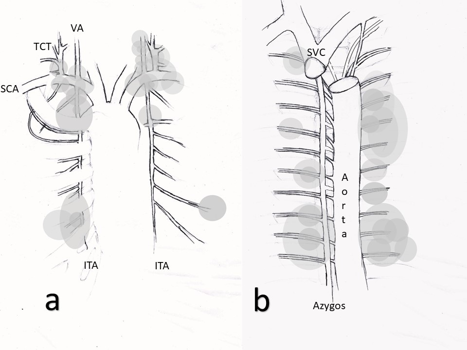
The source of bleeding was identified as the intercostal artery in 18 patients,10,11,13-15,17-26 and the subclavian artery or internal thoracic branch in 23 patients5,7,9,16,27-43 (Figure). Several patients experienced hemothorax attributed to hypervascularized intrathoracic tumors, notably neurofibromas.44-47 In total, 11 patients were found to have intrathoracic tumors, with neurofibromas being a common manifestation. These tumors, although often small in size, were discerned visually during thoracotomy and were not consistently visible on chest CT scans. There were also a few cases of dissecting intramural hematoma with a breakthrough into the pleural cavity,48 arteriovenous malformation,11,17 and arteriovenous fistula19,23 (Table 1).
We categorized the patients into 4 groups based on the immediate treatment administered upon hemothorax detection:
- Nine patients who underwent chest tube insertion
- Eight patients who underwent thoracoscopy followed by chest tube insertion
- Twenty patients who received angiography with endovascular embolization followed by chest tube insertion
- Fourteen patients who underwent open thoracotomy followed by chest tube insertion.
Overall, cardiac arrest was reported in 16 out of 52 patients. Rebleeding incidents were observed in 12 patients within 7 days following the initial operation, while 11 patients succumbed immediately and 6 experienced severe consequential effects. These effects leading to disability included 2 cases of paraplegia following open thoracic surgery,11,18 1 instance of post-resuscitative encephalopathy,37 1 occurrence of brainstem stroke,41 1 case of blindness,28 and 1 incident involving a giant intrathoracic pseudomeningocele and respiratory failure, which ultimately led to death.13
Of the 52 patients, 51 underwent surgery and 1 patient did not survive to reach the operating table (Table 2). We conducted comparisons among patients based on various parameters including age, affected side, presence of scoliosis, intrathoracic tumor presence, and chronic diseases. Then we analyzed group differences in terms of cardiac arrest occurrence, rebleeding in the immediate or delayed postoperative period, redo interventions, mortality rates, and postoperative disabilities (Table 2).
Table 2. Comparison of treatment effectiveness of different approaches to tension hemothorax in patients with neurofibromatosis type 1
| Chest tube | Thoracoscopy + chest tube | Endovascular embolization + chest tube | Thoracotomy + chest tube | P value | |
| Total N of patients | 9 | 8 | 20 | 14 | - |
| Age (yy, median, range) | 38 (14-45) | 53 (31-59) | 44 (30-72) | 46 (30-63) | .05 |
| Scoliosis (N, %) | 7 (78%) | 2 (25%) | 4 (20%) | 6 (43%) | .02 |
| Tumors in the mediastinum (N, %) | 2 (22%) | 1 (13%) | 2 (10%) | 7 (50%) | .04 |
| Chronic pulmonary diseases (N, %) | 1 (11%) | 2 (25%) | 3 (15%) | 1 (7%) | .71 |
| Chronic renal diseases (N, %) | 1 (11%) | - | - | 1 (7%) | .45 |
| Side Dexter | 4 (44%) | 5 (63%) | 4 (20%) | 7 (50%) | .24 |
| Sinister | 5 (56%) | 3 (37%) | 15 (75%) | 7 (50%) | .24 |
| 2-side | - | - | 1 (5%) | - | - |
| Cardiac arrest (N,%) | 3 (33%) | 1 (13%) | 9 (45%) | 2 (14%) | .18 |
| Rebleeding during 7 days (N, %) | 1 (11%) | 1 (13%) | 6 (30%) | 4 (29%) | .60 |
| Redo treatment for rebleeding shortly after initial treatment (N, %) | 1 (11%) | 1 (13%) | 6 (30%) | 4 (29%) | .60 |
| Death during 1 month after incident (N, %) | 3 (33%) | 1 (13%) | 5 (25%) | 1 (7%) | .40 |
| Severe disability after incident (N, %) | - | - | 3 (20%) | 3 (23%) | .37 |
| Follow-up (month, median, range) | 10 (1-19) | 1 (1-12) | 1 (1-60) | 6 (1-120) | .61 |
| Rebleeding after discharge (N, %) | 1 (17%) | - | - | 1 (8%) | .39 |
| Death due to rebleeding after initial discharge (N, %) | 1 (17%) | - | - | - | .12 |
| Death from other reasons (N, %) | - | - | 2 (13%) | - | .32 |
Patients in the thoracoscopy group were slightly older than the other groups (P = .05). In the group of patients who underwent only chest tube insertion, scoliosis was more common (P = .02). In the group of patients who underwent thoracotomy as the initial method of treatment, the presence of visible intrathoracic tumors was more often noted, but it is worth noting that the tumors were often detected intraoperatively (P = .04). Otherwise, there were no significant differences in treatment outcomes between the groups (Table 2).
Discussion
NF1 is a relatively rare autosomal dominant disorder associated with chromosome 17q11.2, defect neurofibromin synthesis (GTPase-activating protein), and impaired intracellular Ras regulation pathway. Neurofibromin expression has been identified in both endothelial and smooth muscle cells within blood vessels, suggesting a potential role of abnormal neurofibromin function in NF1 vasculopathy. Decreased neurofibromin expression could disrupt the response of endothelial or smooth muscle cells to growth suppressive signals, leading to excessive proliferation or compromising endothelial integrity and triggering secondary smooth muscle cell migration and proliferation in vessel walls.
Another possibility is that local vessel wall injury contributes to lesion development, leading to intimal hyperplasia due to dysregulated repair processes.49 The spectrum of NF1 vasculopathy encompasses vessels of various sizes, ranging from the proximal aorta to small arterioles. Symptoms can include vascular narrowing, blockages, aneurysms, pseudoaneurysms, ruptures, or fistula formation.
Among symptomatic cases, renal arteries are commonly affected, often resulting in hypertension.49 Diagnosis of NF1-associated vascular disease may occur in both children and adults, with hemorrhage being a significant contributor to NF1-related mortality.
The pathogenesis of arterial lesions in NF1 is complex, involving factors such as the proliferation of spindle cells within vessel walls, fibrous thickening, cellular nodularity compromising vessel wall integrity (particularly in small vessels), Schwann cell proliferation within large vessel arterial walls, and dysplastic smooth muscle.49 These abnormalities increase the vulnerability of arteries in patients with NF1, potentially leading to life-threatening hemorrhages such as tension hemothorax.
Various predisposing factors can contribute to hemorrhages, including pregnancy-related changes such as increased blood volume, hemodilution, decreased oncotic pressure, peripheral edema, and gestational hypertension. Additionally, arterial hypertension (whether renal artery-dependent, pheochromocytoma-dependent, or primary), renogenic edema, and even allergic reactions (such as type I hypersensitivity reactions and histamine-dependent increases in vascular permeability leading to edema) can exacerbate the risk of hemorrhage.
Surgical procedures involving the thoracic vertebrae or intrathoracic surgeries, such as vertebral resection or correction of deformities, can inevitably lead to tense hemothorax, particularly in patients with neurofibromatosis. Surgical interventions on the thoracic vertebrae or intrathoracic surgeries carry inherent risks of hemothorax in patients with NF1, especially in the presence of apical or paravertebral neurofibromas. It is probable that postsurgical hemothorax is directly linked to the surgical site. This scenario considerably streamlines the process of identifying the bleeding source.
In cases of spontaneous hemothorax, pinpointing the bleeding source is not always feasible. However, the most frequent culprits are branches of the subclavian artery, as well as paravertebral segments of intercostal vessels spanning the entirety of the thoracic spine on either side.
When bleeding emanates from the branches of the subclavian artery, it originates at the uppermost section of the pleural cavity. Often, this type of bleeding is accompanied by radiating pain in the neck and arm, along with swelling of the neck. The blood ingress into the deeper neck structures occurs above the lung apex, but its spread is confined to the neck fascia. In certain scenarios, blood can forcefully enter the pleural cavity, such as when neurofibromas are present in the apical pleura region, or when severe arterial bleeding exerts significant hematoma pressure.
According to the literature review and our clinical experience, the presence of tension hemothorax in patients with NF1 correlates strongly with elevated risks of cardiac arrest, mortality, and recurrent bleeding, both in the immediate and long-term postoperative periods.
Traditional open thoracotomy offers several advantages and disadvantages. Among its strengths is that it proves highly effective in managing profuse bleeding from arteriovenous malformations or neurofibromas. It also provides a broad field of view and facilitates various methods of controlling bleeding simultaneously, such as ligation/clipping, coagulation, tamponade, and the application of different hemostatic agents such as oxidized cellulose, fibrin-thrombin hemostats, and collagen sponges. Additionally, it allows for plastic surgery of the vessel wall and the performance of vascular anastomosis. However, drawbacks include the potential for trauma and the formation of pleural adhesions, which may necessitate thoracoscopic decortication in the future. Difficulty accessing extrathoracic segments of the subclavian arteries and vertebral arteries, particularly if the source of bleeding is localized there, is another limitation.
Thoracoscopy presents itself as a viable alternative to thoracotomy but demands specialized skills. It also comes with restrictions in visualizing and accessing extrathoracic segments of the subclavian arteries and vertebral arteries. Furthermore, when facing relentless bleeding and considering pressure hemostasis as an option, it is essential to note potential complications for thoracotomy and thoracoscopy approaches. In numerous instances of bleeding from intercostal arteries, the source is often localized in the region of dystrophically dilated intervertebral foramina. Notably, there have been reported cases of sponge migration into the canal and subsequent spinal cord injury, along with several instances of inadvertent durotomy and subsequent formation of intrathoracic pseudomeningocele.
Endovascular treatment offers significant advantages as a minimally invasive approach with excellent access to the arteries at the apex of the lung. However, it faces limitations such as dealing with multiple vascular lesions, arteriovenous shunts and malformations, diffuse bleeding from tumor tissue, small vessel size, and the fragility of the vascular wall in neurofibromatosis. In addition, endovascular treatment is usually associated with a longer operation time, which is less preferable in urgent cases.
Challenges persist in managing arterial complications in patients with NF1. Fatal hemorrhage remains a significant concern, highlighting the critical need for fast, careful preoperative planning and timely intervention.
In summary, arterial complications in NF1 represent a complex clinical challenge requiring a multidisciplinary approach for effective management. Continued research and clinical experience are essential for refining treatment strategies and improving outcomes for patients with NF1 and associated arterial complications.
Conclusion
Our research sheds light on the incidence, etiology, and treatment outcomes of tension hemothorax in patients with NF1. The findings underscore the importance of vigilance and prompt intervention when managing this rare but potentially life-threatening complication in patients with NF1.Due to the variability in presentation and treatment approaches, our study highlights the need for a multidisciplinary approach involving fast and careful patient assessment, timely diagnostic imaging, and tailored treatment strategies. n
Affiliations and Disclosures
Olga M. Sergeenko (Pavlova), MD; Anastacia R. Gabrielyan, MD; Dmitry M. Savin, MD; Polina V. Ochirova, MD; and Alexey V. Evsyukov, MD are from the Division of Spinal Surgery, Ilizarov Center, Kurgan, Russia. Alexander V. Burtsev, PhD, MD, is from the Ilizarov Center, Kurgan, Russia
Address for correspondence: Olga M. Sergeenko, MD, Division of Spinal Surgery, Ilizarov Center, 6, M.Ulyanova St., Kurgan, 640014, Russia. Email: pavlova.neuro@mail.ru
Disclosure: The authors report no financial relationships or conflicts of interest regarding the content herein.
Manuscript accepted July 17, 2024.
REFERENCES
1. Walker JA, Upadhyaya M. Emerging therapeutic targets for neurofibromatosis type 1. Expert Opin Ther Targets. 2018;22(5):419-437. doi:10.1080/14728222.2018.1465931
2. Raborn J, McCafferty BJ, Gunn AJ, et al. Endovascular management of neurofibromatosis type 1-associated vasculopathy: a case series and brief review of the literature. Vasc Endovascular Surg. 2020;54(2):182-190. doi:10.1177/1538574419885257
3. Hamilton SJ, Friedman JM. Insights into the pathogenesis of neurofibromatosis 1 vasculopathy. Clin Genet. 2000;58(5):341-344. doi:10.1034/j.1399-0004.2000.580501.x
4. Atici Y, Balioglu MB, Kargin D, Mert M, Albayrak A, Kaygusuz MA. Analysis of complications following posterior vertebral column resection for the treatment of severe angular kyphosis greater than 100°. Acta Orthop Traumatol Turc. 2017;51(3):201-208. doi:10.1016/j.aott.2017.02.015
5. Gil Vázquez PJ, Arroyo Tristán ADA, Martínez Baños J, Torres Lanzas J. Massive spontaneous hemothorax in patients with neurofibromatosis type 1. Emergencias. 2018;30(3):190-193.
6. Degbelo FDA, Cito G, Guendil B, Christodoulou M, Abbassi Z. Spontaneous hemothorax in a patient with von Recklinghausen's disease: a case report and review of the literature. Am J Case Rep. 2019; 20:674-678. doi:10.12659/AJCR.915810
7. Abdulrazeq HF, Goldstein IM, Elsamna ST, Pletcher BA. Vertebral artery aneurysm rupture and hemothorax in a patient with neurofibromatosis Type-1: a case report and review of the literature. Heliyon. 2019;5(8):e02201. doi:10.1016/j.heliyon.2019.e02201
8. Fdil S, Bouchikhi S, Bourkadi JE. [Spontaneous hemothorax: a rare complication of neurofibromatosis type 1]. Article in French. Pan Afr Med J. 2017;28:85. doi:10.11604/pamj.2017.28.85.13820
9. Rodriguez-Guzman M, Gallegos-Carrera B, Vicente-Antunes S, Fernandez-Ormaechea I, Zapatero-Gaviria J, Villar-Alvarez F. Spontaneous hemothorax in a patient with von Recklinghausen's disease. J Clin Med Res. 2014;6(2):149-152. doi:10.14740/jocmr1692w
10. Hashimoto K, Nomata Y, Fukui T, Takada A, Narita K. Massive hemothorax in a pregnant patient with neurofibromatosis type 1. J Cardiothorac Surg. 2021;16(1):116. doi:10.1186/s13019-021-01504-z
11. Narasimman S, Govindasamy H, Seevalingam KK, Paramasvaran G, Ramasamy U. Spontaneous massive haemothorax in the peri-partum period of an undiagnosed neurofibromatosis type 1 patient - a surgical perspective. Med J Malaysia. 2019;74(1):99-101.
12. Urso S, Carballo J, Greco E, et al. Ruptured aneurysm of the mammary artery in a patient with neurofibromatosis type I. J Card Surg. 2007;22(3):229-231. doi:10.1111/j.1540-8191.2007.00396.x
13. Kaneda H, Saito T, Konobu T, Saito Y. Chest wall bleeding with giant intrathoracic meningocele in neurofibromatosis type 1. Interact Cardiovasc Thorac Surg. 2011;12(2):328-330. doi:10.1510/icvts.2010.252676
14. Föhrding LZ, Sellmann T, Angenendt S, et al. A case of lethal spontaneous massive hemothorax in a patient with neurofibromatosis 1. J Cardiothorac Surg. 2014;9:172. doi:10.1186/s13019-014-0172-y
15. Yusuf AS, Pillai A, Menon SK, Panikar D. Massive spontaneous hemothorax, giant intrathoracic meningocele, and kyphoscoliosis in neurofibromatosis type 1. J Surg Tech Case Rep. 2014;6(1):33-36. doi:10.4103/2006-8808.135151
16. Faruque MO, Davidson F. Clinical challenges and serious vascular complications in neurofibromatosis. Emerg Med Australas. 2009;21(6):518-520. doi:10.1111/j.1742-6723.2009.01236.x
17. Kim HJ, Seon HJ, Choi S, Jang NK. Ruptured aneurysm of intercostal arteriovenous malformation associated with neurofibromatosis type 1: a case report. Cardiovasc Intervent Radiol. 2011;34 Suppl 2:S74-S77. doi:10.1007/s00270-010-9965-0
18. Aizawa K, Iwashita C, Saito T, Misawa Y. Spontaneous rupture of an intercostal artery in a patient with neurofibromatosis type 1. Interact Cardiovasc Thorac Surg. 2010;10(1):128-130. doi:10.1510/icvts.2009.222125
19. Arai K, Sanada J, Kurozumi A, Watanabe T, Matsui O. Spontaneous hemothorax in neurofibromatosis treated with percutaneous embolization. Cardiovasc Intervent Radiol. 2007;30(3):477-479. doi:10.1007/s00270-006-0056-1
20. Attia M, Gharsalli H, Rmili H, et al. Spontaneous hemothorax and neurofibromatosis type 1: how to explain it, how to explore it and how to treat it? Monaldi Arch Chest Dis. 2019;89(2). doi:10.4081/monaldi.2019.1038
21. Bommart S, Bourdin A, Solovei L, Canaud L, Kovacsik H. Chest wall vasculopathy in a patient with type 1 neurofibromatosis. Am J Respir Crit Care Med. 2015;192(3):e42-e43. doi:10.1164/rccm.201502-0229IM
22. Dominguez J, Sancho C, Escalante E, Morera JR, Moya JA, Bernat R. Percutaneous treatment of a ruptured intercostal aneurysm presenting as massive hemothorax in a patient with type I neurofibromatosis. J Thorac Cardiovasc Surg. 2002;124(6):1230-1232. doi:10.1067/mtc.2002.124264
23. Hongsakul K, Rookkapan S, Tanutit P, Pakdeejit S, Songjamrat A, Sungsiri J. Spontaneous massive hemothorax in a patient with neurofibromatosis type 1 with successful transarterial embolization. Korean J Radiol. 2013;14(1):86-90. doi:10.3348/kjr.2013.14.1.86
24. Misao T, Yoshikawa T, Aoe M, Ueda Y, Yodoya M, Sakurai J. Recurrent rupture of intercostal artery aneurysms in neurofibromatosis type 1. Gen Thorac Cardiovasc Surg. 2012;60(3):179-182. doi:10.1007/s11748-011-0806-0
25. Miura T, Kawano Y, Chujo M, Miyawaki M, Mori H, Kawahara K. Spontaneous hemothorax in patients with von Recklinghausen's disease. Jpn J Thorac Cardiovasc Surg. 2005;53(12):649-652. doi:10.1007/BF02665078
26. Pantazopoulos I, Papazoglou G, Strataki K, Chalkias A. Spontaneous hemothorax complicating von Recklinghausen disease: case report and treatment algorithm. J Emerg Med. 2020;58(2):e63-e66. doi:10.1016/j.jemermed.2019.10.012
27. Baldó X, Ortiz MR, Sebastián F, Bernadó L. Fatal right spontaneous haemothorax in von Recklinghausen's disease. Interact Cardiovasc Thorac Surg. 2003;2(1):35-37. doi:10.1016/S1569-9293(02)00092-0
28. Fedoruk LM, English J, Fradet GJ. Spontaneous hemothorax and neurofibromatosis: a review of a lethal combination. Asian Cardiovasc Thorac Ann. 2007;15(4):342-344. doi:10.1177/021849230701500417
29. Lafleur J, Rutenberg A. An unusual cause of chest pain in a 33 year old male: neurofibromatosis. Am J Emerg Med. 2020;38(9):1963.e1961-1963.e1963. doi:10.1016/j.ajem.2020.05.003
30. Miyamoto K, Nakamura M, Suzuki K, et al. Diagnosis of neurofibromatosis type 1 after rupture of aneurysm and consequent fatal hemothorax. Am J Emerg Med. 2020;38(7):1543.e3-1543.e5. doi:10.1016/j.ajem.2020.04.004
31. Pezzetta E, Paroz A, Ris HB, Martinet O. Spontaneous hemothorax associated with von Recklinghausen's disease. Eur J Cardiothorac Surg. 2003;23(6):1062-1064. doi:10.1016/s1010-7940(03)00145-3
32. Mydin MIM, Sharma A, Zia Z, Hawari M, Jadoon M, Majewski A. A novel approach in managing right-sided haemothorax in neurofibromatosis type 1. Asian Cardiovasc Thorac Ann. 2015;23(5):573-575. doi:10.1177/0218492314522636
33. Negreira KE, Lichtenberger JP III, Allais B, Alhaddad A, Bernetich M, Jain V. Subclavian artery branch pseudoaneurysm rupture with massive hemothorax in a patient with neurofibromatosis type 1. Chest. 2020;157(4):e103-e105. doi:10.1016/j.chest.2019.11.001
34. Yoshida K, Tobe S. Dissection and rupture of the left subclavian artery presenting as hemothorax in a patient with von Recklinghausen's disease. Jpn J Thorac Cardiovasc Surg. 2005;53(2):117-119. doi:10.1007/s11748-005-0014-x
35. Bidad R, Hall C, Blohm E. Fatal tension hemothorax combined with exsanguination: a rare complication of neurofibromatosis. Clin Pract Cases Emerg Med. 2019;3(4):364-368. doi:10.5811/cpcem.2019.7.43432
36. Conlon NP, Redmond KC, Celi LA. Spontaneous hemothorax in a patient with neurofibromatosis type 1 and undiagnosed pheochromocytoma. Ann Thorac Surg. 2007;84(3):1021-1023. doi:10.1016/j.athoracsur.2007.04.024
37. Hieda M, Toyota N, Kakizawa H, et al. Endovascular therapy for massive haemothorax caused by ruptured extracranial vertebral artery aneurysm with neurofibromatosis Type 1. Br J Radiol. 2007;80(952):e81-e84. doi:10.1259/bjr/47379807
38. Kim SJ, Kim CW, Kim S, et al. Endovascular treatment of a ruptured internal thoracic artery pseudoaneurysm presenting as a massive hemothorax in a patient with type I neurofibromatosis. Cardiovasc Intervent Radiol. 2005;28(6):818-821. doi:10.1007/s00270-004-0067-8
39. Neto AF, Ferreira EG Junior, de Melo Freire Golveia Silveira L, Gusmão F, Saad KR, Saad PF. Spontaneous bilateral hemothorax in neurofibromatosis type 1 due to internal thoracic artery aneurysm: case report. Int J Surg Case Rep. 2020;68:36-38. doi:10.1016/j.ijscr.2020.02.026
40. Sandhu C, Sabharwal T. Spontaneous hemothorax in patients with neurofibromatosis treated with percutaneous embolization. Cardiovasc Intervent Radiol. 2008;31(6):1260-1261. doi:10.1007/s00270-007-9274-4
41. Han KS, Lee KM, Kim BJ, Kwun BD, Choi SK, Lee SH. Life-threatening hemothorax caused by spontaneous extracranial vertebral aneurysm rupture in neurofibromatosis type 1. World Neurosurg. 2019;130:157-159. doi:10.1016/j.wneu.2019.07.007
42. Kwon OY, Kim GJ, Oh TH, Lee YO, Lee SC, Cho JY. Staged management of a ruptured internal mammary artery aneurysm. Korean J Thorac Cardiovasc Surg. 2016;49(2):130-133. doi:10.5090/kjtcs.2016.49.2.130
43. Hoonjan B, Thayur N, Abu-Own A. Aneurysmal rupture of the costo-cervical trunk in a patient with neurofibromatosis type 1: a case report. Int J Surg Case Rep. 2014;5(2):100-103. doi:10.1016/j.ijscr.2013.12.023
44. Jeong SC, Kim JJ, Choi SY, Kim YH, Kim IS. Successful surgical treatment of massive spontaneous hemothorax due to intrathoracic secondary degeneration of a neurofibroma from mediastinal involvement of type 1 neurofibromatosis. J Thorac Dis. 2018;10(3):E203-E206. doi:10.21037/jtd.2018.01.170
45. Pulivarthi S, Simmons B, Shearen J, Gurram MK. Spontaneous hemothorax associated with neurofibromatosis type I: a review of the literature. J Neurosci Rural Pract. 2014;5(3):269-271.
46. Chen J, Bassin L. Salvage thoracotomy for atraumatic tension haemothorax in a patient with neurofibromatosis type 1: an Australian experience. ANZ J Surg. 2021;91(6):E419-E420. doi:10.1111/ans.16458
47. Vaziri M, Mehrazma M. Massive spontaneous hemothorax associated with von Recklinghausen's disease. Ann Thorac Surg. 2006;82(4):1500-1501. doi:10.1016/j.athoracsur.2006.02.010
48. Pomara C, Bello S, D'Errico S, Greco M, Fineschi V. Sudden death due to a dissecting intramural hematoma of the esophagus (DIHE) in a woman with severe neurofibromatosis-related scoliosis. Forensic Sci Int. 2013;228(1-3):e71-e75. doi:10.1016/j.forsciint.2013.02.005
49. Friedman JM, Arbiser J, Epstein JA, et al. Cardiovascular disease in neurofibromatosis 1: report of the NF1 Cardiovascular Task Force. Genet Med. 2002;4(3):105-111. doi:10.1097/00125817-200205000-00002
