


Calciphylaxis and Skin Ulcers: A Diagnostic and Therapeutic Challenge
Abstract
Introduction. Calciphylaxis, also known as calcific uremic arteriolopathy, is a rare and severe disorder that presents with skin ischemia and necrosis. Diagnosis is challenging, and even if the condition is diagnosed in the early stages, the mortality rate is exceptionally high, ranging from 45% to 80%. Case Report. A 55-year-old male with chronic kidney disease secondary to diabetic nephropathy presented with painful, severe, necrotic ulcers in the lower legs and underwent treatment with sodium thiosulfate, debridement of necrotic tissue, and topical oxygen therapy. Complete healing of the ulcers was achieved within 3 months. Conclusion. This case report raises awareness of this rare condition and details successful treatment in 1 patient.
Introduction
Calciphylaxis is a rare and severe disorder that was first described by Selye1 in 1962. It is characterized histologically by calcification of arterioles and capillaries in the dermis and subcutaneous adipose tissue, which results in intensely painful and ischemic skin lesions in areas with increased adiposity, such as the lower extremities and trunk.2-4
Calciphylaxis most commonly affects patients with end-stage renal disease, with an estimated incidence of 1% annually in up to 4% of patients undergoing dialysis, especially females and patients with obesity or diabetes.2,5,6 Even if the condition is diagnosed in the early stages, the mortality rate is exceptionally high, reaching up to 80%.6
The rarity of calciphylaxis in addition to the challenging diagnosis and treatment course contribute to the lack of evidence-based clinical practice guidelines for this condition to date. In addition, only a few case reports have been published.7
The current case report discusses the successful treatment of calciphylaxis in 1 patient.
Case Report
A 55-year-old White male with a medical history of chronic kidney disease secondary to diabetic nephropathy in hemodialysis as well as nephrolithiasis, hypertension, dyslipidemia, obesity, and chronic heart failure was referred to the emergency department with chronic necrotic ulcers on the right lower extremity, an irregular violaceous plaque on the left lower extremity, intense pain in both lower extremities, and a fever (38ºC). Laboratory tests revealed an elevated C-reactive protein level. Blood cultures were negative.
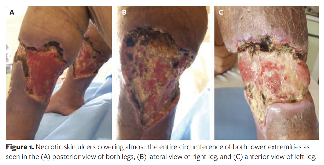
The patient was admitted with a diagnosis of necrotizing fasciitis. Empiric antibiotic therapy and analgesia were started, and surgical debridement of necrotic tissue was performed, followed by application of occlusive dressings with silver sulfadiazine. Despite initial treatment, the lesions progressed to necrosis, resulting in extensive ulceration of almost the entire circumference of the lower legs (Figure 1).
Irregularity of the soft tissue and vascular calcification from the medial region of the lower leg to the foot was shown on plain radiographs (Figure 2).

Additional laboratory tests were performed and revealed normal calcium levels, a normal parathyroid hormone value of 148 pg/mL, and normal tumor markers. A multidisciplinary team of specialists in general surgery, plastic surgery, and vascular surgery made the diagnosis of calciphylaxis based on clinical and radiologic data.
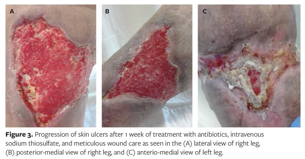
The patient was treated with antibiotics and intravenous sodium thiosulfate. Wound care consisted of collagenase, an enzymatic debriding agent, and polyhexanide gel to clean and decontaminate the wound. Tissue trauma and excessive manipulation were avoided, and impressive results were achieved after just 1 week of treatment (Figure 3).
After successful management of the infection, topical oxygen therapy was started. The authors of the current case report have previously successfully used this therapy to manage chronic wounds.
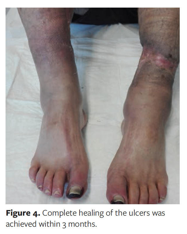
Complete healing of the ulcers was observed within 3 months (Figure 4).
Discussion
Calciphylaxis, also known as calcific uremic arteriolopathy, is a rare, complex, life‑threatening condition that occurs mostly in populations with end-stage chronic kidney disease, usually in dialysis.2,8 The pathogenesis of calciphylaxis remains unclear; however, a state resulting in the accumulation of calcium content in the medial wall of small blood vessels with fibrotic changes in intima has been described.4
Several risk factors have been associated with the development of calciphylaxis, including obesity, diabetes, female sex, and dependence on dialysis for more than 2 years.3 Other risk factors include hyperphosphatemia, use of medications such as warfarin and calcium-based phosphate binders, hypercoagulable states such as protein C and S deficiency or antiphospholipid antibody syndrome, hypoalbuminemia, inflammatory or autoimmune conditions, and recurrent skin trauma.3
A high index of clinical suspicion is necessary for diagnosis, because many medical conditions may mimic calciphylaxis.8 The differential diagnosis includes cellulitis, vasculitis, cholesterol embolization, antiphospholipid syndrome, and warfarin-associated skin necrosis.9
Calciphylaxis is characterized by extremely painful, violaceous, mottled skin lesions (livedo reticularis) that may progress to tissue necrosis and nonhealing ulcers, frequently in areas with increased adiposity, such as the lower extremities and trunk.2,3,8,9 There are no specific laboratory or radiologic tests to confirm the diagnosis. While hyperphosphatemia, hypercalcemia, and hypo- and hyperparathyroidism are risk factors for calciphylaxis, many patients present with normal values.10 Plain radiography and 3-phase nuclear bone scan can be important diagnostic tools, showing vascular calcification and calcified areas in the dermis and subcutaneous fat.2,10,11
Skin biopsy was not performed in the patient discussed in the current case report. Although there is ongoing discussion about whether skin biopsy may worsen the clinical course owing to intensive manipulation, the benefits of skin biopsy include ruling out other conditions that mimic calciphylaxis.2,4,7
Currently, owing to the paucity of evidence from randomized controlled trials, there is no standard treatment
for calciphylaxis.7,12 Management is focused on local wound care and metabolic control to prevent local and systemic infection.
The primary goals of wound care are to remove exudate and necrotic tissue. Surgical debridement can enlarge the necrotic area because the wound bed is poorly vascularized. In addition, these lesions are extremely painful, and consultation with a pain management specialist is necessary. In the current authors’ experience, for noninfected lesions the use of debriding agents (ie, hydrocolloid or hydrogel dressings) seems to be preferred for management of necrotic wounds as well as pain management.
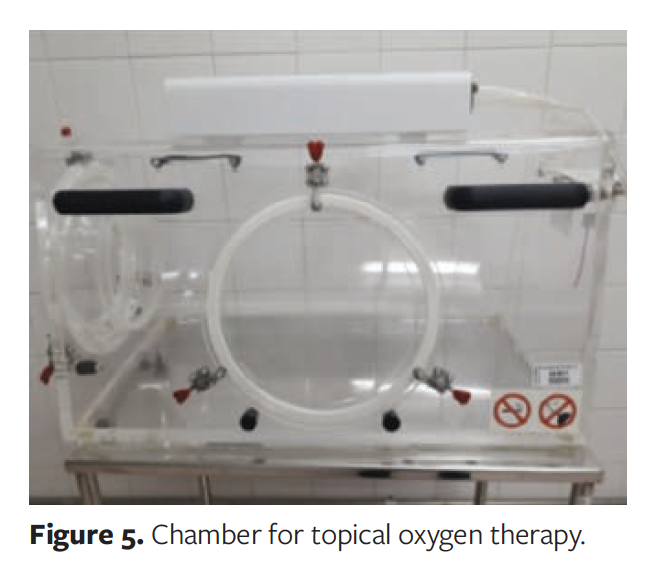
Options for adjunct topical treatment include negative pressure wound therapy (for infected wounds and large necrotic areas), hyperbaric oxygen therapy (available in only a small number of specialized centers), and topical oxygen therapy.13 In the authors’ hospital, topical oxygen therapy used in an airtight chamber to the affected area (Figure 5) has led to promising results for chronic wounds, especially diabetic foot ulcers.
Studies have demonstrated the effects of topical oxygen therapy in improving wound tissue perfusion, reducing hypoxia, downregulating inflammatory cytokines, and promoting fibroblast proliferation, collagen production, and angiogenesis. It also aids in eradicating difficult-to-treat soft tissue and bone infections by killing microorganisms and enhancing the effect of antimicrobials.14-16
Medications such as sodium thiosulfate and bisphosphonates have also been proposed as effective in the management of calciphylaxis. Sodium thiosulfate is an inorganic salt that is postulated to have vasodilatory and antioxidant properties. In an in vitro study, it was found to block the ability of adipocytes to induce calcification of vascular smooth muscle cells. Depending on the patient’s kidney function and dialysis modality, dosing varies from 5 g to 25 g given intravenously over 1 hour after high-flux hemodialysis 3 times a week.2,13 The optimal duration of treatment is not known.
Even if diagnosed in the early stages, calciphylaxis has a very poor prognosis, with a 1-year mortality rate that reaches up to 80%.6 Septicemia is the main reason for death, typically in the first 10 months after diagnosis.9
Limitations
This report is limited by its single case as well as the small number of publications on the condition.
Conclusion
Calciphylaxis is a rare, complex, life‑ threatening condition that occurs mostly in populations with chronic kidney disease or in patients on dialysis. Early diagnosis and a multidisciplinary approach are key to the management of this condition. Therapies that have shown promise include sodium thiosulfate, bisphosphonates, and oxygen therapy (an adjunctive topical treatment option). Establishing specific guidelines for the diagnosis and treatment of calciphylaxis will inform efforts to provide uniform clinical care and improve the prognosis of affected patients.
Acknowledgments
Authors: Raquel Barros Pereira, MD1; Aline Gomes, MD1; Emanuela Francisco2; and Carlos Casimiro, MD1
Affiliations: 1General Surgery Department, Tondela-Viseu Hospital Centre, Av. Rei Dom Duarte, Viseu, Portugal; 2Wound Care Nursing Department, Tondela-Viseu Hospital Centre, Av. Rei Dom Duarte, Viseu, Portugal
Disclosure: The authors disclose no financial or other conflicts of interest.
Correspondence: Raquel Barros Pereira, MD; Tondela-Viseu Hospital Centre, Av. Rei Dom Duarte, Viseu, 3504-509, Portugal; raquelmbpereira@gmail.com
References
1. Selye H. Calciphylaxis. University of Chicago Press; 1962.
2. Nigwekar SU, Thadhani R, Brandenburg VM. Calciphylaxis. N Engl J Med. 2018;378(18):1704-1714. doi:10.1056/NEJMra1505292
3. Nigwekar SU, Kroshinsky D, Nazarian RM, et al. Calciphylaxis: risk factors, diagnosis, and treatment. Am J Kidney Dis. 2015;66(1):133-146. doi:10.1053/j.ajkd.2015.01.034
4. Baby D, Upadhyay M, Joseph MD, et al. Calciphylaxis and its diagnosis: a review. J Fam Med Prim Care. 2019;8(9):2763-2767. doi:10.4103/jfmpc.jfmpc_588_19
5. Angelis M, Wong LL, Myers SA, Wong LM. Calciphylaxis in patients on hemodialysis: a prevalence study. Surgery. 1997;122(6):1083-1090. doi:10.1016/s0039-6060(97)90212-9
6. Roncada EV, Abreu MA, Pereira MF, Oliveira CC, Nai GA, Soares DF. Calciphylaxis, a diagnostic and therapeutic challenge: report of a successful case. An Bras Dermatol. 2012;87(5):752-755. doi:10.1590/s0365-05962012000500014
7. Brandenburg VM, Evenepoel P, Floege J, et al. Lack of evidence does not justify neglect: how can we address unmet medical needs in calciphylaxis? Nephrol Dial Transplant. 2016;31(8):1211-1219. doi:10.1093/ndt/gfw025
8. Kodumudi V, Jeha GM, Mydlo N, Kaye AD. Management of cutaneous calciphylaxis. Adv Ther. 2020;37(12):4797-4807. doi:10.1007/s12325-020-01504-w
9. Westphal SG, Plumb T. Calciphylaxis. In: StatPearls. StatPearls Publishing; August 8, 2022.
10. Chang JJ. Calciphylaxis: diagnosis, pathogenesis, and treatment. Adv Skin Wound Care. 2019;32(5):205-215. doi:10.1097/01.ASW.0000554443.14002.13
11. Burdorf BT. Calciphylaxis: the potential diagnostic role of radiologists. Radiol Case Rep. 2020;16(3):415-418. doi:10.1016/j.radcr.2020.11.033
12. Udomkarnjananun S, Kongnatthasate K, Praditpornsilpa K, Eiam-Ong S, Jaber BL, Susantitaphong P. Treatment of calciphylaxis in CKD: a systematic review and meta-analysis. Kidney Int Rep. 2018;4(2):231-244. doi:10.1016/j.ekir.2018.10.002
13. Erfurt-Berge C, Renner R. Management of patients with calciphylaxis: current perspectives. Chronic Wound Care Manag Res. 2019;6:109-115. doi:10.2147/cwcmr.s182417
14. Cole W, Yoder CM, Coe S. The use of topical oxygen therapy to treat a calciphylaxis wound during a global pandemic: a case report. Wounds. 2020;32(11):294-298.
15. Sayadi LR, Banyard DA, Ziegler ME, et al. Topical oxygen therapy & micro/nanobubbles: a new modality for tissue oxygen delivery. Int Wound J. 2018;15(3):363-374. doi:10.1111/iwj.12873
16. Wilmer WA, Voroshilova O, Singh I, Middendorf DF, Cosio FG. Transcutaneous oxygen tension in patients with calciphylaxis. Am J Kidney Dis. 2001;37(4):797-806. doi:10.1016/s0272 6386(01)80129-3


