


Charcot Neuroarthropathy Versus Osteomyelitis: A Case Series
Abstract
Introduction. Patients with diabetes and peripheral neuropathy have a 25% risk of developing a foot ulcer, and these can lead to soft tissue infections that worsen and result in osteomyelitis. While Charcot neuroarthropathy is not as common as osteomyelitis, it is often misdiagnosed as osteomyelitis. Case Reports. Three patients presented with diabetes, neuropathy, and foot ulcers. They underwent prophylactic surgery but later developed swelling at the surgical sites. Radiographs showed fragmentations that caused concern about osteomyelitis. The authors maintained diagnoses of Charcot neuroarthropathy and treated the patients with immobilization and offloading. All patients resolved the fragmentations without antibiotics or surgery. Conclusion. While Charcot neuroarthropathy and osteomyelitis have similar signs and symptoms, understanding the similarities and differences between the conditions can aid providers in appropriate wound management.
Abbreviations
BMI, body mass index; CAM, controlled ankle motion; CN, Charcot neuroarthropathy; COPD, chronic obstructive pulmonary disease; CT, computed tomography; DFU, diabetic foot ulcer; GERD, gastroesophageal reflux disease; MTPJ, metatarsophalangeal joint; MRI, magnetic resonance imaging; OM, osteomyelitis; PTB, probe to bone.
Introduction
According to the 2016 World Health Organization data, an estimated 422 million adults live with diabetes globally.1 Diabetes prevalence is also increasing rapidly among the US population. The Centers for Disease Control and Prevention’s 2019 estimates were that 28.7 million people of all ages, or 8.7% of the US population, are diagnosed with diabetes.2
Soft tissue ulceration in the feet is a widespread complication for patients with diabetes. The lifetime risk of DFU is 19% to 34%.3 These ulcerations can become infected if not treated quickly or adequately. One study found that soft tissue infections advanced to OM in 20% of cases.4 Patients present with swelling, redness, pain, and warmth in such instances, and the ulcer may have purulent discharge.
A lesser complication is CN, seen in 0.12% to 0.3% of patients with diabetes.5 That risk increases to 16% if patients have peripheral neuropathy.6 Patients with CN present signs similar to those of OM: swelling, redness, and warmth. However, they do not present with ulcers or a history of injury. Acute CN often gets misdiagnosed as OM.7
The authors report 3 cases of post-surgical patients who developed CN after healing from their surgery for chronic, non-infected ulcers. These patients presented with red, warm, and swelling feet with no new ulcers or history of trauma. The radiographic exams were read as concerning for OM. The authors maintained the findings as changes due to CN rather than OM.
Materials and Methods
The Institutional Review Board of Boston Medical Center and Boston University approved this study as exempt. All patients in this report were seen in the outpatient clinic of Boston Medical Center Podiatry. All clinicians involved in these reports are with Boston Medical Center, where interdisciplinary teamwork is valued. Electronic health records were used to obtain patients’ history, physical examination, progress notes, and radiographic results.
Results
Case 1: CN developed after Keller arthroplasty
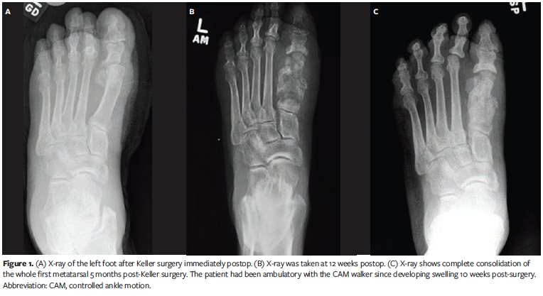
A 55-year-old male with a history of type 2 diabetes presented to the podiatry clinic with a foot ulcer lasting 3 months. Medical history included peripheral neuropathy, foot ulcers, hypertension, atrial fibrillation, and hyperlipidemia. He had palpable pedal pulses but lacked protective sensation. The DFU was located on the left plantar hallux interphalangeal joint, measuring 3 cm × 1 cm × 0.1 cm. It had mild edema and erythema but did not probe to the bone. The patient underwent weekly debridement, moist wound dressings, and offloading with felted foam in a postoperative shoe, but the DFU did not show improvement after 4 weeks. The patient underwent a left hallux Keller arthroplasty (Figure 1A). He was outfitted with a CAM boot and instructed to offload the left foot. The DFU healed in 2 weeks, but the patient developed dehiscence on the dorsal incision site. The wound was superficial and did not probe deep. The patient continued receiving wound care and offloading. The dehisced wound healed in 2 weeks. The patient returned to the clinic 2 months later, 10 weeks after the Keller procedure. The surgical site was closed but had mild swelling. Radiographs were not taken during this visit. The patient returned in 2 weeks, concerned that the left hallux had more swelling. He did not have an ulcer. The radiographic examination was read as having diffuse erosive and bony proliferative change concerning OM (Figure 1B). Still, the authors maintained that the clinical presentation was more consistent with a neuropathic fracture of CN. The patient was treated with strict offloading in a CAM boot, and the fragmented bone consolidated after 5 months (Figure 1C).
Case 2: CN developed after hallux arthroplasty
A 62-year-old male with a history of type 2 diabetes presented with an ulcer on the left hallux interphalangeal joint. The DFU had recurred several times over the past 2 years. Medical history included a cerebral vascular accident, hypertension, obesity (BMI, 50.4 kg/m2), hyperlipidemia, GERD, and epilepsy. He had palpable pedal pulses and a loss of protective sensation up to the ankle. The ulcer was measured at 1.8 cm × 1.5 cm × 0.2 cm. There was no sign of infection, and the bone was not exposed. He was treated with wound debridement, dressing changes, and offloading with a CAM boot.
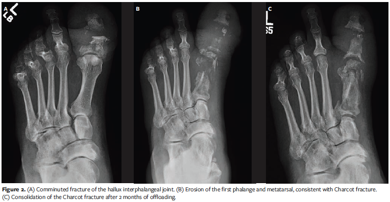
After the ulcer healed, a left hallux interphalangeal arthroplasty was performed. The patient was instructed to ambulate with a surgical shoe and walking cane, only putting weight on the heel. One month after surgery, he developed a dehiscence, approximately 0.4 cm × 2 cm in size, with no clinical signs of infection. He was treated and instructed to offload. The dehisced wound healed in 2 months. The patient presented 3 months later with swelling and redness of the foot. He had no ulcer and denied any trauma. Radiographs showed a comminuted fracture of the hallux interphalangeal joint (Figure 2A). The radiographic reports did not mention OM or CN. He was placed in a CAM boot and instructed not to bear weight on the foot. The patient returned in 2 weeks, but the swelling had not improved. Repeat radiographs at this visit showed more bone fragments of the left hallux proximal phalange and first metatarsal concerning OM (Figure 2B). The current study’s authors maintained that the changes were consistent with CN rather than OM and continued to treat the patient with offloading and a CAM boot. He was scheduled to follow up every 2 weeks and was advised to return sooner if there was a wound. The swelling subsided after 2 months, and new radiographs showed consolidation of the fragmented bone (Figure 2C).
Case 3: CN developed after MTPJ resection
A 57-year-old male with type 2 diabetes presented with an ulcer on the dorsum of his right first MTPJ with a duration of 2 months. Medical history included peripheral neuropathy, COPD, alcohol use, and DFU. The ulcer had been treated with debridement, wound care, and oral antibiotics at a community clinic before the patient presented to the authors’ institution. The patient had palpable pulses in the right dorsalis pedis and posterior tibial arteries and absent epicritic sensation via 10 g mono-filament testing.
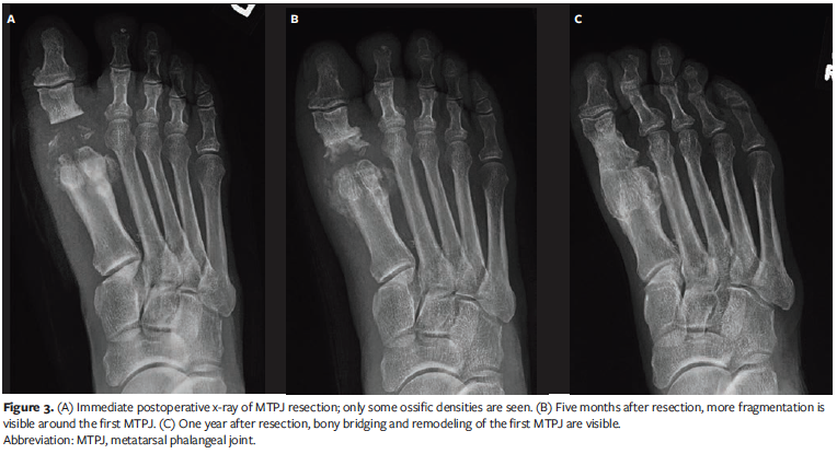
The ulcer measured 1 cm × 1.1 cm × 0.3 cm. The ulcer base had fibrogranular tissue with periwound erythema and edema. The ulcer did not probe to the bone. Deep wound cultures grew methicillin-resistant Staphylococcus aureus. The patient was admitted for intravenous antibiotic treatment and wound care. He underwent incision and drainage of the ulcer, resection of the right first MTPJ, and extensor tenotomy (Figure 3A). The patient was discharged on an oral antibiotic when the infection improved. He was placed in a CAM boot and instructed to offload. He returned 1 week later with a smaller ulcer. The ulcer worsened over the course of 2 weeks, but there were no signs of infection. The patient was admitted, restarted on antibiotics, and discharged after 5 days. The ulcer healed in 1 month.
The patient returned to the clinic 3 months later. The foot was swollen, warm, and erythematous, but there was no ulcer. He denied any trauma. Radiographs at this visit showed bone erosion around the base of the proximal phalange and the head of the first metatarsal concerning OM (Figure 3B). The authors maintained that the radiographic changes were consistent with CN, and the patient was placed on a CAM boot and advised to offload. The swelling, warmth, and redness improved in 2 months. When the patient returned to the clinic 6 months later, the radiographs showed complete coalescence of the first MTPJ (Figure 3C).
Discussion
Diagnosis
OM and CN can present with similar clinical features (redness, swelling, warmth, pain) and radiographic findings (bone and soft tissue edema).8 An OM diagnosis should be based on clinical signs of infection supported by laboratory, microbiological, and radiological evaluation. A PTB test can be used to evaluate OM. A prospective study of 1666 patients with diabetes found that the PTB test has a sensitivity of 87% and a specificity of 91%.9 An acute CN diagnosis should also be based on clinical signs similar to OM. However, acute CN is not associated with ulcers. Patients with CN have good pedal pulses that may not be present in OM, and CN is often misdiagnosed as cellulitis and OM. CN and OM are destructive processes, but painless, and glycosylated hemoglobin can be elevated in both conditions. An ulcer usually precedes OM, while CN can form without an ulcer. Trauma can lead to OM and CN, but patients may not recall any injury due to neuropathy. Delaying and misdiagnosing CN can lead to devastating health outcomes for the patient.10 A delayed Charcot foot diagnosis was associated with 10.8% greater inpatient costs and a 12.1% longer stay.9 More patients underwent lower extremity amputation when a CN diagnosis was delayed, resulting in a 30.4% increase in costs and a 31.6% longer length of stay.11
Imaging
Plain radiographs show clear signs of OM once 30% to 50% of the bone becomes involved, usually 2 to 3 weeks after infection sets in.12 The OM-involved bones are characterized by osteopenia, erosion of cortical bone, cortical lysis, osteolysis, periosteal thickening, and bone sequestration.10 MRI, ultrasonography, CT scans, and nuclear scintigraphy may be required to diagnose OM. MRI should be recommended when radiographs are inconclusive for OM and CN.13
Pathogenesis
While soft tissue infection precedes OM, there are several theories on how CN develops. A combination of mechanical, vascular, and biological factors trigger CN. There is no singular cause for the development of CN, but there are factors that predispose to its development, as well as several likely precipitating events.14 Several diseases must be considered for the differential diagnosis of CN, often related to the acute phase, including gout, ankle sprain, inflammatory arthritis, cellulitis, venous thrombosis, and osteomyelitis.14 Diabetic peripheral neuropathy is a risk for CN; other factors include a history of foot ulceration, retinopathy, nephropathy, renal failure, rheumatoid arthritis, iron deficiency anemia, and obesity.14
There are many theories on the development of CN.15,16 The neurotraumatic theory states that peripheral neuropathy causes sensory loss in the feet, which lose protective sensation and become vulnerable to an increased risk of unrecognized trauma. The neurovascular theory suggests that increased blood flow to the bones due to damage to the “trophic nerves” results in bone resorption and weakening, ultimately leading to fractures and deformities. Other theories relate to the association between diabetes and osteoporosis,17 significant weight loss,18 and kidney transplantation.19
Prevalence
OM is a common DFU infection in 10% to 15% of moderate and 50% of severe infections.20 CN has an incidence ranging from 0.1% to 10% among people with diabetes,21 but it is rare to see CN in both feet.22
Classification
Classification and scoring systems can help the clinician manage DFUs and communicate with others in a multidisciplinary approach.23 The Meggitt-Wagner and University of Texas diabetic foot classification systems are the most commonly used in DFUs.24,25 The Meggitt-Wagner system allows the classification of wound depth and the presence of gangrene; it was modified to include ulcers that extend to the bone. The University of Texas classification categorizes DFUs using 4 grades and 4 stages.
The Eichenholtz and Sanders classifications are the most frequently used for CN. Eichenholtz’s classification26 has 3 stages based on radiologic changes. Shibata et al added a stage 0 in 1990 to the Eichenholtz classification for no radiographic change.27 The anatomical classification proposed by Sanders and Frykberg28 describes 5 different patterns depending on the foot areas involved. Wukick et al showed that the Sanders-Frykberg system is most helpful for physician communication.29
Treatment
The standard of care for DFUs is sharp debridement, daily wound care dressings to keep the wound moist, offloading, keeping blood glucose under control, having adequate blood flow, and infection control.30 A multidisciplinary approach to care for the diabetic foot is recommended, which includes annual assessments by a primary care physician and referral to a podiatrist and vascular surgeon. If the standard of care does not heal the ulcer after 4 weeks, it is recommended to consider a surgical option. A meta-analysis of comparative studies by Yammine and Assi shows that surgery can lead to faster healing than the standard of care.31 No study directly compares surgical and antibiotic regimens in OM.32 Lázaro-Martínez et al reviewed a prospective randomized controlled trial to compare antibiotics versus conservative surgery for OM.33 Still, there were only 18 patients included, and the criteria for OM were weak. The ideal duration of antibiotic administration for patients treated conservatively is not well studied,34 and pharmacological options have not been shown effective in treating CN.6 Offloading is essential to heal and manage both DFU and CN.35
Removing the infected bone is necessary for OM, while the indication for surgical treatment of CN is a severe deformity.36 OM and CN can lead to amputation if not treated adequately, and treatment gets more complicated when OM occurs in the same foot as CN. A case report by Aragón-Sánchez et al indicated that CN is triggered and complicated by OM.37 Berli et al reported in a retrospective cohort study that CN and OM could occur separately or concurrently.38
Summary
All 3 patients in this case series had a history of chronic ulcers around the first metatarsal, presented with DFUs but not OM, and underwent prophylactic surgery. Shortly after the patients healed from the surgery, they presented to the clinic with foot swelling, warmth, and redness. None of the patients had open wounds or recent injuries. The initial radiographs showed erosion and destruction of the first metatarsal joints and concern for OM. Hematogenous OM is rare in adults. The current study’s authors maintained that the change in x-rays was more consistent with CN because of the lack of open wounds. All 3 patients were treated with immobilization and offloading in CAM boots. The follow-up x-rays showed coalescence and consolidation of the bones. These patients showed a pattern of CN as described by Sanders and Frykberg’s classification of zone I: metatarsophalangeal and interphalangeal joints, occurring in 15% of patients.28
Limitations
This case series has limitations, including a small number of cases. In addition, patients missed many follow-up visits. Also, MRI was not done to evaluate the changes in radiographs. Photography of wound progression and healing would improve this report.
Conclusion
While only a few cases are reported here, the authors want to highlight the need to distinguish OM from CN before making a surgical decision. CN and OM can easily be mistaken for each other, given similar signs and symptoms, laboratory results, and radiographic findings. Early diagnosis of CN is essential, as failure to treat CN early and adequately can lead to deformity, leading to OM. While CN deformity can be reconstructed, OM requires amputation. The clinician should know how to recognize and manage both CN and OM. These disease processes can lead to devastating patient consequences, and a prospective study to evaluate OM and CN would be beneficial. Understanding all aspects of CN and OM is essential. One must focus on the patient, not laboratory or radiographic studies alone. The patients in this case series did not need antibiotics, the CN consolidated with immobilization, and amputation was avoided.
Acknowledgments
Authors: Hau T. Pham, DPM1,2; Elizabeth Sanders, DPM1,2; Ewald R. Mendeszoon, DPM1,2; and Wei Tseng, DPM1,2
Affiliations: 1Boston Medical Center, Boston, MA; 2Boston University Chobanian & Avedisian School of Medicine, Boston, MA
Disclosure: The authors disclose no financial or other conflicts of interest.
Correspondence: Hau T. Pham, DPM; Boston Medical Center Division of Podiatry, 732 Harrison Ave 5th floor, Boston, MA 02118; Hau.pham@bmc.org
References
1. World Health Organization. https://www.who.int/news-room/fact-sheets/detail/diabetes. 5 April 2023.
2. Centers for Disease Control and Prevention. National Diabetes Statistics Report website. https://www.cdc.gov/diabetes/data/statistics-report/index.html. June 29, 2022.
3. McDermott K, Fang M, Boulton AJM, Selvin E, Hicks CW. Etiology, epidemiology, and disparities in the burden of diabetic foot ulcers. Diabetes Care. 2023;46(1):209-221. doi:10.2337/dci22-0043
4. Lavery LA, Peters EJ, Armstrong DG, Wendel CS, Murdoch DP, Lipsky BA. Risk factors for developing osteomyelitis in patients with diabetic foot wounds. Diabetes Res Clin Pract. 2009;83(3):347-352. doi:10.1016/j.diabres.2008.11.030
5. Gratwohl V, Jentzsch T, Schöni M, et al. Long-term follow-up of conservative treatment of Charcot feet. Arch Orthop Trauma Surg. 2022;142(10):2553-2566. doi:10.1007/s00402-021-03881-5
6. Cavanagh PR, Young MJ, Adams JE, Vickers KL, Boulton AJ. Radiographic abnormalities in the feet of patients with diabetic neuropathy. Diabetes Care. 1994;17(3):201-209. doi:10.2337/diacare.17.3.201
7. Jude E. Charcot foot: what’s new in pathogenesis and medical management? In: Boulton AJM, Cavanagh PR, Rayman G, eds. The Foot in Diabetes. John Wiley & Sons, Ltd; 2006:265-273.
8. Botek G, Figas S, Narra S. Charcot neuroarthropathy advances: understanding pathogenesis and medical and surgical management. Clin Podiatr Med Surg. 2019;36(4):663-684. doi:10.1016/j.cpm.2019.07.002
9. Lavery LA, Armstrong DG, Peters EJ, Lipsky BA. Probe-to-bone test for diagnosing diabetic foot osteomyelitis: reliable or relic? Diabetes Care. 2007;30(2):270-274. doi:10.2337/dc06-1572
10. Diacogiorgis D, Perrin BM, Kingsley MIC. Factors impacting the evidence-based assessment, diagnosis and management of acute Charcot


