


Misdiagnosis of Cutaneous Epithelioid Angiosarcoma as Diabetic Foot Ulcer: A Case Study
© 2024 HMP Global. All Rights Reserved.
Any views and opinions expressed are those of the author(s) and/or participants and do not necessarily reflect the views, policy, or position of Wounds or HMP Global, their employees, and affiliates.
Abstract
Background. AS is a malignant tumor that originates from vascular endothelial cells and is known for a high rate of local recurrence and metastasis. Case Report. A 48-year-old male presented with cutaneous epithelioid AS. Cutaneous AS of the foot is quite rare, especially in the absence of predisposing factors, and in this patient it was previously misdiagnosed as a DFU. Conclusion. Physicians should be aware of this rare presentation of cutaneous AS. The authors of the current report advise regular clinical reassessment of chronic ulcers and biopsies of nonhealing wounds, even when adequate wound treatment has been administered, with the goal of identifying ulcerated skin malignancies and preventing delay in providing appropriate treatment.
Abbreviations
AS, angiosarcoma; DFU, diabetic foot ulcer; ETS, E26 transformation-specific; STS, soft tissue sarcoma.
Introduction
AS is a rare tumor that may develop in the subcutaneous tissues of various anatomic areas, including the head and neck or chest; however, it rarely develops in the limbs.1 It is one of the most common tumors caused by therapeutic radiation, often following treatment for breast cancer or Hodgkin lymphoma.2 Cutaneous AS is highly aggressive clinically, with reported 5-year survival rates ranging from 12% to 24%.2 The majority of patients present with locally advanced disease, regional lymph node involvement, or distant metastases at initial diagnosis, all of which are associated with a poor prognosis.3 Severe malignant skin tumors present as chronic leg or foot ulcers, which may be misdiagnosed as diabetic ulcers.4
The current report discusses a case of cutaneous epithelioid AS that was initially misdiagnosed as a DFU.
Case Report
A 48-year-old Moroccan male with a medical history of diabetes who had been taking metformin for 3 years presented to the dermatology department at the authors’ institution with a nonhealing wound of the left foot of 2 years’ duration. At this presentation, the patient’s HbA1c value was 5.7%. The patient was initially diagnosed with a DFU 2 years prior and was given a course of oral and topical antibiotics and dressings; however, the wound did not respond to treatment.
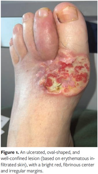
In the 3 months prior to presentation to the department of the authors of the current report, the lesion enlarged and became increasingly painful. On physical examination, the lesion was found to be ulcerated, oval-shaped, and well-confined (based on erythematous infiltrated skin), with a bright red, fibrinous center and irregular margins. It bled easily to the touch; secreted a purulent, foul-smelling fluid; and was painful on palpation. The lesion measured 7.5 cm in diameter and was located on the dorsal aspect of the left foot (Figure 1). Dactylitis of the left second toe with restricted mobility of the first and second toes was noted as well. A punch biopsy of the lesion was positive for cutaneous epithelioid AS.
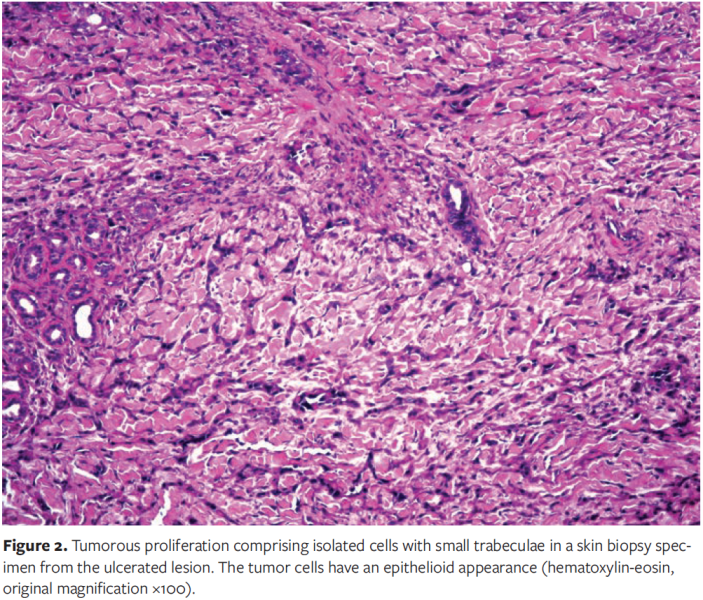
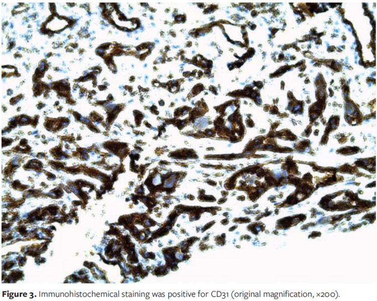
Microscopic examination revealed a tumorous proliferation of atypical mesenchymatous appearance, composed of isolated cells with small trabeculae; tumor cells sometimes border vascular slits of varying size, which can be anfractuous. Tumor cells have an epithelioid appearance with abundant acidophilic cytoplasm, sometimes with a vacuole; they have ovoid or irregular hyperchromatic and anisokaryotic nuclei, sometimes multilobulated with some multinucleated cells (Figure 2). Immunohistochemical staining was negative for CD34, cytokeratin, and ERG, but was positive for CD31 (Figure 3). No necrosis was identified in the specimen, and the mitotic count was 0 to 9 mitoses per 10 high-power fields. Magnetic resonance imaging of the foot revealed a tumor measuring 62 mm × 42 mm × 44 mm that had invaded the first, second, and third metatarsals; the first phalanx of the second toe; the first 2 joint spaces of the first, second, and third metatarsals and the muscles of the sole of the foot; and had reached the fourth metatarsal. Fluorodeoxyglucose-positron emission tomography was negative for hypermetabolic lesions.
After a multidisciplinary consultation meeting, the therapeutic decision was to amputate the foot and forego adjuvant radiation therapy. Unfortunately, the patient died 6 months after amputation.
Discussion
AS is a malignant tumor that originates from vascular endothelial cells and is known for a high rate of local recurrence and metastasis.¹ One-third of such tumors occur in the skin, 25% occur in soft tissue, and the remainder occur in other areas. In the majority of STS cases, no definitive cause is clearly identified. However, several associated or predisposing factors have been recognized, including radiation therapy, vascular insufficiency, chronic lymphedema, trauma, and sun exposure.5 In the current case, no predisposing factor was found.
Typically, AS is a disease of older adults, with a mean age of onset of 65 to 70 years and a male-to-female ratio of 2:1. More than 90% of cutaneous AS cases occur on the head and neck; other, noncutaneous regions affected include the chest and liver.¹ AS of the lower extremity without lymphedema, as in the current case, is exceptional.6 The vast majority of lower limb AS is associated with chronic lymphedema, which can lead to Stewart-Treves syndrome; this syndrome accounts for 5% of AS cases, with approximately 400 cases of the syndrome reported in the literature as of 2015.7
Clinically, patients with AS most commonly report a gradually enlarging, painless mass. Some patients report pain or symptoms related to compression caused by the mass, including paresthesia or edema in an extremity. However, a variable clinical presentation may be observed that may include bruise-like lesions, raised purplish-red papules, and rosacea-like lesions.8 AS may also appear as nodules, papules, plaques, and exophytic tumors on the surface of progressive lesions. In some cases, advanced lesions may resemble epithelial neoplasms such as squamous cell carcinoma, keratoacanthoma, basal cell carcinoma, and melanoma.8 This variability in appearance can lead to a delay in diagnosis, as in the current case.
Toussaint et al4 evaluated the incidence of malignant skin tumors presenting as chronic leg or foot ulcers and analyzed the frequency and nature of initial clinical misdiagnoses. Of the 673 cases in that study, 26 (3.9%) were identified with a total of 27 malignant tumors that presented as chronic lower leg or foot ulcers. Of these 27 tumors, 7 were diagnosed as melanoma, 8 as squamous cell carcinoma, and 12 as basal cell carcinoma. There are case reports of other, less common skin cancers that mimic chronic leg ulcers, such as AS.4 Furthermore, as noted by Toussaint et al,4 "[a] German S3 guideline for the local treatment of chronic wounds from 2013 recommends biopsy in cases of atypical wounds and differential-diagnostic evaluation in cases of absence of healing tendency after 6 weeks of treatment, according to the guidelines." This S3 guideline was created in accordance with generally recognized quality criteria under the aegis of the German Association for Wound Healing and Wound Treatment.
Cutaneous AS may be well or poorly differentiated histopathologically. Well-differentiated lesions are composed of a proliferation of irregular and sometimes anastomosing vascular structures in an ill-defined lesion. Under higher magnification, the malignant endothelial cells appear pleomorphic and hyperchromatic, and often bulge into the vascular lumina. Poorly differentiated cutaneous AS can be indistinguishable from carcinoma and melanoma because of the presence of sheets of mitotically active pleomorphic cells with ill-defined, intervening vascular structures. The presence of irregular vessels at the periphery of the tumor, intracytoplasmic vacuoles, small areas of hemorrhage, and erythrocytes in vascular lumens may serve as diagnostic clues for vascular origin.6,8
Cutaneous epithelioid AS, as in the current case report, is characterized by the predominance of round or polygonal cells with abundant eosinophilic cytoplasm and vesicular nuclei. Nodular, micronodular, syncytial, and diffuse growth patterns have also been described.6,8 Zones of necrosis are often observed.6,8 Immunohistochemistry is used to diagnose cutaneous AS. CD31 and CD34 are the most commonly used markers; however, these markers are also expressed by other cell lineages, such as hematopoietic and fibrohistiocytic cells. CD31 is more specific for AS, but it also may be expressed in macrophages, histiocytes, and plasma cells, leading to possible diagnostic pitfalls.6,8 Nuclear immunohistochemical markers from the ETS transcription family, including ERG and FLI1, are highly sensitive for cutaneous AS. In contrast to conventional cutaneous AS, epithelioid AS may express cytokeratins and epithelial membrane antigen.6,8 In the patient in the current case report, immunohistochemical staining was negative for CD34, but positive for CD31, cytokeratin, and ERG.
The objectives for managing extremity STSs are to minimize local recurrence, perioperative morbidity, and mortality while maximizing function and long-term survival. The primary approach for managing localized STS involves surgical resection. For most patients with high-grade primary extremity STS, however, a combination of radiation therapy and wide surgical resection is recommended, with some exceptions. The role of chemotherapy in the management of any STS remains a subject of ongoing research and discussion.9 In rare cases in which it is deemed necessary, primary amputation may be considered for patients with large, locally advanced or recurrent extremity STS that cannot be addressed with resection, or for patients with significant medical comorbidities.10 Long-term survival following surgical management of extremity STS depends on histologic grade and the presence of metastatic disease.10
STS of the hand and foot tends to have a slightly better prognosis than does more proximal extremity STS. The 5-year survival rate for patients with STS of the hand and foot is approximately 80%, which is better than for proximal extremity disease.1
Limitations
This case report has limitations. Cutaneous epithelioid AS of the foot is rare, and only 1 new case is reported.
Conclusion
Cutaneous AS of the foot is quite rare, especially in the absence of predisposing factors. In the patient reported herein, the tumor had previously been misdiagnosed as a DFU. The aim of the current case report is to increase awareness of this rare presentation of cutaneous AS. Additionally, the authors advise conducting regular clinical reassessment of chronic ulcers and biopsies of nonhealing wounds, even when adequate wound treatment has been administered, with the goal of identifying ulcerated skin malignancies and preventing delay in providing the appropriate therapy.
Acknowledgments
Authors: Yousef Almheirat, MD1; Lamis Elyamani, MD1; Ouissal Hormi, MD1; Nassiba Zerrouki, Prof1,2; Nada Zizi, Prof1,2; and S. Dikhaye, Prof1,2
Affiliations: 1Department of Dermatology, Mohammed VI University Hospital of Oujda, Medical School of Oujda, Mohammed First University of Oujda, Oujda, Morocco; 2Department of Epidemiology, Clinical Research and Public Health Laboratory, Medical School of Oujda, Mohammed First University of Oujda, Oujda, Morocco
Disclosure: The authors disclose no financial or other conflicts of interest.
Correspondence: Yousef Almheirat, MD; Department of Dermatology, Mohammed VI University Hospital (Oujda), Boulevard Allal el Fassi, Oujda, Oujda 60000 Morocco; joudehyousef94@gmail.com
Manuscript Accepted: December 14, 2023
References
1. Ryan CW, Meyer J. Clinical presentation, histopathology, diagnostic evaluation, and staging of soft tissue sarcoma - UpToDate. Accessed June 1, 2023. https://www.uptodate.com/contents/clinical-presentation-histopathology-diagnostic-evaluation-and-staging-of-soft-tissue-sarcoma
2. Albores-Saavedra J, Schwartz AM, Henson DE, et al. Cutaneous angiosarcoma. Analysis of 434 cases from the surveillance, epidemiology, and end results program, 1973-2007. Ann Diagn Pathol. 2011;15(2):93-97. doi:10.1016/j.anndiagpath.2010.07.012
3. Toll A. Cutaneous angiosarcoma: the importance of clinical suspicion. Article in Spanish, English. Actas Dermosifiliogr. 2017;108(5):394. doi:10.1016/j.ad.2017.03.002
4. Toussaint F, Erdmann M, Berking C, Erfurt-Berge C. Malignant tumours presenting as chronic leg or foot ulcers. J Clin Med. 2021;10(11):2251. doi:10.3390/jcm10112251
5. Le Corre Y, Avenel-Audran M, Croué A, Steff M, Verret JL. Cutaneous angiosarcoma of the leg without lymphoedema. Article in French. Ann Dermatol Venereol. 2008;135(6-7):488-491. doi:10.1016/j.annder.2007.08.006
6. Tenjarla S, Sheils LA, Kwiatkowski TM, Chawla S. Cutaneous angiosarcoma of the foot: a case report and review of the literature. Case Rep Oncol Med. 2014;2014:657876. doi:10.1155/2014/657876
7. Veiga RRG da, Nascimento BAM do, Carvalho AH, Brito AC de, Bittencourt M de JS. Stewart-Treves syndrome of the lower extremity. An Bras Dermatol. 2015;90(3 Suppl 1):232-234. doi:10.1590/abd1806-4841.20153926
8. Shustef E, Kazlouskaya V, Prieto VG, Ivan D, Aung PP. Cutaneous angiosarcoma: a current update. J Clin Pathol. 2017;70(11):917-925. doi:10.1136/jclinpath-2017-204601
9. Bi S, Zhong A, Yin X, Li J, Cen Y, Chen J. Management of cutaneous angiosarcoma: an update review. Curr Treat Options Oncol. 2022;23(2):137-154. doi:10.1007/s11864-021-00933-1
10. Requena C, Alsina M, Morgado-Carrasco D, et al. Kaposi sarcoma and cutaneous angiosarcoma: guidelines for diagnosis and treatment. Article in English, Spanish. Actas Dermosifiliogr. 2018;109(10):878-887. doi:10.1016/j.ad.2018.06.013


