


Outcomes of Layered Closure and Adjunctive Adhesive Retention Suture Device Use Following Ankle Fracture Open Reduction and Internal Fixation: A Single Center, Retrospective Cohort Study
Abstract
Introduction. Wound edge necrosis, dehiscence, infection, and need for reoperation can occur following ankle fracture ORIF. An assistive retention suture device has been developed to serve as an adjunct to skin closure to help mitigate the potential for wound dehiscence and subsequent complications. Objective. This single-center retrospective review compared early postoperative healing and functional outcomes following ankle fracture ORIF in 39 patients in which layered closure alone (n = 20) and layered closure with use of a DAC (n = 19) was performed. Results. Adjunctive assistive retention suture device use with layered closure following ankle fracture ORIF resulted in a 60% decrease in the time to healing, an 80% reduction in postoperative dehiscence, a 90% reduction in the need for reoperation, and a 53% reduction in the time in which use of an ambulatory assistive device, brace, or orthoses was required compared to layered closure alone. Conclusions. The results of this study suggest that use of this assistive retention suture device minimizes potential postoperative healing complications, decreases need for revisional surgery, and reduces time until return to activity.
Abbreviations
DAC, device for assisted closure; IDDM, insulin-dependent diabetes mellitus; IRB, institutional review board; ORIF, open reduction and internal fixation.
Introduction
Early postoperative soft tissue complications—including wound dehiscence, necrosis, infection, and swelling—have been documented to occur in up to 40% of patients following ankle fracture ORIF.1 These complications occur secondary to various patient- and injury-related factors combined with the limited and vulnerable soft tissue coverage in this area.2 Avoidance of wound dehiscence is paramount to mitigating these complications. Layered closure is commonly performed to help reduce tension along the incision line, reducing the potential for wound dehiscence and improving aesthetic outcomes.3 However, the evidence supporting these functions of deep/dermal closure combined with superficial skin closure is lacking.4,5
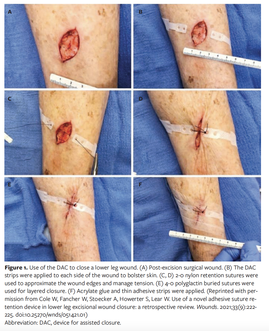
Lateral approach and minimally invasive techniques,6-9 different suture closure techniques,10-14 and closure assistive devices15-18 have been used to reduce the risk of postoperative incision dehiscence. Variations in suture closure techniques and use of closure assistive devices both require multiple full-thickness puncture holes for suture placement and pulling tension applied across the incision site for closure.10-18 Disadvantages of these techniques include the inability to obtain full primary closure, misalignment of the skin edges, and skin tearing and tissue strangulation, which can result in tissue loss due to the multiple suture puncture sites and tension placed on the skin for closure.19 One novel DAC (HEMIGARD; SUTUREGARD Medical Inc.) was designed to sit above the incision site and be held in place with a single full-thickness percutaneous or subcutaneous retention suture. Stress-relaxation response of the skin and soft tissue allows for reapproximation of the skin edges for layered closure to be performed. The ability to achieve full primary closure with fewer full-thickness puncture sites and less pulling tension for closure reduces the potential for skin tearing, tissue strangulation, and tissue loss compared to the previously mentioned closure techniques (Figure 1).13,15 Use of the DAC in patients with fragile skin and wounds in areas of limited cutaneous laxity has been shown to assist in primary closure and reduced post-procedural dehiscence rates.20-22 The aim of the current study was to determine if supplementation of layered closure with the DAC would reduce surgical incision dehiscence following ankle fracture ORIF compared to layered closure alone.
Materials and Methods
This was a single-center, retrospective cohort study comparing rates of postoperative dehiscence prior to and following the use of DAC on patients who underwent ankle fracture ORIF between 03/2019 and 11/2021 by 2 senior surgeons (A.K., Principal Investigator, and E.H.). Patients included in the study were 18 years of age or older. All patients provided written informed consent for the use of deidentified images and medical and surgical history for educational purposes. IRB approval was not required by the author’s institution as this was a retrospective review of deidentified patient data to be used for educational purposes. The consent forms were maintained within each respective patient’s medical record. The ankle fracture injury types were designated as trimalleolar, bimalleolar, pilon, or lateral malleolar fracture. The study included both open and closed ankle fractures. While the risk for postoperative healing complications can differ between these ankle fracture patterns and open versus closed fractures, all ankle fracture types—including those requiring delta frame application—were included in this study to allow for the greatest number of patients for review.
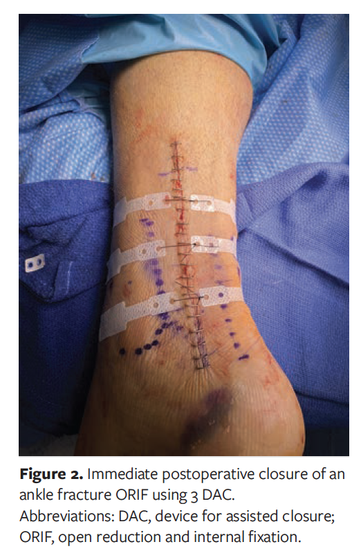
Ankle fracture ORIF was performed on the day of injury, or within 7 to 10 days following injury if edema reduction or fracture blister management was required. Delta frame application was performed when deemed necessary by the senior surgeons, typically due to a grossly unstable ankle fracture or soft tissue compromise. For patients for whom application of a delta frame was deemed necessary, time to the final ORIF procedure ranged from 7 to 28 days. Layered closure was performed in all patients. This closure consisted of 3-0 polyglactin 90 for deep closure and 4-0 polyglactin 90 for subcutaneous closure in simple interrupted fashion, followed by 4-0 nylon using horizontal sutures or skin staples for skin closure. For patients in which a DAC was used, subcutaneous and skin closure techniques were the same as noted above. The difference in this group was placement of one DAC every 3 cm along the incision using a 2-0 nylon following subcutaneous closure and prior to skin closure. Once approximation of the wound edges was achieved, skin closure was completed with either horizontal sutures using 4-0 nylon or skin staples (Figure 2).
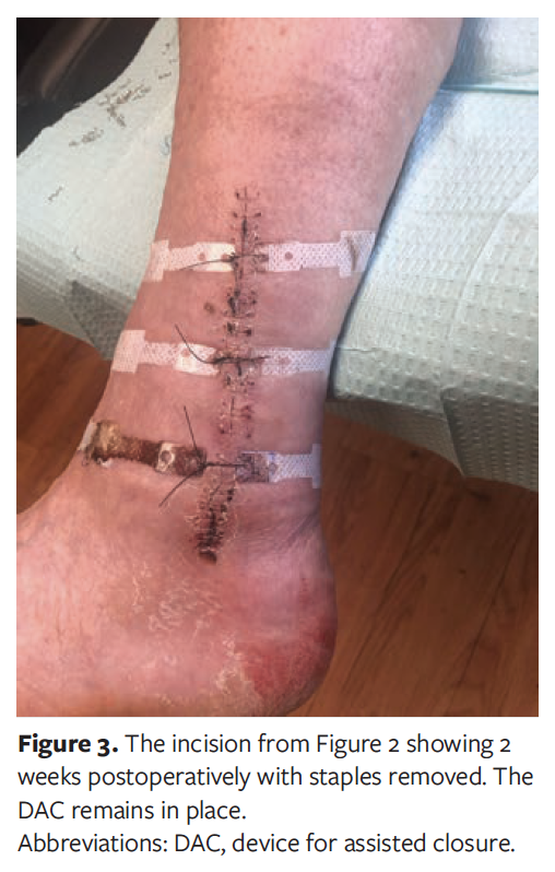
The postoperative treatment regimen was outlined by the same 2 senior surgeons (A.K. and E.H.). Patients were allowed to begin non-weightbearing ankle range-of-motion exercises at 1 week postoperative. Sutures or staples for skin closure were removed at 2 weeks postoperative in patients without diabetes (Figure 3) and 3 weeks postoperative in patients with diabetes. Patients were allowed to begin partial weightbearing in a removable walking boot at this time, if deemed clinically appropriate. If concern existed that starting partial weightbearing may precipitate potential complications, this was delayed. The DAC device was removed at 4 weeks postoperative. Removal of the DAC occurred once the incision was fully healed or one of the 2 senior surgeons deemed that the device was no longer providing additional support, ie, was loose or non-functioning.
A healed incision was defined as 100% reepithelization with no drainage or dressing requirement. Edge necrosis was defined as dusky tissue appearance resulting in tissue loss along the incision edges. Postoperative dehiscence was defined as the partial or total separation of previously approximated wound edges. Postoperative infection was defined as local signs and symptoms of infection appearing at the surgical site requiring topical and/or systemic antibiotic therapy within the first 30 days postoperative. Readmission and reoperation rates were recorded for the first 30 days postoperative. Full weightbearing in an ankle brace was allowed at 6 weeks postoperative. Complication assessment was performed by the same 2 senior surgeons (A.K. and E.H.).
Statistical plan
Time to incision healing; time to suture removal; rates of edge necrosis; postoperative dehiscence; postoperative infection; need for readmission; need for reoperation; time to partial weightbearing and complete weightbearing; time needed for continued use of ambulatory assistive devices, braces, or orthoses; and the need for physiotherapy were compared between subjects in which layered closure alone was performed (control) and those in which layered closure was supplemented with DAC (DAC group). Patient demographics and wound characteristics were abstracted from the patients’ medical records by E.S. Descriptive statistics were used for data analysis, with numerical data reported as mean ± standard deviation and categorical data reported as percentages of the total group.
Results
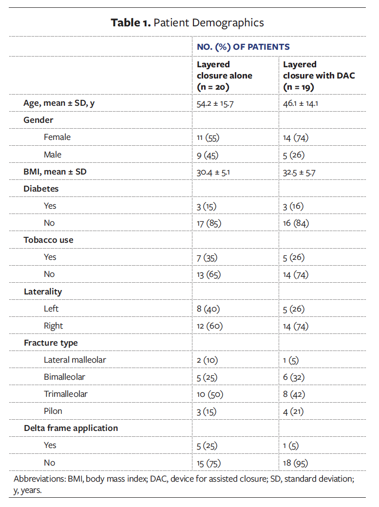
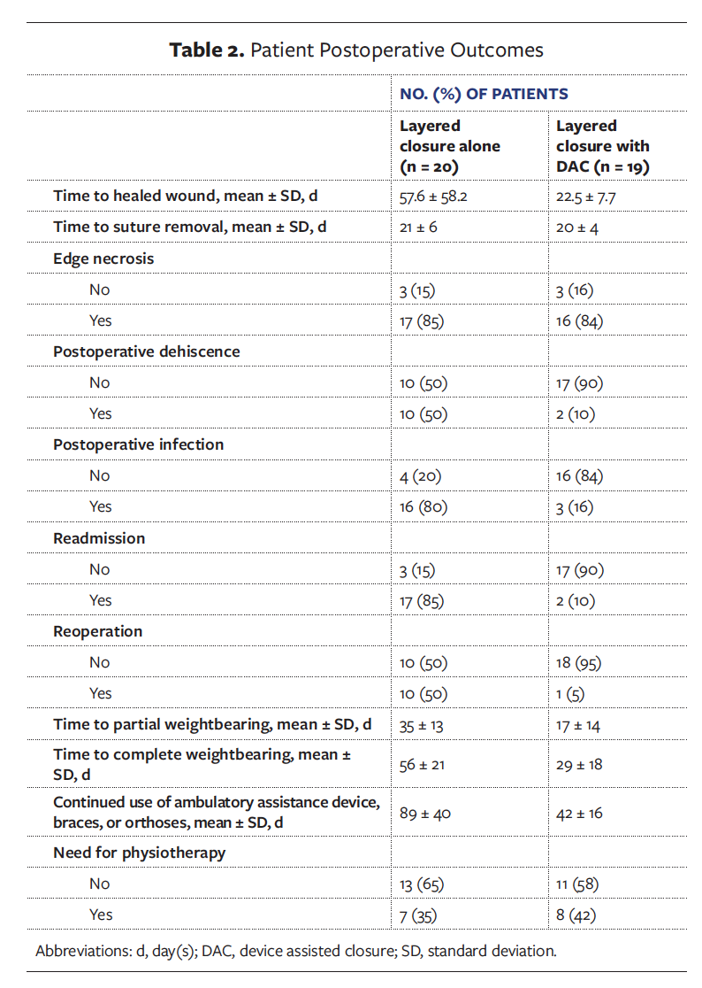
Thirty-nine patients underwent ankle fracture ORIF. Twenty (51%) patients underwent layered closure alone, and 19 (49%) patients underwent layered closure with DAC use. The groups were treated subsequently. The average patient age for the control group was 54.2 ± 15.7 years (range, 24-77). The average patient age for the DAC group was 46.1 ± 14.1 years (range, 21-77). The average body mass index for the control group was 30.4 ± 5.1 kg/m2 (range, 20.5–40.7). The average body mass index for the DAC group was 32.5 ± 4.7 kg/m2 (range, 23.7–39.5). Patients with diabetes accounted for 15% of patients in the layered closure alone group and 16% of patients in the layered closure supplemented with DAC group. Active tobacco users accounted for 35% of patients in the control group and 26% in the DAC group. The left leg was affected in 40% of patients in the control group and 26% of patients in the DAC group. Ankle fracture injury types are listed in Table 1. Delta frame application was required in 25% of patients in the control group and 5% of patients in the DAC group, as more trimalleolar fractures were present in the control group. The average time from initial delta frame application to ORIF was 14 days. Patient demographics for each group are listed in Table 1. Time to a healed incision was reduced in patients treated with layered closure and DAC compared to those who had layered closure alone (22.5 ± 7.7 days vs 57.6 ± 58.2 days) (Table 2). In regards to the control and the DAC group, the incidence of wound dehiscence (50% vs 10%, respectively) and need for reoperation (50% and 5%, respectively) was also reduced in patients where the DAC was applied compared to those who received layered closure alone. All reoperations performed were for the sake of infection management. Additional procedures performed included incision and drainages; bone biopsies and hardware removal were performed if concern for osteomyelitis was clinically or radiographically noted. There was a delay in time to partial weightbearing for 17 (85%) patients in the control group compared to 10 (58.8%) patients in the DAC group. Patients with DAC use also had a significantly shorter time requirement for use of ambulatory assistive devices, braces, or orthoses postoperatively compared to patients in which layered closure alone was performed.
Discussion
In this study, use of the DAC following ankle fracture ORIF resulted in a reduced rate of postoperative dehiscence; need for reoperation; and time of need for ambulatory assistive device, brace, or orthoses use compared to layered closure alone. Historically reported rates of surgical incision dehiscence following ankle fracture ORIF range from 0% to 25%.8,9,23-26 Several patient- and procedural-related factors also increase the risk of postoperative complications. Patient-related factors include the mechanism of injury, fracture type, condition of the skin envelope, age over 50 years, the presence of IDDM, tobacco use, illicit drug use, and congestive heart failure.9,27-32 Procedural-related factors include surgeon experience and length of procedure.26,27,31,33 An 11-year review of the American College of Surgeons National Surgical Quality Improvement Program database found that every 15-minute increase in operative time was associated with a 20% increased risk of wound dehiscence.33
Variations in surgical technique, suturing techniques in deep and superficial skin closure, and use of various closure assistive devices have been employed in attempts to mitigate healing complications following ankle ORIF.6-18 Use of a lateral approach with maintenance of full-thickness skin flaps for closure has conflicting reports on its ability to reduce incisional complication rates.6,7 While a lower rate of dehiscence has been reported with minimally invasive surgical techniques,8,9 these techniques are not always possible, particularly when anatomic reduction cannot be obtained. Use of buried absorbable sutures for deep closure is thought to provide additional support to the tissue and assist in preventing dehiscence,4,5 but evidence to support this belief is lacking. An animal study noted no additional strength at the incision site 10 and 42 days following the use of buried absorbable sutures for layered closure.4 Use of buried sutures is also associated with a higher risk of surgical site infections and incisional pain.3,5,34 The use of pulley and modified pulley suturing methods for skin closure have been shown to require the least amount of pulling force for incisional closure compared to simple interrupted, vertical mattress, horizontal mattress, and double butterfly suturing techniques.11,18
This reduction in the pulling force, however, does not translate to the ability of the closed incision to withstand strain and tension forces that can be applied in the postoperative recovery period, which is of particular importance when early mobilization following ankle fracture ORIF is desired. The patient must also have repeated visits for progressive tightening to be performed.12-14 Disadvantages of this suturing technique can include tissue strangulation, erosion, tearing, and wound edge necrosis, particularly in incision sites greater than 2.0 cm2.18,22,27 Prolonged retention of superficial skin closure has also been used to help combat postoperative dehiscence, but may have a contraindicatory effect on reduced time to healing.15 Use of zip-type skin-closing devices has been demonstrated to reduce time to wound closure and surgical skin infection.16,17 This has been theorized to be due to use of staples instead of sutures, minimizing puncturing of the skin, and the ability to evenly distribute closing forces along the incision site. However, a recent systematic review and meta-analysis17 found no difference in the incidence of wound dehiscence with use of the zip-type skin-closing device compared to layered closure alone. As noted by Xie et al, “The zip-type skin-closing device has the potential for adverse reactions, including skin peeling, skin discoloration, epidermolysis, blisters, allergic reaction to the adhesive tape area, pruritus in patient[s] with dry skin, and exfoliation of the device.”17 The disadvantages of these various suturing techniques and devices for assisted closure, as well as continued postoperative healing complications, lend to the need for a device that can assist in incisional closure for efficient healing while minimizing additional trauma to the skin.
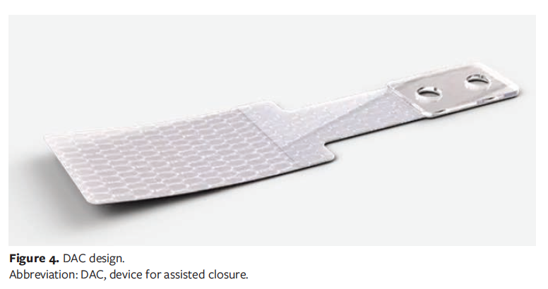
The DAC used in this study was designed to exploit this load-dependent skin behavior to reduce tension and allow for primary closure of soft tissue defects in areas of limited laxity or in patients at risk for postoperative dehiscence. The DAC consists of 3 zones: Zone A, a water-resistant, rigid, elevated zone with 2 holes to accept and withstand forces of various suture types, including high-tension, percutaneously or subcutaneously placed, absorbable, simple, interrupted, or vertical; Zone B, a water-resistant, less rigid, non-elevated zone placed to the side of the wound that transmits the force of closure away from the wound edge; and Zone C, the least rigid zone, composed of a monolayer of stretchy, nonwoven polyester to reduce shear forces applied to high-tension wound closures (Figure 4).21,22 Elevation of Zone A keeps sutures for skin closure elevated above the skin, which helps to prevent associated erosion or pressure injury.21,22 Use of a DAC has been shown to increase perfusion at the incision site by 25%, thereby reducing the potential of local ischemia and subsequent necrosis.35 Improved perfusion along the incision line is theorized to be due to the reduced force needed to maintain closure at the individual suture sites. The device can also be left in place for several weeks postoperative to provide continued support against strain and tension forces at the incision site, as was noted in this study.22
Limitations
Limitations of this study include the retrospective cohort design, more severe injury sustained in the patients who had layered closure alone, the small sample size, and lack of cost analysis related to use of the DAC. The rate of postoperative complications observed in the patients that underwent layered closure alone is what prompted trial of the DAC as closure supplementation to determine if its use would reduce these complications. Due to the low prevalence of ankle fractures annually in the United States (approximately 134 642, based on an estimated 5-year total of 673 214),36 no limitations were placed on patient inclusion. This resulted in the inclusion of various ankle fracture patterns, open and closed fractures, and lack of matching between groups. Matching between groups was not possible due to the low number of included patients seen overall prior to and following implementation of the DAC. The incidence of postoperative complications was reduced with use of the DAC, with no device-related complications occurring, including adhesive-related complications. These results are similar to other reports of use of the same DAC device in patients at high-risk for dehiscence, including elderly patients with fragile skin and patients with diabetes undergoing extensive surgical reconstruction or partial amputation of the foot.22,37,38 A retrospective chart review comparing 6 months preceding and following the addition of the DAC for primary closure in wound excisions on the leg found faster healing rates, significantly less postoperative dehiscence, and a trend towards lower infection rates.38 As the focus of the current study was to determine if a reduced rate of postoperative complications related to healing occurred with use of the DAC in ankle ORIF surgery, a cost analysis was not performed. Future studies with a greater number of patients, groups matched for fracture type and patient comorbidities, use of a blinded adjudicator for healing assessment, and cost analysis with use of the DAC compared to standard closure or other assisted closure techniques and modalities are recommended.
Conclusion
In conclusion, use of the DAC in patients who underwent ankle fracture ORIF in this study reduced rates of postoperative dehiscence, need for reoperation, and duration of patient need for ambulatory assistive device, brace, or orthoses use compared to layered closure alone. These reduced postoperative complication rates were seen in patients with multiple known risk factors, including extensive soft tissue injury, increased age, presence of diabetes, and tobacco use. Reduced wound dehiscence and infection rates allowed for earlier mobilization and return to normal activities. Results of this study suggest that use of the DAC should be considered in patients with known risk factors for wound dehiscence following ankle fracture ORIF. Further studies are needed to determine if these findings are maintained and if they translate to use in other surgical procedures for acute wound closure.
Acknowledgments
Authors: Emily Stefanski, DPM1; Arun Karwal, DPM2; and Erik Haniuk, DPM2
Affiliations: 1Foot and Ankle Surgery, Kent Hospital, Warwick, RI; 2Landmark Medical Center, Woonsocket, RI
ORCID: Stefanski, 0000-0002-9771-5429
Disclosure: A.K. is a consultant for SUTUREGARD and stock owner of the study product. E.H. is a consultant for SUTUREGARD.
Correspondence: Emily Stefanski, DPM; Kent Hospital, Foot and Ankle Surgery, 115 Cass Avenue, Warwick, RI 02895; emilybstefanski@gmail.com
References
1. Sun Y, Wang H, Tang Y, et al. Incidence and risk factors for surgical site infection after open reduction and internal fixation of ankle fracture: a retrospective multicenter study. Medicine (Baltimore). 2018;97(7):e9901. doi:10.1097/MD.0000000000009901
2. Akgun U, Canbek U, Kilinc CY, Acan AE, Karalezli N, Aydogan NH. Efficacy of pie-crusting technique on soft tissues in distal tibia and fibula fractures. J Foot Ankle Surg. 2019;58(3):497-501. doi:10.1053/j.jfas.2018.09.027
3. Joo JS, Zhuang AR, Tchanque-Fossuo C, et al. Dermal suture only versus layered closure: a randomized, split wound comparative effectiveness trial. J Am Acad Dermatol. 2019;81(6):1346-1352. doi:10.1016/j.jaad.2019.08.040
4. Townsend KL, Lear W, Robertson BL, Kruzic JJ. Buried absorbable polyglactin 910 sutures do not result in stronger wounds in porcine full thickness skin incisions. J Mech Behav Biomed Mater. 2016;63:386-389. doi:10.1016/j.jmbbm.2016.06.033
5. Anderson ER, Gates S. Techniques and materials for closure of the abdominal wall in caesarean section. Cochrane Database Syst Rev. 2004;2004(4):CD004663. doi:10.1002/14651858.CD004663.pub2
6. Grose A, Gardner MJ, Hettrich C, et al. Open reduction and internal fixation of tibial pilon fractures using a lateral approach. J Orthop Trauma. 2007;21(8):530-537. doi:10.1097/BOT.0b013e318145a227
7. Hu C, Zhu W, Chahal K, et al. Open reduction and internal fixation of Gustilo type-I and type-II open pilon fractures using a lateral approach. J Orthop Surg (Hong Kong). 2019;27(3):2309499019864722. doi:10.1177/2309499019864722
8. Abdelgaid SM, Moursy AF, Elgebaly EAA, Aboelenien AM. Minimally invasive treatment of ankle fractures in patients at high risk of soft tissue wound healing complications. J Foot Ankle Surg. 2018;57(3):557-571. doi:10.1053/j.jfas.2017.11.041
9. Bazarov I, Kim J, Richey JM, Dickinson JD, Hamilton GA. Minimally invasive plate osteosynthesis for treatment of ankle fractures in high-risk patients. J Foot Ankle Surg. 2018;57(3):494-500. doi:10.1053/j.jfas.2017.11.004
10. Austin BR, Henderson RA. Buried tension sutures: force-tension comparisons of pulley, double butterfly, mattress, and simple interrupted suture patterns. Vet Surg. 2006;35(1):43-48. doi:10.1111/j.1532-950X.2005.00110.x
11. Marsidi N, Vermeulen SAM, Horeman T, Genders RE. Measuring forces in suture techniques for wound closure. J Surg Res. 2020;255:135-143. doi:10.1016/j.jss.2020.05.033
12. Malone CH, McLaughlin JM, Ross LS, Phillips LG, Wagner RF Jr. Progressive tightening of pulley sutures for primary repair of large scalp wounds. Plast Reconstr Surg Glob Open. 2017;5(12):e1592. doi:10.1097/GOX.0000000000001592
13. Croley JA, Malone CH, Hirshburg JM, Wagner RF Jr. Reconstruction of medium to large scalp defects by progressive tightening of pulley sutures for staged primary closure. Dermatol Surg. 2020;46(6):837-839. doi:10.1097/DSS.0000000000001946
14. Saeed S, Ahmed SK, Chinoy MA, Khan MA. The pulley suture: a reliable option for closure of selected soft tissue defects under tension- three years experience of a tertiary care hospital. J Pak Med Assoc. 2015;65(11 Suppl 3):S35-S39.
15. Townsend KL, Akeroyd J, Russell DS, Kruzic JJ, Robertson BL, Lear W. Comparing the tolerability of a novel wound closure device using a porcine wound model. Adv Wound Care (New Rochelle). 2018;7(6):177-184. doi:10.1089/wound.2017.0777
16. Ko JH, Yang IH, Ko MS, Kamolhuja E, Park KK. Do zip-type skin-closing devices show better wound status compared to conventional staple devices in total knee arthroplasty? Int Wound J. 2017;14(1):250-254. doi:10.1111/iwj.12596
17. Xie CX, Yu CQ, Wang W, Wang CL, Yin D. A novel zipper device versus sutures for wound closure after surgery: a systematic review and meta-analysis. Int Wound J. 2020;17(6):1725-1737. doi:10.1111/iwj.13460
18. Ferrell K, Fancher W, Lear W. Use of a novel suture retention device to protect skin edges while using pulley suture technique for high-tension wound closure. J Am Acad Dermatol. 2020;83(2):e113-e115. doi:10.1016/j.jaad.2019.04.030
19. Stoecker A, Howerter S, Fancher W, Lear W. A novel suture retention device for intraoperative tissue support. JAAD Case Rep. 2019;5(5):454-457. doi:10.1016/j.jdcr.2019.03.018
20. Roybal LL, Howerter S, Markus B, Young J, Lear W. Use of a novel adhesive suture retention wound closure device to prevent patient follow-up visits during the COVID-19 pandemic. JAAD Case Rep. 2020;6(7):593-597. doi:10.1016/j.jdcr.2020.04.032
21. Roybal LL, Blattner CM, Young J, Lear W. A novel adhesive suture retention device for the closure of fragile skin under tension. JAAD Case Rep. 2020;6(2):109-114. doi:10.1016/j.jdcr.2019.12.010
22. Blattner CM, Perry B, Young J, Lear W. The use of a suture retention device to enhance tissue expansion and healing in the repair of scalp and lower leg wounds. JAAD Case Rep. 2018;4(7):655-661. doi:10.1016/j.jdcr.2018.06.004
23. Lehtonen E, Patel H, Phillips S, Correia Pinto M, Naranje S, Shah A. Staple versus suture closure for ankle fracture fixation: retrospective chart review for safety and outcomes. Foot (Edinb). 2018;37:71-76. doi:10.1016/j.foot.2018.08.003
24. Macera A, Carulli C, Sirleo L, Innocenti M. Postoperative complications and reoperation rates following open reduction and internal fixation of ankle fracture. Joints. 2018;6(2):110-115. doi:10.1055/s-0038-1653949
25. Yasui Y, Shimozono Y, Hung CW, et al. Postoperative reoperations and complications in 32,307 ankle fractures with and without concurrent ankle arthroscopic procedures in a 5-year period based on a large U.S. healthcare database. J Foot Ankle Surg. 2019;58(1):6-9. doi:10.1053/j.jfas.2018.03.030
26. White TO, Guy P, Cooke CJ, et al. The results of early primary open reduction and internal fixation for treatment of OTA 43.C-type tibial pilon fractures: a cohort study. J Orthop Trauma. 2010;24(12):757-763. doi:10.1097/BOT.0b013e3181d04bc0
27. Stoecker A, Howerter S, Young J, Lear W. The use of a suture retention device with punctureless technique for rapid tissue expansion in facial and lower extremity wounds. JAAD Case Rep. 2018;4(9):910-914. doi:10.1016/j.jdcr.2018.09.005
28. McKissack HM, Viner GC, Jha AJ, et al. Comparison of risk factors for postoperative complications across age groups in patients undergoing ORIF of the ankle. Injury. 2019;50(11):2116-2122. doi:10.1016/j.injury.2019.09.014
29. Liu JW, Ahn J, Nakonezny PA, et al. Insulin dependence increases the risk of 30-day postoperative complications following ankle fracture surgery in patients with diabetes mellitus. J Foot Ankle Surg. 2021;60(5):917-922. doi:10.1053/j.jfas.2021.03.011
30. Spek RWA, Smeeing DPJ, van den Heuvel L, et al. Complications after surgical treatment of geriatric ankle fractures. J Foot Ankle Surg. 2021;60(4):712-717. doi:10.1053/j.jfas.2019.12.012
31. Cole W. Wound care update: surgical site dehiscence in the foot: risk factors and prevention. March 2021. Accessed February 1, 2023. https://lermagazine.com/article/wound-care-update-surgical-site-dehiscence-in-the-foot-risk-factors-and-prevention
32. Lynde MJ, Sautter T, Hamilton GA, Schuberth JM. Complications after open reduction and internal fixation of ankle fractures in the elderly. Foot Ankle Surg. 2012;18(2):103-107. doi:10.1016/j.fas.2011.03.010
33. Gowd AK, Bohl DD, Hamid KS, Lee S, Holmes GB, Lin J. Longer operative time is independently associated with surgical site infection and wound dehiscence following open reduction and internal fixation of the ankle. Foot Ankle Spec. 2020;13(2):104-111. doi:10.1177/1938640019835299
34. Huang YH, Chen C, Lee CH, Loh EW, Tam KW. Wound closure after thyroid and parathyroid surgery: a meta-analysis of randomized controlled trials. Scand J Surg. 2019;108(2):101-108. doi:10.1177/1457496918798203
35. Stoecker A, Lear W, Johnson K, Bahm J, Kruzic JJ. Enhanced perfusion of elliptical wound closures using a novel adhesive suture retention device. Health Sci Rep. 2021;4(3):e364. doi:10.1002/hsr2.364
36. Scheer RC, Newman JM, Zhou JJ, et al. Ankle fracture epidemiology in the United States: patient-related trends and mechanisms of injury. J Foot Ankle Surg. 2020;59(3):479-483. doi:10.1053/j.jfas.2019.09.016
37. Cole W, McKanna A. The use of a novel suture retention device to prevent surgical wound dehiscence (SWD). April/May 2021. Accessed February 1, 2023. https://podiatrym.com/pdf/2021/4/Cole421Web.pdf#:~:text=The%20Use%20of%20a%20Novel%20Suture%20Retention%20Device,BY%20WINDY%20COLE%2C%20DPM%20AND%20ALEX%20MCKANNA%2C%20DPM
38. Cole W, Fancher W, Stoecker A, Howerter S, Lear W. Use of a novel adhesive suture retention device in lower leg excisional wound closure: a retrospective review. Wounds. 2021;33(9):222–225. doi:10.25270/wnds/051421.01


