Using Imaging to Improve Stent Visualization in Complex Bifurcation Disease
© 2024 HMP Global. All Rights Reserved.
Any views and opinions expressed are those of the author(s) and/or participants and do not necessarily reflect the views, policy, or position of Cath Lab Digest or HMP Global, their employees, and affiliates.
Images/courtesy Heart Center of Nevada
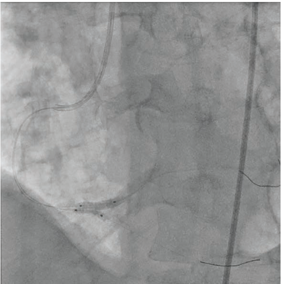
A 53-year-old male with chest pain and shortness of breath was diagnosed with complex bifurcation disease. Coronary angiography revealed a 99% bifurcation stenosis of the distal right coronary artery (RCA), with significant disease extending into the right posterior descending artery (RPDA) and right posterolateral artery (RPL) (Figure 1). Intravascular ultrasound (IVUS) revealed a 4.0 mm distal RCA and RPL, and a 3.0 mm proximal RPDA. Using the double kissing crush (DK crush) technique, .014-inch coronary wires were placed in both vessels. A 3.0 mm x 15 mm drug- eluting stent was placed in the RPDA, with 1-2 mm protrusion into the distal RCA.
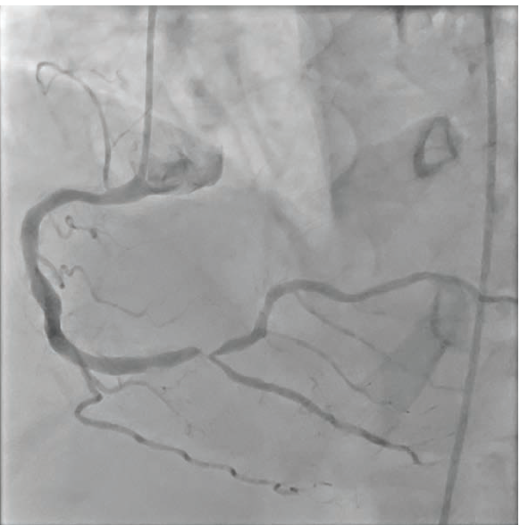
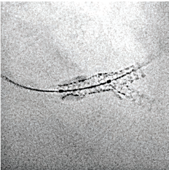
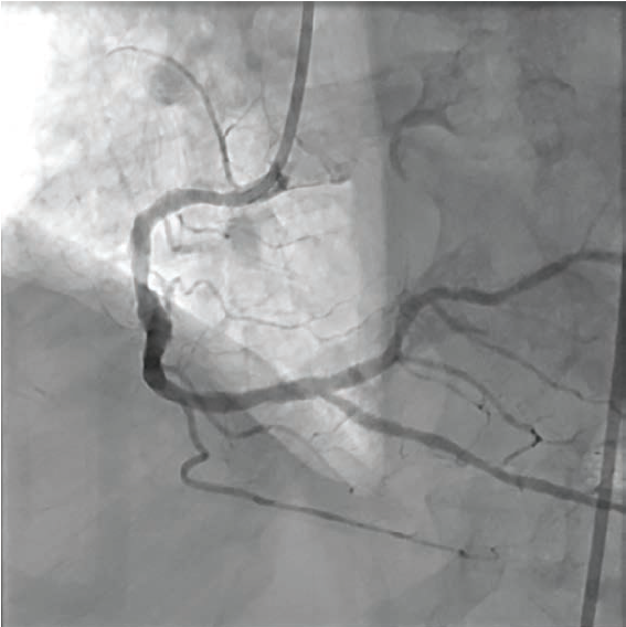
A balloon was inflated in the distal RCA, crushing the stent’s proximal portion. Following balloon inflation, a 4.0 mm x 23 mm drug-eluting stent was deployed in the distal RCA extending into the RPL branch, followed by a second inflation (Figure 2). Stent visualization (Artis icono floor CLEARstent technology from Siemens Healthineers) clearly articulated the stent borders (Figure 3). Proximal optimization of the distal RCA stent was performed with a 4.0 noncompliant (NC) balloon. The final angiogram showed good angiographic result, with thrombolysis in myocardial infarction (TIMI)-3 flow in all vessels (Figure 4).