

Virtual Histology Optical Coherence Tomography Imaging of Orbital Rotational Atherectomy for Calcified Peripheral Arterial Disease
Key words: rotational atherectomy, virtual histology intravascular ultrasound optical coherence tomography, VH-IVOCT
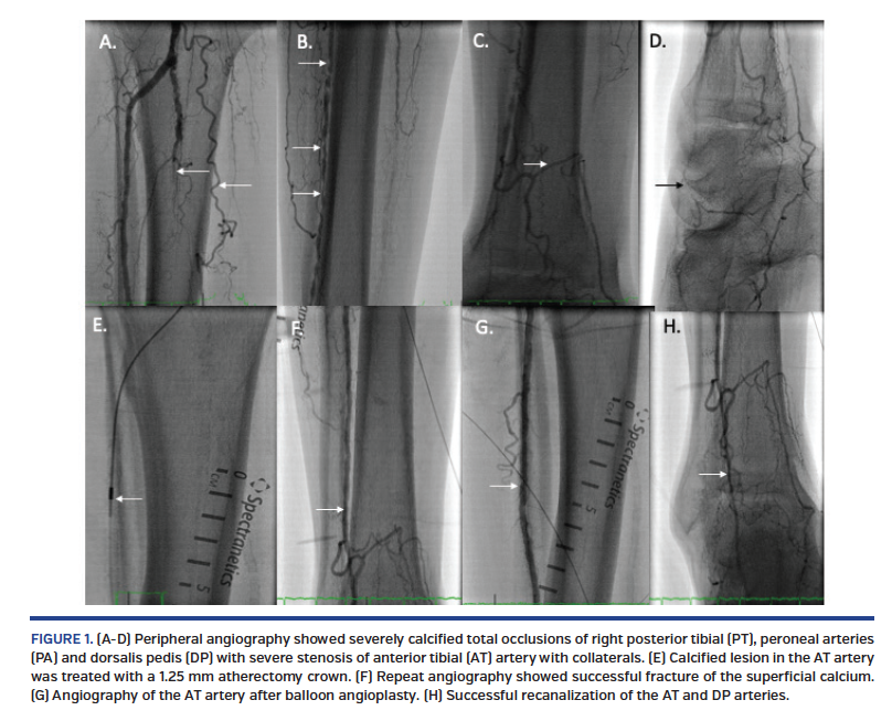
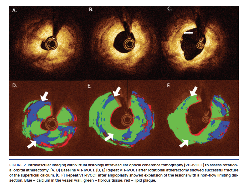
A 72-year-old man with critical limb ischemia of the right lower extremity was referred for revascularization. His peripheral angiography showed severely calcified total occlusions of the right posterior tibial artery, peroneal arteries, and dorsalis pedis, with severe stenosis of the anterior tibial artery with collaterals (Figures 1A-1D). In order to assess the vascular effects of rotational orbital atherectomy, we performed intravascular imaging with virtual histology intravascular optical coherence tomography (VH-IVOCT) (Figures 2A-2F).
We performed rotational orbital atherectomy of the calcified lesions in the anterior tibial artery using a 1.25 mm solid crown. Repeat angiography (Figure 1F) and VH-IVOCT (Figures 2B and 2E) showed successful fracture of the superficial calcium. Percutaneous transluminal angioplasty (PTA) was performed in the anterior tibial artery using a 3.5 mm balloon and in the dorsalis pedis artery using a 2 mm balloon. Final angiography showed successful recanalization of anterior tibial and dorsalis pedis arteries (Figures 1F and 1H). VH-IVOCT showed successful fracture of the calcium, leading to expansion of the lesions with a non-flow limiting dissection (Figures 2C and 2F). Blue color shows calcium in the vessel wall, green color, fibrous tissue and red color for lipid plaque.
From the 1Bahcesehir University, School of Medicine, Istanbul, Turkey; 2UTHSCSA, San Antonio, Texas; and 3UT Austin School of Biomedical Engineering, Austin, Texas and University of California Irvine, School of Medicine, Irvine, California.
Disclosure: The authors have completed and returned the ICMJE Form for Disclosure of Potential Conflicts of Interest. The authors report no conflicts of interest regarding the content herein.
The authors report that patient consent was provided for publication of the images used herein.
Manuscript accepted January 29, 2020.
Address for correspondence: Mehmet Cilingiroglu, MD, FSCAI, FACC, FESC, FAHA, Professor of Medicine and Biomedical Engineering, Bahcesehir University, School of Medicine, Yıldız, Çırağan Cd., 34349 Beşiktaş/Istanbul, Turkey. Email: cilingiroglumehmet@gmail.com


